Glaucoma Surgery
Glaucoma surgery aims to lower intraocular pressure and protect vision when eye drops or laser treatment are not enough. Techniques may include trabeculectomy, drainage implants, or minimally invasive procedures.
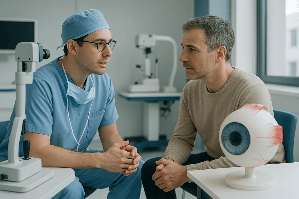
Quick answer
Glaucoma surgery lowers the pressure inside the eye when drops or laser treatment no longer control it. Options include trabeculectomy, drainage implants (tube shunts) and minimally invasive glaucoma surgery (MIGS). Surgery cannot restore vision already lost to glaucoma; its purpose is to reduce stress on the optic nerve and slow or prevent further damage. The right procedure depends on glaucoma type, stage and eye anatomy.
Glaucoma Treatment: Where Surgery Fits In
Glaucoma surgery is a group of operations that lower the pressure inside the eye when drops, tablets or laser treatment are no longer enough to protect the optic nerve. It cannot bring back vision that glaucoma has already taken. Its purpose is to reduce intraocular pressure, ease the ongoing stress on the optic nerve, and slow or prevent further damage to your visual field.
Glaucoma is often called a silent disease because many people notice nothing until the optic nerve has already been affected. If you have been told your eye pressure remains too high despite medication or laser treatment, the suggestion of surgery can feel unsettling. You may be asking how serious your condition really is, whether an operation can preserve the vision you still have, what recovery involves, and how to weigh one procedure against another. This page works through those questions in order.
Glaucoma treatment has one proven, modifiable target: intraocular pressure. Everything in modern care, drops, laser, surgery, exists to bring pressure down to a level your optic nerve can tolerate. Surgery is one part of glaucoma treatment, not a replacement for it. Because glaucoma is a lifelong condition, an operation is best understood as a step within a continuing care plan rather than a one-time fix. Monitoring continues afterwards, and some patients still need medication.
A brief note on spelling: you will sometimes see the condition written as glucoma online. The correct spelling is glaucoma, and both searches lead to the same disease. Whichever way you found this page, the information below applies.
There is no single operation that suits every eye. The right glaucoma treatment depends on the type and severity of disease, the anatomy of the eye, previous treatments, current medications, your general health and your visual needs. At experienced ophthalmology centres, the decision follows detailed diagnostic testing and a frank discussion of expected benefits, limitations, risks and follow-up requirements.
What Is Glaucoma Surgery?
Glaucoma surgery covers several procedures that either improve the outflow of aqueous humour, the clear fluid the eye constantly produces and drains, or reduce how much of that fluid the eye makes. When fluid leaves the eye more easily, or less is produced, intraocular pressure falls. Lower pressure is the only intervention proven to protect the optic nerve in glaucoma, which is why every surgical option ultimately serves that single goal.
What types of glaucoma surgery are there?
There are four broad categories of glaucoma surgery, and each occupies a different position on the spectrum between pressure-lowering power and risk.
- Trabeculectomya filtering operation that creates a new drainage pathway out of the eye. It offers substantial pressure lowering and is often chosen for more advanced disease or eyes that need a particularly low target pressure.
- Glaucoma drainage implants (tube shunts)a small device guides fluid from inside the eye to a plate positioned under the conjunctiva. Often used in complex or previously operated eyes.
- Minimally invasive glaucoma surgery (MIGS)microscopic devices or tissue-sparing techniques that enhance the eye’s natural drainage. Gentler, quicker to recover from, but typically less powerful than filtering surgery.
- Laser and cyclodestructive procedureslaser applied to the drainage tissues to improve outflow, or to the fluid-producing tissue (the ciliary body) to reduce production.
Combined cataract and glaucoma surgery is a further option for patients who have both conditions, addressing lens clouding and pressure control in a single planned operation. A patient with early glaucoma and a visually significant cataract may need an entirely different plan from a patient with advanced glaucoma, previous eye surgery, or rapidly progressing optic nerve damage. The choice is always individual.
What is trabeculectomy?
Trabeculectomy is the longest-established filtering operation for glaucoma. The surgeon creates a controlled opening in the wall of the eye so that aqueous fluid can drain from the inside to a small reservoir under the conjunctiva: the thin, transparent tissue covering the white of the eye. This reservoir, called a bleb, usually sits discreetly under the upper eyelid, where it slowly absorbs fluid into the surrounding tissue.
Because the body naturally tries to heal any new opening, medications that moderate scarring may be applied during or after the operation; healing that is too aggressive can close the new pathway and undo the pressure-lowering effect. The surgeon closes the tissues with very fine sutures, balancing adequate drainage against the risk of pressure dropping too low. That balancing act continues into the postoperative weeks, which is why trabeculectomy demands more frequent follow-up than most eye operations, sometimes including suture adjustment or minor office-based interventions to guide healing.
What is MIGS surgery for glaucoma?
MIGS stands for minimally invasive glaucoma surgery: a family of procedures performed through very small incisions, using microscopic instruments and, in many cases, tiny implanted devices to improve the eye’s own drainage pathways. Depending on the technique, the procedure may bypass the trabecular meshwork, remove a small strip of drainage tissue, widen a natural drainage canal, or place a micro-stent that keeps fluid moving outward.
MIGS procedures are generally associated with shorter recovery and a lower risk profile than trabeculectomy or tube surgery. The trade-off is honesty about power: MIGS may not lower pressure enough for every patient, particularly in advanced disease or where a very low target pressure is needed. It is most commonly considered in mild to moderate glaucoma, and frequently combined with cataract surgery when both conditions are present: the surgeon is already working inside the eye, and adding a MIGS step extends the operation only slightly.
Glaucoma drainage implants (tube shunt surgery)
Glaucoma drainage implant surgery places a small tube into the front chamber of the eye, or another suitable location, connected to a plate fixed on the outer surface of the eye beneath the conjunctiva. Fluid travels along the tube to the plate, where surrounding tissue absorbs it. Some implants are designed to restrict flow in the early weeks, until healing forms a capsule around the plate; this staged approach reduces the risk of pressure falling too low immediately after surgery.
Tube shunts are often recommended for patients with complex glaucoma: scarring from previous surgery, certain secondary glaucomas such as neovascular or uveitic disease, or eyes in which trabeculectomy is judged less likely to succeed. They are a workhorse of difficult glaucoma, chosen precisely when the more conventional routes have been exhausted or are unsuitable.
Laser and cyclodestructive procedures
Laser-based options sit alongside incisional surgery. Some laser treatments are applied to the drainage tissues to improve outflow and are often tried before an operation is considered. Cyclodestructive procedures take the opposite approach: rather than improving drainage, they apply controlled laser energy to the ciliary body, the tissue that produces aqueous fluid, reducing how much fluid the eye makes. These procedures are typically reserved for eyes where other surgery has failed or is not feasible, although gentler, more targeted versions are increasingly used earlier in the disease course. Your ophthalmologist will explain where laser fits in your particular plan.
Who May Need Glaucoma Surgery?
Glaucoma surgery is considered when intraocular pressure stays above target despite drops, laser treatment or other medical management. It may also be recommended when a patient cannot tolerate their medications, cannot use drops reliably, shows progressive visual field loss, or needs a degree of pressure reduction that non-surgical methods are unlikely to reach.
Many people with glaucoma have no symptoms at first. As the disease advances, they may notice reduced peripheral vision, difficulty seeing in dim light, bumping into objects, trouble driving, or a sense that parts of the visual field are simply missing. Acute angle-closure glaucoma behaves differently: pressure can rise suddenly, producing eye pain, redness, blurred vision, halos around lights, headache, nausea and vomiting. Ophthalmology treats this form as an emergency because pressure can climb quickly enough to damage the nerve within hours to days.
Patients referred for surgery usually arrive there after months or years of monitoring. Some have open-angle glaucoma that is slowly worsening. Others have narrow-angle or angle-closure disease, pseudoexfoliative or pigmentary glaucoma, neovascular or uveitic glaucoma, congenital glaucoma, or pressure problems after trauma or previous eye surgery. In each case, the surgical plan is adapted to the underlying mechanism of pressure elevation and the condition of the individual eye.
What is the major cause of glaucoma?
The major cause of glaucoma is impaired drainage of aqueous fluid, which raises pressure inside the eye and gradually damages the optic nerve. In the most common form, primary open-angle glaucoma, the trabecular meshwork: the eye’s natural drain, becomes less efficient over time, often without any identifiable trigger. Recognised risk factors include older age, family history, certain ethnic backgrounds, high short-sightedness or long-sightedness, previous eye injury, long-term steroid use and some systemic conditions such as diabetes.
It is worth knowing that pressure is not the whole story. In normal-tension glaucoma, the optic nerve deteriorates even though pressure readings sit within the statistically normal range, which suggests that nerve vulnerability and blood supply also play a role. Even in these patients, however, lowering pressure remains the treatment with proven benefit.
What are the 5 stages of glaucoma?
Glaucoma is commonly described in five stages, based on how much the optic nerve and visual field have been affected. First is the glaucoma suspect stage, where pressure is raised or the nerve looks suspicious but no definite damage is measurable. Second is mild or early glaucoma, with early structural nerve changes and minimal visual field defects. Third is moderate glaucoma, where field loss is measurable and may start intruding on daily awareness. Fourth is advanced or severe glaucoma, with substantial field loss, often approaching central vision. Fifth is end-stage glaucoma, where only a small island of vision, or no useful vision, remains.
Staging matters because it drives the target pressure and the choice of procedure. A suspect may need only monitoring; a patient with advanced disease may need filtering surgery to reach a low target quickly. Because staging depends on repeated measurements over time, a single visit rarely tells the whole story.
What does a glaucoma eye test involve?
A glaucoma eye test is not one test but a set of examinations built up into a complete picture. Your ophthalmologist measures intraocular pressure, examines the optic nerve under magnification, assesses the drainage angle with a technique called gonioscopy, measures corneal thickness (which influences how pressure readings should be interpreted), and images the retina and optic nerve fibre layer with high-resolution scanning. Visual field testing maps areas of functional vision loss, the practical consequence of nerve damage, while structural imaging can detect change before you notice anything yourself.
These tests are repeated at intervals, because glaucoma management depends less on any single pressure reading than on whether the disease is stable or progressing. Two patients with the same pressure can need entirely different treatment if one is stable and the other is losing field year on year. If further tests are recommended beyond the standard set, it helps to understand why; our guide on how we explain additional tests before surgery or treatment describes the reasoning process patients can expect.
Conditions and Indications Treated With Glaucoma Surgery
The most common indication is primary open-angle glaucoma, a chronic condition in which drainage through the trabecular meshwork declines gradually. When medication or laser cannot hold pressure at target, surgery helps reduce the risk of further optic nerve injury.
Angle-closure glaucoma occurs when the drainage angle is narrow or blocked. Many patients are treated with laser first, but surgery may be needed if pressure remains high, if the angle has been extensively damaged, or if the natural lens is contributing to crowding inside the eye.
Secondary glaucomas arise from another eye condition or systemic problem: pseudoexfoliative glaucoma, pigmentary glaucoma, neovascular glaucoma linked to diabetes or retinal vascular disease, inflammatory glaucoma associated with uveitis, steroid-induced glaucoma, and glaucoma after trauma. These cases tend to be more complex, because inflammation, abnormal blood vessels, scarring or previous surgery influence both procedure choice and healing behaviour.
Normal-tension glaucoma may need surgical attention if nerve damage progresses despite apparently normal pressures. Here the target pressure may need to be particularly low, and the decision requires careful weighing of benefit against risk.
Paediatric and congenital glaucoma require specialised evaluation. Children may need angle surgery or other procedures designed for developing eyes, with long-term follow-up covering vision, eye growth, refractive change and pressure control throughout childhood.
Does cataract surgery fix narrow angle glaucoma?
Cataract surgery can improve narrow angle glaucoma in selected patients, but it does not fix every case. Removing the natural lens, which thickens with age and crowds the front of the eye, often opens the drainage angle and lowers pressure. For some patients with angle-closure disease, lens removal is genuinely the most effective single intervention, sometimes combined with a glaucoma procedure in the same operation.
The honest limits: cataract surgery does not reverse optic nerve damage that has already occurred, cannot reopen an angle destroyed by long-standing scarring, and does not guarantee that drops become unnecessary. Whether lens surgery alone is enough, or needs pairing with a glaucoma procedure, depends on gonioscopy findings, pressure history and the state of the nerve: a judgement your surgeon makes case by case.
Eye Drops for Glaucoma and Other Non-Surgical Treatment
Eye drops for glaucoma remain the most common first-line treatment worldwide, and understanding them puts surgery in context. Different drop classes work in different ways: some improve fluid outflow, others reduce fluid production, and many patients use more than one class to reach their target pressure. Laser treatments offer another non-surgical route and can reduce or delay the need for drops in suitable eyes.
Drops have real limitations, and these limitations are often what leads to a surgical conversation. Some patients experience irritation, redness, allergy or systemic side effects. Others struggle with the practical burden of instilling several drops on schedule every day for years, and missed doses translate directly into uncontrolled pressure. Still others use their medication faithfully and progress anyway, because the achievable pressure simply is not low enough for their nerve.
None of this means drops are abandoned after an operation. Many patients continue to need glaucoma eye drops after surgery, sometimes fewer than before, sometimes the same regimen while healing settles. Any change to your drops, adding, stopping or switching, is a decision for your treating ophthalmologist, made against measured pressures and nerve status, never something to adjust on your own.
How Glaucoma Surgery Is Performed
The surgical journey begins with confirming that an operation is genuinely the right next step. Before recommending a procedure, the ophthalmologist reviews your glaucoma history, pressure trends, visual field tests, optic nerve imaging, previous laser or surgical treatments, current medications, allergies and general medical conditions.
Preparation typically includes repeat pressure measurement, gonioscopy, optic nerve and retinal nerve fibre layer imaging, visual field testing, corneal thickness measurement and cataract evaluation. Your physician will explain which of your regular medications to continue and which to pause temporarily; patients taking blood thinners, diabetes medication, immune-suppressing drugs or steroids may need coordination with their other doctors. Infection-prevention measures, anti-inflammatory treatment or pressure adjustments may be prescribed beforehand. For patients who could be pregnant, standard safeguards such as pregnancy testing are completed before anaesthesia or imaging.
On the day itself, most glaucoma operations follow a common sequence:
- Step 1: Anaesthesia. Most procedures are performed under local anaesthesia with sedation; the exact approach depends on the procedure, your medical status, your preference, and whether cataract surgery is being combined. The anaesthetic is designed to control pain during the operation; patients may still sense light, touch or gentle pressure.
- Step 2: Preparation of the eye. The eye is numbed, cleaned meticulously and protected with sterile drapes.
- Step 3: The procedure itself. Depending on the plan: a trabeculectomy opening with a bleb, placement of a drainage tube and plate, a MIGS device or tissue technique through micro-incisions, or laser application, each as described in the sections above.
- Step 4: Closure and early control. Fine sutures, flow-restricting elements or intraoperative medication are used to keep early pressure within a safe range.
- Step 5: Monitoring. You are observed before going home. Some patients stay in hospital briefly, depending on the procedure, general health and the surgeon’s recommendation.
Modern glaucoma surgery leans heavily on precise diagnostic and microsurgical technology: high-resolution imaging of the nerve, macula and anterior chamber; visual field analysers that quantify functional loss; surgical microscopes, microsurgical instruments, controlled fluid systems and specialised implants or stents. The value of this technology is not just technical accuracy: it helps the surgeon choose the right procedure for the right eye, anticipate risk, and monitor healing afterwards. Operating time varies by technique and complexity; many procedures are completed within a short theatre session, while combined or complex cases take longer.
Recovery After Glaucoma Surgery
Recovery is an active period of care rather than passive waiting. Postoperative drops control inflammation and reduce infection risk. Pressure is checked closely, and the surgeon evaluates the drainage pathway, wound healing, cornea, anterior chamber and optic nerve. After trabeculectomy, follow-up may involve suture adjustment, massage instructions or minor office-based interventions to steer healing. After tube surgery, pressure shifts as the device begins to function fully. After MIGS, recovery is usually faster, but monitoring remains essential: the disease has not gone away.
| Time Period | What Patients Can Expect |
|---|---|
| Day 1 | The operated eye may feel scratchy, watery or mildly uncomfortable. Vision is often blurry. Your surgeon checks pressure, the incision and early healing. |
| First week | Postoperative drops are used on a strict schedule. Activity is limited to protect the eye. Visits may be frequent, especially after trabeculectomy or tube surgery. |
| First month | Inflammation settles gradually. Pressure may fluctuate as healing develops. Medication adjustments or minor office procedures may be needed to optimise drainage. |
| Longer term | Most patients return to normal routines, but glaucoma monitoring continues indefinitely: visual fields, nerve imaging and pressure checks remain part of care. |
How long does blurred vision last after glaucoma surgery?
Blurred vision after glaucoma surgery commonly lasts days to a few weeks, though the timeline varies by procedure and by individual healing. After MIGS, vision often clears relatively quickly. After trabeculectomy or tube surgery, blur can persist for several weeks, because inflammation, pressure changes, drops and shifts in the shape of the eye’s surface all affect focus while healing continues; your glasses prescription may also change temporarily. The follow-up schedule after surgery is designed to track exactly this: the surgical team watches whether vision is settling as expected and adjusts the plan if healing takes an unusual course.
What are the do’s and don’ts if I have glaucoma?
The single most important “do” is consistency: use your prescribed drops exactly as directed and attend every monitoring appointment, because glaucoma damages silently between visits. Other sensible habits during the surgical recovery period and beyond:
- Do protect the operated eye as instructed, shields at night, no rubbing.
- Do keep dust, soap and unclean water out of the eye while it heals.
- Do resume reading, screen use, walking and light daily activities gradually, following your surgeon’s timeline.
- Don’t swim, lift heavy weights or do strenuous exercise in the early healing period.
- Don’t adjust, skip or stop any medication on your own: that decision belongs to your treating doctor.
- Don’t assume stable vision means stable disease; peripheral loss is easy to miss without testing.
Why Acting Early Matters
Glaucoma-related vision loss happens because optic nerve fibres die, and current treatment cannot reliably bring them back. Modern glaucoma treatment therefore concentrates on preserving the vision that remains. If pressure stays too high for too long, or the nerve keeps deteriorating despite treatment, the risk of permanent visual field loss grows.
Delay is especially risky when glaucoma is progressing quickly, when field loss is already advanced, or when central vision is under threat. People adapt to peripheral loss without realising how much function has changed; by the time daily life becomes obviously difficult, the disease may be further along than expected. Early surgical evaluation does not always mean immediate surgery. It means understanding whether your current treatment is sufficient, whether your target pressure is right, and whether another approach could better protect your nerve. For some patients, surgery mainly reduces dependence on multiple medications; for others, it exists to reach a pressure that drops and laser cannot safely achieve.
How do you stop glaucoma from getting worse?
The only proven way to slow glaucoma is to keep intraocular pressure at or below the target your ophthalmologist has set for your nerve, through drops, laser, surgery or a combination, and to verify with regular testing that the target is actually working. That second part matters: a pressure that once seemed adequate may prove too high if fields keep deteriorating, in which case the target is lowered and treatment escalated. Beyond pressure control, general health measures such as managing diabetes and blood pressure, and avoiding unsupervised steroid use, support the overall picture. What does not work is waiting for symptoms; by design, effective glaucoma care acts before you notice anything.
Benefits of Glaucoma Surgery
The potential benefits depend on the type and stage of disease, but every benefit flows from the same source: lower pressure protecting remaining vision.
| Benefit | What It Means for You |
|---|---|
| Lower intraocular pressure | Reduced pressure decreases stress on the optic nerve, which may slow or prevent further glaucoma-related vision loss. |
| Protection of remaining vision | Surgery is intended to preserve functional vision, especially when glaucoma is progressing despite drops or laser treatment. |
| Reduced medication burden in selected patients | Some patients need fewer drops after surgery, improving comfort and adherence, though medication may still be required. |
| Individualised procedure choice | Different techniques let the surgeon match treatment to disease severity, eye anatomy, previous procedures and lifestyle. |
| Option to combine with cataract surgery | Where cataract and glaucoma coexist, combined surgery can address vision clouding and pressure control in one planned operation. |
What Influences a Good Outcome?
A good result in glaucoma surgery is not measured by the pressure number on a single day. It is measured by sustained pressure control, protection of the optic nerve, safe healing, preserved visual function and your ability to continue long-term monitoring. Several factors shape that result.
Disease stage. Eyes with early or moderate glaucoma carry more optic nerve reserve than eyes with advanced disease. In advanced glaucoma, even small pressure fluctuations may matter, and the target pressure often needs to be lower, which in turn influences which operation is chosen.
Type of glaucoma. Open-angle, angle-closure, inflammatory, neovascular and post-traumatic glaucoma behave differently. Some forms are more prone to scarring, bleeding, inflammation or pressure swings, which affects both procedure selection and how intensively follow-up must be scheduled.
Eye anatomy and previous surgery. Conjunctival scarring, prior cataract or retinal surgery, trauma, corneal disease or narrow angles can all change the approach. The presence of cataract matters too, since lens surgery influences pressure and may be combined with a glaucoma procedure.
Healing response. This is one of the most decisive variables. Filtering surgery depends on controlled healing: enough to close tissues safely, not so much that scarring shuts the drainage pathway. Postoperative visits exist precisely so that timely adjustments can keep the pathway working as intended.
Medication adherence. Using postoperative drops correctly and attending follow-up appointments are non-negotiable parts of recovery, and some patients continue pressure-lowering medication long term.
General health. Diabetes, autoimmune disease, vascular disease, sleep apnoea, steroid use and blood pressure patterns can all influence glaucoma progression and surgical recovery. Bringing complete medical records and a current medication list allows the team to plan safely.
Surgeon experience and technique selection. A procedure that suits one patient may be wrong for another. The best plan balances pressure-lowering power, risk profile, recovery time and the realistic likelihood of long-term control for your specific eye.
What is the success rate of glaucoma surgery?
There is no single success rate that honestly applies to all glaucoma surgery, because “success” itself is defined differently across procedures and studies, sometimes as reaching a target pressure, sometimes as pressure control without medication, sometimes as avoiding further field loss. Outcomes vary with the operation chosen, the type and stage of glaucoma, previous surgery, healing response and length of follow-up. Filtering procedures can also lose effect over years as tissues scar, which is why long-term monitoring continues even after a technically successful operation. The meaningful question is not a universal statistic but what your surgeon expects for your eye, with your diagnosis, using the specific technique proposed, and that is a conversation worth having in detail before you consent.
Long-Term Outlook After Glaucoma Surgery
A successful operation is the beginning of a new phase of care, not the end of it. In the early months, visits are frequent while the drainage pathway matures and pressure settles. As the eye stabilises, the schedule stretches out, but periodic pressure checks, visual field tests and optic nerve imaging remain part of life with glaucoma indefinitely. Comparison over years: this year’s field against last year’s, this scan against the baseline, is the heart of glaucoma monitoring, which is why keeping copies of previous test results and bringing them to appointments genuinely improves the quality of your care.
It also helps to know what can happen down the line, so that later developments do not come as a shock. Filtering surgery can lose some of its effect over time as tissue heals and scars around the drainage pathway; if pressure begins to creep upward, options include restarting or adding drops, office-based procedures such as bleb revision, or, in some eyes, a further operation. After tube shunt surgery, the capsule that forms around the plate continues to mature for months, and pressure may shift during that period before finding its long-term level. After MIGS, the disease can still progress in some eyes, and escalation to a filtering procedure or tube remains possible, one advantage of the minimally invasive approach is that it rarely closes off future surgical options.
Practically, most patients return to reading, screen work, ordinary exercise and daily routines once healing is complete. Driving depends on meeting visual field standards, which your ophthalmologist can assess formally. Finally, remember that glaucoma usually affects both eyes, even if unevenly: the fellow eye needs its own monitoring and may need treatment on its own timeline. An operation on one eye never removes the need to watch the other.
Moving Forward With Informed Glaucoma Treatment
Being advised to consider glaucoma surgery is a difficult moment, but it can also be the point where your care becomes genuinely protective rather than reactive. The most useful step is to understand your own case precisely: which type of glaucoma you have, how much nerve damage exists, whether the disease is progressing, what pressure target your nerve needs, and which surgical option fits your eye’s anatomy and history.
Weighing surgery against continued medication is rarely a quick decision, and it should not be. Approached with detailed testing, clear explanation and a personalised plan, glaucoma surgery becomes a decision you can make with clarity rather than fear: not a promise that vision will improve, but a considered, evidence-based effort to keep the vision you have.
Preparation
- Before glaucoma surgery, the ophthalmologist measures eye pressure, checks the optic nerve, and reviews current medications. Blood thinners or certain eye drops may need adjustment before the procedure. Patients should arrange transportation, as vision may be blurred after surgery.
Aftercare
- After surgery, antibiotic and anti-inflammatory eye drops are usually prescribed, and follow-up visits are essential to monitor eye pressure and healing. Patients should avoid rubbing the eye, heavy lifting, swimming, and dusty environments until cleared by the doctor. Vision may fluctuate during recovery, and urgent care is needed for severe pain or sudden vision loss.
Frequently Asked Questions
What affects the cost of glaucoma surgery?
The main factors are the glaucoma type and severity, the chosen procedure, whether an implant is needed, hospital and surgeon fees, diagnostic testing, medications, and the amount of follow-up required. Travel, accommodation, and interpreter support may also affect the overall budget for international patients.
How can I get a personalised quote?
A personalised quote is prepared after a specialist reviews your eye reports, pressure history, visual field tests, imaging, current medications, and previous treatments. You can request a free consultation to understand the likely treatment plan and what may be included.
Is the cheapest glaucoma surgery option always appropriate?
No. The safest option depends on your eye anatomy, glaucoma stage, target eye pressure, previous surgery, and general health. A glaucoma specialist should decide suitability rather than choosing a procedure based only on cost.
What is usually included in an international patient package?
Packages may include specialist consultation, diagnostic eye tests, the operation, standard hospital services, routine medications, interpreter support, transfer assistance, and planned follow-up. Exact inclusions should be confirmed before travel.
Will I need follow-up after glaucoma surgery?
Yes. Follow-up is an important part of glaucoma surgery because eye pressure, healing, medications, and vision need monitoring. Your care team will explain which reviews should take place in Turkey and which may be continued with your local ophthalmologist.
Medically reviewed by the Acıbadem International Medical Board September 1, 2026
See our medical review board →
Update history
- PublishedJune 8, 2026
- Medical review approvedSeptember 1, 2026
- Last content updateSeptember 1, 2026
Trusted care for international patients
From the Acibadem Blog

Trabeculectomy, Drainage Implants or MIGS: How the Main Glaucoma Operations Compare
Trabeculectomy, glaucoma drainage implants and MIGS all lower eye pressure by giving fluid a new way out of the eye, but they sit at…

Glaucoma Surgery Recovery: Frequent Pressure Checks, Drop Changes and Lifting Limits
Glaucoma surgery recovery usually spans several weeks, with the first month watched most closely. Expect frequent eye-pressure checks, a changed drop routine that often…