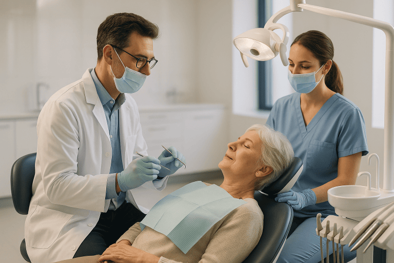
Impacted Tooth Treatment
Impacted tooth treatment manages a tooth that cannot erupt normally, often a wisdom tooth or canine. Care may include monitoring, surgical exposure, orthodontic guidance, or extraction.
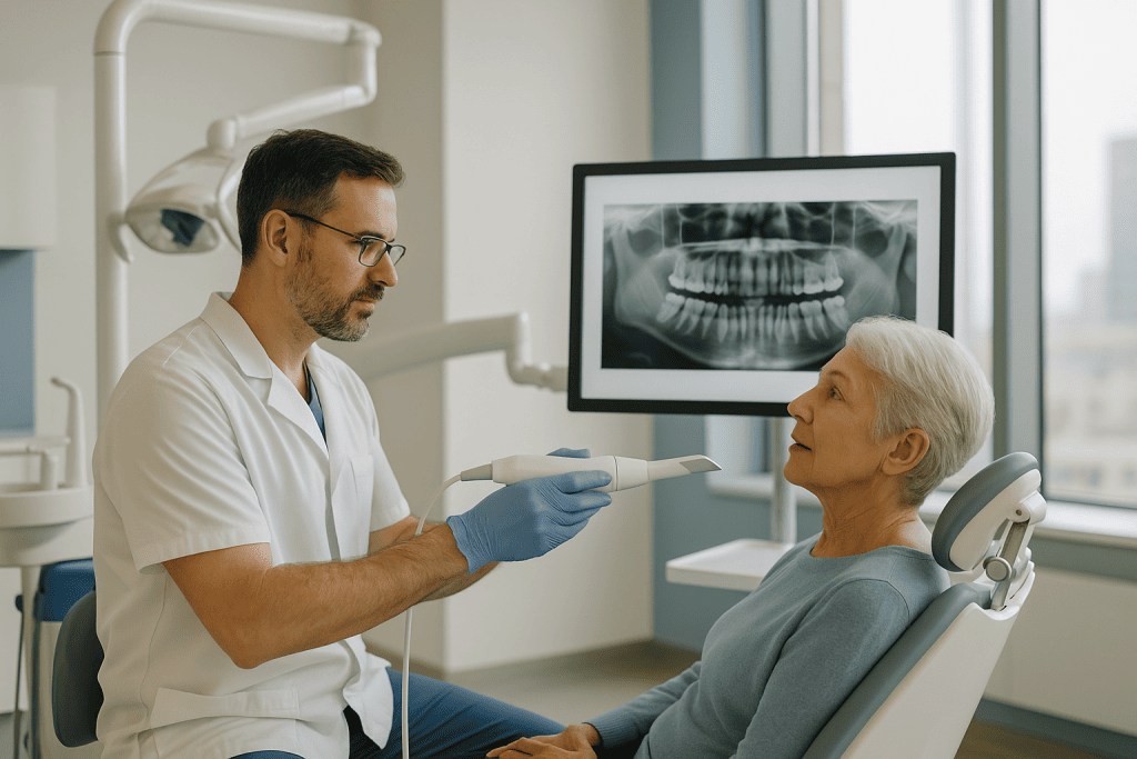
Quick answer
An impacted tooth is a tooth that cannot erupt into its normal position because bone, gum tissue or another tooth blocks its path. Wisdom teeth, called akal teeth in some languages, are affected most often. Treatment ranges from planned monitoring to surgical extraction, or surgical exposure with orthodontic traction when the tooth is worth preserving, such as an upper canine.
Impacted Tooth: When a Tooth Cannot Come Through
An impacted tooth is a tooth that cannot erupt into its normal position in the mouth because bone, gum tissue or another tooth blocks its path. Treatment ranges from planned monitoring to surgical removal, or, when the tooth is worth keeping, surgical exposure combined with orthodontic traction that guides it gradually into place. The teeth affected most often are the third molars: the wisdom teeth, known in several languages as akal teeth.
If you arrived here after searching for akal teeth, you are reading about the right subject. The name comes from words for wisdom and maturity, akal daad is the Urdu term for a wisdom tooth, and similar names exist across South Asia and the Middle East. Akal teeth and wisdom teeth are the same teeth: the third molars at the very back of each side of the jaw, usually the last teeth to appear and the ones with the least room in which to do it.
An impacted tooth is easy to underestimate at first. It may not hurt at all. Many are noticed only on a dental X-ray, during an orthodontic consultation, or when a teenager or adult begins to feel pressure at the back of the jaw. For other patients the problem announces itself: swelling, gum tenderness, repeated infection, crowding, or pain that comes and goes. Whether the tooth is a wisdom tooth, an upper canine, a premolar or another tooth, the central issue is the same: the tooth cannot take up its normal position, and a decision has to be made about it.
If you are weighing up treatment, your questions are probably practical. Is surgery necessary? Can the tooth be saved? Will treatment affect the bite, appearance or long-term dental health? How long does recovery take? Is it reasonable to travel around the time of treatment? These are fair questions, especially when the tooth sits close to the jaw nerve, the sinus, neighbouring roots or teeth that matter for the smile. This page sets out how impacted teeth are assessed, what each treatment option involves, and what recovery genuinely looks like.
What does an impacted tooth mean?
An impacted tooth means a tooth that is physically prevented from erupting into its normal place in the dental arch. The blockage can be bone, thick gum tissue, a neighbouring tooth, a retained baby tooth or, occasionally, a cyst. A tooth may be fully impacted, remaining completely within the bone or under the gum, or partially impacted, meaning part of the crown has emerged while the rest stays trapped. The distinction matters. A partially erupted tooth creates a channel between the mouth and the tissue around the buried crown, which is why partial impaction is more often associated with inflammation, infection and decay than a tooth that stays fully buried and quiet.
What is an impacted wisdom tooth?
An impacted wisdom tooth is a third molar that lacks the space or the angle to erupt normally, so it stays trapped in the jaw, tilted against the tooth in front, lying horizontally in the bone, or only partly emerged through the gum. Third molars are the most commonly impacted teeth of all, simply because they arrive last, when the rest of the arch is already occupied. Impacted wisdom teeth can stay symptom-free for years and then cause trouble abruptly: a flap of gum over a partly erupted crown traps food and bacteria, the area swells, and chewing or even opening the mouth becomes uncomfortable. You can read more about the teeth themselves on the wisdom teeth page; this page focuses on what happens when they, or any other teeth, become impacted.
Which teeth become impacted most often?
Wisdom teeth and upper canines top the list. The third molars become impacted because they erupt last into a jaw that often has no room left for them. The maxillary canines, the upper “eye teeth”, are the second most familiar case, and they matter differently: canines are important for the smile line and for guiding the bite, so the aim with an impacted canine is often to save it rather than remove it. Premolars, second molars and other teeth can also become impacted, sometimes because a baby tooth never fell out, sometimes because the permanent tooth developed in an unusual position. Each situation calls for its own plan.
What Is Impacted Tooth Treatment?
Impacted tooth treatment is not one single procedure. It is a personalised care pathway that manages a tooth unable to erupt normally into the dental arch. In some cases, observation is the right answer. In others, the tooth is surgically removed to prevent infection or damage to nearby teeth. When the impacted tooth is important for function and appearance, an upper canine is the classic example, treatment may combine surgical exposure with orthodontic guidance to bring the tooth gradually into position.
The main options are four. Monitoring means the tooth is followed over time with clinical examinations and periodic imaging, watching for change rather than intervening immediately. Surgical exposure means a specialist opens the gum, and sometimes removes a small amount of bone, to uncover the impacted tooth. Orthodontic traction, most often used for impacted canines, applies gentle, controlled force over months to move the exposed tooth into the dental arch. Extraction removes the impacted tooth when it is unlikely to erupt normally, is causing disease, or poses a meaningful risk to neighbouring structures.
Which option fits depends on the tooth involved, your age, your symptoms, the anatomy of your jaw, any orthodontic goals, and the realistic risk of future problems. For wisdom teeth, treatment usually focuses on preventing or resolving pain, infection, cyst formation, decay of the adjacent molar, gum disease or jaw complications. For impacted canines, the emphasis shifts towards preserving your natural tooth, protecting the roots of the front teeth, and building a stable, functional bite. Because the goals differ so much between teeth, careful diagnosis has to come before any decision about care.
Why do teeth become impacted?
Impaction happens for several reasons, and often more than one at a time. There may simply be too little space in the jaw. The tooth may be angled incorrectly, so that it points into the tooth in front rather than upwards into the arch. A dense layer of bone or thick gum tissue may block eruption. A neighbouring tooth, a cyst, a retained baby tooth or a developmental variation may stand in the way. None of these causes is anyone’s fault, and none can be corrected by brushing harder or waiting longer, once the eruption window has passed, an impacted tooth rarely finds its own way out.
How is an impacted tooth positioned?
Surgeons describe impacted teeth by their angle and depth, because both shape the treatment. A tooth may be tilted forwards against the neighbouring molar (mesial impaction), standing upright but unable to break through (vertical), lying flat within the bone (horizontal), or angled backwards (distal). Depth matters too: a tooth trapped only under gum tissue is a different proposition from one partly covered by bone, or one buried completely within it. Broadly, the deeper and more horizontal the tooth, and the closer it lies to the nerve canal or sinus, the more planning the procedure needs. This is exactly what imaging is for.
Wisdom Tooth Pain: What an Impacted Tooth Feels Like
Wisdom tooth pain is usually the symptom that brings an impacted tooth to attention. It tends to sit at the very back of the jaw, behind the last visible molar, and it often builds when a flap of gum over a partly erupted crown becomes inflamed: a condition called pericoronitis. The gum swells, the area is tender to touch and to chewing, there may be a bad taste, and opening the mouth can become limited. Episodes typically flare, settle, and return.
People describe the same problem in different words, wisdom molar pain, an aching jaw, pressure behind the teeth, but the teeth pain wisdom teeth cause usually comes down to a small set of mechanisms: inflammation of the gum flap, infection trapped around the crown, decay in the impacted tooth or the molar beside it, or pressure within the bone. Because akal teeth erupt in the late teens and twenties, this is also the age at which these symptoms most commonly begin, although impacted teeth can become symptomatic at any age.
Two useful cautions. First, wisdom teeth pain can radiate: patients sometimes feel it in the ear, the temple or along the jaw, which is one reason facial pain deserves proper assessment rather than assumption: the orofacial pain page explains how jaw and facial pain is evaluated more broadly. Second, the absence of pain does not mean the absence of a problem. A fully buried tooth can be slowly damaging the root of its neighbour, or developing a cyst, without causing any sensation at all. That is why impacted teeth are assessed on imaging, not on symptoms alone.
Who May Need Impacted Tooth Treatment?
You may need impacted tooth treatment when a tooth remains unerupted beyond the expected time, causes symptoms, affects surrounding teeth, or is likely to create problems in the future. The decision is not based on age alone. A healthy young adult with a deeply impacted wisdom tooth may need removal because of recurrent infection or pressure on the adjacent molar. A teenager with an impacted canine may need coordinated surgical and orthodontic care to guide the tooth into its proper position. An older adult with a quiet, deeply buried tooth may need nothing more than periodic review.
Common symptoms include pain or pressure in the jaw, swelling of the gum, a bad taste or drainage, difficulty opening the mouth, headaches related to jaw inflammation, and discomfort when chewing. A partially erupted wisdom tooth can trap food and bacteria under the gum flap, causing the repeated inflammation of pericoronitis described above. Some patients notice teeth shifting or becoming crowded, although crowding has multiple possible causes and should be evaluated carefully rather than attributed automatically to a wisdom tooth.
Many impacted teeth are found before symptoms develop. A dentist or orthodontist may identify the issue on a panoramic dental X-ray taken during routine assessment, orthodontic planning, or evaluation of delayed eruption. In more complex cases, three-dimensional imaging may be recommended to show the exact position of the tooth relative to nerves, roots, bone and sinus spaces. This is particularly valuable when planning wisdom tooth removal near the inferior alveolar nerve, or canine exposure near the roots of the front teeth.
Diagnosis usually includes a clinical examination of the mouth and jaw, a review of dental history, evaluation of symptoms, and radiographic imaging. The specialist assesses whether there is adequate space for the tooth, whether it is damaging neighbouring roots, and whether there is infection, cystic change, decay or gum disease. Recent imaging is valuable at this stage, because it lets the specialist estimate the likely pathway in advance and plan any additional views before a decision is made.
Conditions and Indications Treated
Impacted tooth treatment covers a range of dental and oral surgical situations. The plan is shaped by three questions: is the tooth causing active disease, can it be guided into a useful position, and would leaving it untreated create avoidable risk?
- Impacted wisdom teeth: Third molars that are trapped in the jaw, partially erupted, tilted, or causing pain, infection, decay, gum problems or pressure on the adjacent teeth.
- Impacted upper canines: Canines that fail to erupt normally and may need surgical exposure and orthodontic traction, because of their importance for appearance and bite function.
- Delayed eruption: A permanent tooth that has not come in at the expected time, especially when its counterpart on the other side has already erupted.
- Over-retained baby teeth: Primary teeth that remain in place because the permanent tooth behind them is blocked or displaced.
- Dental crowding or lack of space: Insufficient room in the jaw can contribute to impaction and may require orthodontic planning before or after surgery.
- Recurrent gum infection: Repeated swelling, tenderness or discharge around a partially erupted tooth, most commonly around lower wisdom teeth.
- Cysts or bone changes: Some impacted teeth are associated with cyst-like spaces or other pathology that must be assessed and, where needed, treated and examined.
- Damage to adjacent teeth: An impacted tooth can contribute to root resorption, periodontal defects, or tooth decay in the neighbouring molar, sometimes in an area too awkward to clean or to restore well.
- Orthodontic treatment planning: Impacted teeth may need exposure, removal or monitoring as part of a broader plan to align the teeth and the bite.
Not every impacted tooth must be removed. Some deeply buried teeth that are symptom-free and low risk are best monitored with periodic imaging. But when a tooth is associated with infection, pathology, progressive damage or a high likelihood of future complications, active treatment is usually the more protective choice. The honest answer for any individual tooth comes from examination and imaging, not from a general rule.
How Impacted Tooth Treatment Is Performed
Initial Assessment and Treatment Planning
The process begins with a detailed consultation. The specialist reviews your symptoms, prior dental treatment, medical conditions, medications, allergies and any previous imaging. For patients with heart conditions, bleeding disorders, diabetes, immune suppression or a history of difficult anaesthesia, medical coordination may be arranged before treatment.
Clinical examination looks at the tooth position, gum condition, jaw opening, bite, oral hygiene and any signs of infection. Imaging then defines the anatomy. Panoramic radiographs give an overview of the jaws and tooth positions. Three-dimensional dental imaging is used when the tooth lies close to nerves, sinus cavities or adjacent roots, or when surgical exposure and orthodontic guidance require precise planning. This is what lets the surgeon choose the safest access route, estimate complexity, and explain the risks specifically rather than generically.
For impacted canines and other orthodontic cases, the oral surgeon and orthodontist plan together. The orthodontist may create space in the arch before exposure. In some cases a small attachment is bonded to the impacted tooth during surgery, so controlled traction can begin after healing. This coordination matters: moving an impacted tooth too quickly, or in the wrong direction, increases the risk of root damage and gum problems.
Preparation Before the Procedure
Preparation depends on the chosen treatment. Monitoring needs only periodic examinations and imaging. Surgical treatment comes with instructions on eating and drinking before anaesthesia, oral hygiene, and arranging transport home after sedation or general anaesthesia. Decisions about any regular medication around the time of surgery, blood thinners are the obvious example, belong to the treating doctor together with the physician who prescribed it; nothing about a medication schedule should change without that conversation. If there is active infection, antibiotics or local care may be used before definitive surgery, although antibiotics alone never correct the underlying impaction.
Tell the care team about blood thinners, supplements, bisphosphonate or other osteoporosis medications, chemotherapy history, radiation treatment to the jaws, pregnancy, smoking, and any previous complications with dental procedures. These details influence timing, the choice of anaesthesia, healing expectations and post-operative precautions.
Travel timing deserves a mention. It is generally sensible not to schedule long-haul travel immediately after oral surgery, particularly after a complex wisdom tooth extraction or multiple extractions. Surgical teams routinely advise when flying is reasonable and how follow-up can be organised, so recovery does not have to be improvised.
Monitoring Without Immediate Surgery
If the impacted tooth is not causing symptoms and appears low risk, the recommended plan may be observation. Monitoring means periodic dental examinations and repeat imaging at intervals the specialist sets, watching for early signs of change: cyst development, root resorption, decay, or movement towards critical structures. Observation is an active decision, not “doing nothing”. It is the right choice when the risk of surgery would outweigh its likely benefit at that point in time, and it comes with the understanding that the plan can change if the imaging does.
What does monitoring actually involve in practice? At each review, the dentist examines the gum over the area, checks the health of the neighbouring molar and asks about any new symptoms, then compares fresh imaging with the earlier films. The specialist is looking for specific changes: widening of the tissue space around the buried crown, early cystic change, decay beginning on the adjacent root surface, or drift of the tooth towards the nerve canal or sinus. The interval between reviews is set individually, based on the tooth’s position and the findings so far. If any of these warning signs appears, the plan converts from observation to treatment while the problem is still small and the surgery still comparatively simple.
Surgical Exposure and Orthodontic Guidance
Surgical exposure is used mainly for impacted canines and other teeth that are worth preserving and can realistically be moved into a useful position. Under local anaesthesia, sedation or general anaesthesia depending on the case, the surgeon opens the gum over the impacted tooth. A small amount of bone may be removed to uncover the crown. The surgeon may bond a small orthodontic attachment to the tooth, usually connected to a fine chain or a similar guide, and the gum is repositioned and sutured to support healing.
Once the surgical site has healed, the orthodontist applies gentle traction to bring the tooth into alignment gradually. This phase takes months, and sometimes longer, depending on the tooth’s starting position, your age, the space available and how the tissues respond. The aim throughout is threefold: preserve the tooth, protect the roots of the neighbouring teeth, and finish with a stable, functional position in the arch. It is slower than extraction, deliberately so, because speed is what damages roots.
Extraction of an Impacted Tooth
Extraction is the usual recommendation for impacted wisdom teeth that are painful, infected, decayed, impossible to clean, damaging the adjacent molar, or associated with cystic change. It is also chosen for other impacted teeth that cannot be guided into position or would compromise the bite or the orthodontic plan. The procedure follows a consistent sequence:
- Anaesthesia is given, local anaesthesia, sedation or general anaesthesia, matched to the complexity of the tooth and your needs.
- Once the area is fully numb, the surgeon makes a small incision in the gum to reach the tooth.
- Bone around the tooth is removed carefully where necessary.
- In many cases the tooth is sectioned into smaller pieces, so it can come out through a smaller opening with less pressure on the jaw.
- The socket is cleaned, and the gum is closed with sutures when needed.
The choice of anaesthesia is part of the plan rather than a formality. Local anaesthesia alone suits many single extractions; sedation helps patients who are anxious or facing a longer procedure; general anaesthesia is considered for complex impactions, multiple teeth treated in one session, or patients whose medical situation makes it the safer setting. Each option carries its own preparation and recovery requirements, which is why it is agreed in advance rather than decided on the day.
Modern oral surgery relies on careful imaging, magnification, precise instruments and controlled bone removal to reduce trauma to the surrounding tissues. Three-dimensional planning identifies the relationship between the roots and the jaw nerve or the sinus before the first incision, which is where much of the safety in this surgery actually lives.
When the Tooth Sits on the Nerve: Coronectomy
When a lower wisdom tooth lies very close to the inferior alveolar nerve, the surgeon may discuss a modified approach in selected cases: coronectomy, in which only the crown of the tooth is removed and the roots are deliberately left in place to reduce the risk of nerve injury. It is not suitable for every patient, an infected or mobile tooth, for instance, changes the calculation, but it illustrates why individualised planning matters more than any standard technique. The right operation for your anatomy is a decision, not a default.
Typical Duration and Immediate Recovery
Treatment time varies. A straightforward extraction can be completed in a short appointment; deeply impacted teeth, multiple extractions, surgical exposure or complex anatomy take longer. Orthodontic guidance after exposure is a longer journey again, because the tooth moves gradually over months. It helps to keep the two timescales separate in your mind: the surgical appointment is one afternoon; the full treatment pathway may not be.
After surgery you stay under observation until you are ready to leave. Instructions cover bleeding control, swelling reduction, pain relief, eating and drinking, oral hygiene and activity limits. Soft foods, cold compresses and careful rinsing are commonly recommended. Smoking, spitting forcefully, drinking through a straw and vigorous exercise are usually restricted in the early days, because each can increase bleeding or dislodge the blood clot protecting the socket.
Oral hygiene after surgery follows a simple logic: keep the mouth clean without disturbing the healing site. Brushing continues normally away from the surgical area from the first day, the socket itself is left alone, and gentle salt-water rinsing usually begins the day after surgery rather than immediately, so the protective clot is not washed away. The team explains when normal brushing can resume near the site and whether an antiseptic rinse is appropriate for your case.
Expect some swelling, bruising, limited jaw opening and discomfort after impacted tooth surgery, particularly with lower wisdom teeth. These symptoms typically improve over days. Structured pain control is part of the plan rather than an afterthought: the guide on how pain is controlled after surgery and invasive procedures at Acibadem describes what that looks like in practice.
Why Acting Early Matters
Early evaluation does not always mean immediate surgery, but it lets you make an informed decision before complications make the decision for you. Impacted teeth can stay quiet for years and then become painful at the least convenient moment, during travel, pregnancy, military service, exams or a demanding stretch at work. A partially erupted wisdom tooth can trap bacteria repeatedly, leading to swelling, infection and difficulty opening the mouth, and recurrent infection becomes harder to manage if it spreads into the deeper spaces of the face.
Delay also has a cost for the neighbouring teeth. An impacted tooth can create a pocket where decay forms on the adjacent molar, sometimes in a spot that is difficult or impossible to restore well. It can contribute to gum defects, bone loss or root resorption. Impacted canines can damage the roots of the front teeth if they erupt in an unfavourable direction, and in orthodontic patients a late diagnosis can make treatment more complex and reduce the chance that the tooth can still be guided into a good position.
Age shapes the picture too. Younger patients often heal efficiently, and roots that are still developing can make some extractions less complex. In older adults, impacted teeth may be surrounded by denser bone, and medical considerations or slower recovery are more likely to feature. None of this means older patients cannot be treated safely: it means the plan is adapted to anatomy, health status and risk, which is precisely what a plan is for.
Benefits of Impacted Tooth Treatment
The benefits depend on which tooth is impacted and which approach is chosen, but they group into three themes: prevention of disease, relief of symptoms, and protection of long-term oral health. For wisdom teeth, the benefit is usually the end of a cycle of flare-ups; for canines, it is often keeping a tooth that matters for decades to come.
| Benefit | What It Means for You |
|---|---|
| Relief from pain and recurrent inflammation | Treatment can reduce episodes of jaw pain, gum swelling, bad taste and tenderness caused by a partially erupted or infected tooth. |
| Protection of adjacent teeth | Removing or repositioning an impacted tooth may help prevent decay, root damage, gum defects or bone loss around neighbouring teeth. |
| Improved orthodontic outcome | For selected impacted canines or premolars, surgical exposure and orthodontic guidance can bring an important tooth into the dental arch. |
| Lower risk of cysts or related pathology | When an impacted tooth is associated with cystic change or other concerning findings, treatment addresses the source and allows tissue evaluation where needed. |
| Clearer long-term planning | A precise diagnosis tells you whether monitoring, extraction or coordinated orthodontic treatment is the appropriate pathway, and why. |
Recovery Timeline After Impacted Tooth Treatment
Recovery varies with the procedure, the anaesthesia, the number of teeth treated and your own healing, but most patients follow a recognisable pattern after oral surgery.
| Time Period | What Patients Can Expect |
|---|---|
| Day 1 | Bleeding gradually slows with gentle pressure. Numbness wears off over several hours. Soft, cool foods and pain relief as directed by the team. |
| First Week | Swelling and stiffness may peak in the first few days, then begin to improve. Soft foods, careful oral hygiene and activity limits continue. |
| First Month | Most daily activities are usually resumed. The gum tissue matures and the extraction socket gradually fills in. Orthodontic traction, if planned, begins or continues after healing. |
| Longer Term | Bone remodelling continues beneath the gum. Patients treated with exposure and orthodontic guidance attend ongoing visits until the tooth reaches its planned position. |
A few points make this table more useful. Swelling that peaks around the second or third day and then recedes is the normal shape of healing after impacted tooth surgery: it does not mean something has gone wrong. Jaw stiffness eases with time and gentle use. Sutures are either dissolvable or removed at a follow-up visit, and eating progresses from soft foods back to a normal diet as comfort allows.
What does not fit the normal pattern: pain that worsens after several days of improvement, a persistent bad taste, a sensation of exposed bone in the socket, fever, or swelling that keeps increasing. These can point to infection or to dry socket: a treatable condition in which the protective blood clot is lost from the socket before the tissue beneath has healed. Dry socket typically appears a few days after extraction as a deep, throbbing ache that ordinary pain relief barely touches, and it responds well to a simple dressing placed by the dental team. Timely follow-up is what keeps small complications small.
How long does an extraction socket take to heal?
An extraction socket closes over at the surface within a few weeks, while the bone beneath continues to fill in over several months. In the first days a blood clot seals the socket; soft tissue then grows across it, and most patients find the area comfortable for normal eating well before healing is complete underneath. Deeply impacted teeth leave larger sockets that take longer to consolidate than simple extractions do. The timeline also depends on age, general health, smoking and how carefully the early instructions are followed, which is why the same operation can heal differently in two different patients.
Factors That Influence Outcomes
A good result depends on more than the technical act of removing or exposing a tooth. It begins with diagnosis. The tooth’s depth, angle, root shape, stage of development and proximity to nerves, sinus cavities and neighbouring roots all set the level of complexity. A vertically positioned tooth near the surface is a different operation from a deeply horizontal tooth wrapped around a nerve canal, and three-dimensional imaging becomes important precisely when standard X-rays cannot show that difference clearly enough.
Age and general health matter too. Younger patients tend to have more favourable healing conditions and more flexible bone; adults may have denser bone or existing restorations that need additional care. Diabetes, immune suppression, bleeding disorders and smoking can all slow healing. Anticoagulants and certain bone-modifying drugs require specific planning, planning, not improvisation, and always in coordination with the doctors who manage those medicines.
For impacted canines, the outcome is strongly influenced by the tooth’s position, the space available, the root health of adjacent teeth, the quality of the periodontal tissue, and how well the surgeon and orthodontist coordinate. Some impacted canines can be guided into position; others are ankylosed, severely displaced or associated with damage that makes movement difficult or inadvisable. A realistic explanation of the likely pathway, including the possibility that traction may not succeed, belongs at the start of treatment, not the end.
Your own participation is a genuine factor, not a polite footnote. Following post-operative instructions reduces the risk of bleeding, dry socket, infection and delayed healing. Maintaining oral hygiene, attending follow-up appointments, avoiding smoking and reporting unusual symptoms early all support recovery. In orthodontic cases, consistent attendance and appliance care are essential, because tooth movement depends on controlled force applied steadily over time, miss the visits and the biology simply stops cooperating.
Finally, judgement shapes outcomes as much as technique. Complex impacted teeth demand decisions about when to remove, when to monitor, when to preserve and how to reduce risk. The safest plan is not always the most aggressive one. It is the plan that matches your anatomy, your symptoms, your long-term dental goals and your overall health.
How Acibadem Plans Impacted Tooth Care
At Acibadem, impacted tooth care is planned within a hospital-based environment. For many patients this is a background detail; for those with complex anatomy, medical conditions, anaesthesia concerns or a need for multidisciplinary input, it is the point. The same setting that handles a routine extraction can also support sedation, general anaesthesia, medical consultation and coordination with other specialties when a case requires them.
Care is shaped by evidence-based clinical protocols and individual planning. Oral and maxillofacial surgeons, dentists, orthodontists, radiologists and anaesthesiology teams are involved as the case demands. For an impacted wisdom tooth close to the jaw nerve, the discussion centres on imaging findings, nerve-sparing strategies and honest risk. For an impacted canine, the plan may run through orthodontic space creation, surgical exposure, attachment placement and staged follow-up. Diagnostic pathways, panoramic radiography, three-dimensional dental imaging, digital treatment planning and clinical photography, support the decisions, but they do not replace clinical judgement; they make it better informed and easier to explain.
The treatment plan is defined before any procedure is performed. Some patients need only monitoring and a clear follow-up strategy; others need a single, well-planned surgical appointment; patients undergoing orthodontic guidance need a staged plan in which surgery and tooth movement are sequenced deliberately. Setting this out in advance, including the imaging required, the anaesthesia planned and the follow-up schedule, means decisions are made calmly and with full information, not at the last minute.
Second opinions have a particular place in impacted tooth care, because the honest answer is sometimes “leave it alone” and sometimes “this is more urgent than you were told”. A second opinion can clarify whether an impacted tooth truly needs removal, whether a canine can still be preserved, whether three-dimensional imaging changes the risk assessment, or whether a particular case is better performed in a hospital setting. The aim is a clear, medically grounded recommendation you can understand and weigh calmly.
Making Sense of the Decision
An impacted tooth is not just a finding on an X-ray. It can affect comfort, oral health, orthodontic planning and the stability of the teeth around it, and the right response ranges from simple monitoring to carefully planned extraction to a coordinated surgical and orthodontic pathway that preserves an important tooth. Whether the tooth in question is one of the akal teeth at the back of the jaw or a canine near the front of the smile, the sound decision starts in the same place: accurate diagnosis, a clear explanation of benefits, risks and alternatives, and a realistic picture of what recovery will ask of you. Everything after that is a matter of matching the plan to the person.
Preparation
- A dental examination and panoramic X-ray or 3D imaging may be used to assess the tooth position, nerves, roots, and nearby structures. Tell your dentist about medications, allergies, bleeding disorders, or previous anesthesia reactions. You may be advised to avoid eating for a short period if sedation or general anesthesia is planned.
Aftercare
- Mild swelling, soreness, and limited mouth opening are common after impacted tooth surgery and usually improve within a few days. Follow instructions for prescribed medicines, cold compresses, oral hygiene, and soft foods. Avoid smoking, alcohol, hard foods, and forceful rinsing until your dentist confirms healing.
Frequently Asked Questions
What affects the cost of impacted tooth treatment?
The main factors are the tooth location, depth and angle of impaction, whether extraction or orthodontic guidance is needed, imaging requirements, anesthesia type, surgeon and facility fees, and follow-up needs. A personalised quote is only possible after a specialist reviews your examination and imaging.
How can I get a personalised quote from Acibadem?
You can request a free consultation by sharing your dental records, recent imaging if available, symptoms, medical history, and any orthodontic information. The team can then advise which treatment pathway may be appropriate and what the package may include.
Is extraction always necessary for an impacted tooth?
No. Some impacted teeth can be monitored, while others may be surgically exposed and guided into place with orthodontic treatment. The decision depends on symptoms, tooth position, risks to nearby structures, and long-term dental goals.
Will imaging change the quote?
Yes. Imaging can show whether the impacted tooth is close to nerves, sinuses, adjacent roots, or cystic changes. These findings may affect surgical complexity, anesthesia planning, specialist involvement, and the final treatment cost.
What is usually included in an international patient package?
A package may include consultation, imaging review, treatment planning, the procedure, standard medications, and follow-up guidance. Interpreter support and appointment coordination may also be available, but inclusions should be confirmed before travel.
Is this information a medical or financial recommendation?
No. This is general educational information. A dental specialist or oral and maxillofacial surgeon should assess your case, explain suitable options, and provide a personalised quote before you make a decision.
Medically reviewed by the Acıbadem International Medical Board September 13, 2026
See our medical review board →
Update history
- PublishedJune 8, 2026
- Medical review approvedSeptember 13, 2026
- Last content updateSeptember 12, 2026
References2
- Impacted Tooth medlineplus.gov
- Wisdom tooth removal nhs.uk
Trusted care for international patients
From the Acibadem Blog
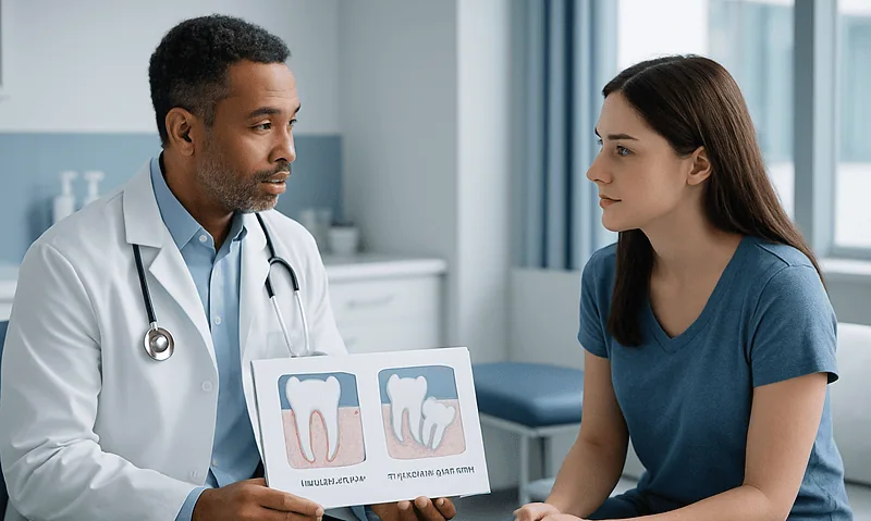
Impacted Canine vs Impacted Wisdom Tooth: Why One Is Often Saved and the Other Removed
An impacted canine is usually saved because it is a cornerstone tooth with the longest root in the mouth, guides the bite and anchors…

Recovering From Impacted Tooth Surgery: Swelling, Limited Mouth Opening and Soft Foods
Recovery from impacted tooth surgery usually takes up to two weeks, according to the NHS. Swelling of the cheek tends to peak around the…
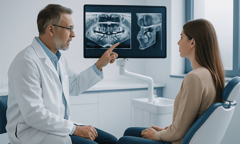
What a Panoramic X-Ray or 3D Scan Shows About an Impacted Tooth: Roots, Nerves and Angle
A panoramic X-ray of an impacted tooth shows, in one flat image, how many roots the tooth has and how they curve, how deeply…