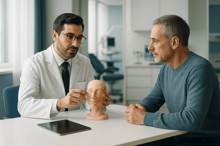
Mole Removal
Mole removal eliminates unwanted or suspicious skin moles using excision, shaving, or laser-based methods after dermatologic evaluation. It can improve appearance and allow pathology testing when needed.
Quick answer
Mole removal is a short outpatient procedure that takes a mole off the skin under local anaesthetic. The main methods are surgical excision with stitches, shave removal for raised moles, and laser for selected superficial cosmetic cases. When a mole looks atypical, the removed tissue is sent to a pathology laboratory to rule out skin cancer. Most people return to light activity the same day.
What Is Mole Removal?
Mole removal is a minor medical procedure that takes a mole, the medical term is nevus, off the skin, either by cutting it out with a small margin, shaving it level with the surface, or, in selected cosmetic cases, treating it with laser. It serves two purposes that often overlap: improving appearance, and obtaining tissue for laboratory diagnosis when a mole looks unusual or has changed. Most mole removal is done under local anaesthetic in a short outpatient appointment, and most people go back to light daily activity the same day.
Moles themselves are common growths made up of pigment-producing cells called melanocytes. They can be flat or raised, light brown, dark brown, skin-coloured, pink or nearly black. Most are benign. Some are present from childhood; others appear later in life. A mole may stay unchanged for decades, or it may alter because of friction, sun exposure, hormonal shifts, ageing or, less often, abnormal cell behaviour. That last possibility is why the decision to remove a mole should always begin with an assessment, not with a choice of technique.
The goal of the procedure depends on the reason for it. If the mole is cosmetically unwanted, the aim is to remove it while keeping the visible scar as small and as well placed as possible. If the mole is suspicious, changing, symptomatic or atypical, the aim shifts: the priority becomes removing adequate tissue for diagnosis and, where appropriate, treating the lesion completely. In those cases the removed tissue is sent to a pathology laboratory, where it is examined under a microscope and the result determines whether anything further is needed.
A precise diagnosis matters because not every dark spot on the skin is a mole. Lesions that look similar to the untrained eye include seborrheic keratoses, dermatofibromas, skin tags, freckles, lentigines, vascular lesions and, occasionally, early skin cancer. Each of these is handled differently. Safe mole removal therefore starts with a professional evaluation of what the lesion actually is, and only then moves to how it should come off.
Can you remove moles?
Yes, almost any mole can be removed, and the procedure is one of the most common minor operations in dermatology. The more useful questions are whether a particular mole should be removed, which method suits it, and whether the tissue needs laboratory analysis afterwards. A raised benign mole on the trunk, a flat changing mole on the back and a small congenital mole on the scalp are three different problems that call for three different plans. The technique is chosen to fit the lesion, not the other way round.
The Main Mole Removal Methods
Three approaches cover the great majority of cases. Each has a defined role, a distinct healing pattern and a different relationship with pathology testing. Understanding them makes the consultation easier to follow and the recommendation easier to weigh.
Surgical excision
Surgical excision removes the entire mole together with a narrow margin of surrounding skin, usually through an elliptical incision planned along natural skin tension lines. The depth is chosen to include the full lesion, which matters because the cells of a mole extend below the surface you can see. The wound is closed with sutures, some dissolve on their own, others are taken out after several days or weeks depending on the body area. Excision is the standard choice when a mole is flat, deeper, recurrent or suspicious, and whenever the pathologist needs a complete, full-thickness specimen. It trades a fine linear scar for the most definitive removal and the most reliable diagnosis.
Shave removal
Shave removal uses a fine surgical blade to take a raised mole down to the level of the surrounding skin, or slightly below it. Bleeding is controlled with pressure, cautery or topical agents, and deep stitches are usually unnecessary. The site heals like a shallow graze over days to weeks. Shave removal suits selected benign-looking raised moles, particularly where a linear scar would be more conspicuous than a flat mark. Its limits are worth stating plainly: because it does not reach the deepest cells, it can occasionally leave residual pigment or allow the mole to regrow, and the healed area may end up lighter, darker or slightly depressed compared with the surrounding skin.
How do you remove a mole, can you use laser?
Laser can be used for certain moles, but only after the lesion has been examined and judged clearly benign. Laser-based treatment directs concentrated light energy at pigment or tissue in superficial lesions, and it can be a reasonable option when the goal is purely cosmetic. The critical limitation is diagnostic: laser destroys or alters the tissue rather than preserving it, so it cannot provide a proper specimen for the pathology laboratory. For that reason it is never the right tool when there is any concern about melanoma or another skin cancer, and a responsible clinician will decline to laser a pigmented lesion that has not been properly assessed. Used on the wrong lesion, laser can also make future evaluation harder by changing the mole’s appearance without removing its deeper cells.
Can I remove my mole without surgery?
Sometimes, but only within narrow limits. Shave removal is technically minor surgery, though it involves no deep cutting and usually no stitches, and laser treatment avoids incisions altogether for suitable superficial lesions. What does not exist is a safe non-procedural route: no cream, plaster, acid pen or freezing kit sold to the public removes a mole reliably or safely, and every method that destroys the tissue on the surface eliminates the possibility of laboratory analysis. If a mole turns out to have been an early melanoma, that lost diagnosis is a serious problem. The honest answer is that avoiding surgery is occasionally possible; avoiding professional evaluation never is.
Who May Need Mole Removal?
People consider mole removal for a mixture of practical, cosmetic and medical reasons. Some are bothered by a mole that rubs against clothing, catches on jewellery, bleeds after shaving or becomes repeatedly inflamed. Others want to remove a mole that draws attention in photographs or affects their confidence in social and professional settings. In many of these cases the mole has already been checked and looks benign, and the patient simply prefers to be rid of it for comfort or appearance. That is a legitimate reason, and the planning then centres on scar quality.
Removal may also be recommended on medical grounds. Dermatologists commonly apply the ABCDE warning signs when assessing pigmented lesions: asymmetry, border irregularity, colour variation, diameter or size change, and evolution over time. A mole that grows, darkens, becomes uneven in colour, changes shape, or develops itching, crusting, pain or spontaneous bleeding warrants prompt examination. So does a new pigmented lesion appearing in adulthood, especially one that looks different from the person’s other moles, dermatologists sometimes call this the “ugly duckling” sign.
Diagnosis begins with a skin examination and a conversation. The dermatologist asks how long the mole has been there, whether it has changed, whether there is a personal or family history of melanoma or other skin cancers, and how much sun exposure or tanning-bed use the skin has had. The lesion is inspected visually and usually with dermoscopy: a magnified, light-based examination that reveals pigment structures beneath the surface which the naked eye cannot see. Dermoscopy substantially sharpens the distinction between lesions that can be left alone, lesions that should be watched and lesions that should come off.
For people with many moles or a history of atypical ones, digital skin imaging or mole mapping can document lesions so that changes can be compared over time rather than judged from memory. If a lesion appears suspicious, removal for biopsy is advised rather than observation or cosmetic treatment. If it appears benign and removal is requested for appearance, the discussion covers the likely scar, the best technique for that site and skin type, and whether sending the tissue for pathology is still sensible, often it is, simply because certainty costs little once the mole is off.
Patients travelling from abroad sometimes have a preliminary review of photographs and medical history before a visit, which helps a clinic anticipate whether dermatology alone or plastic surgery input is likely to be needed and how much time to allow for the procedure and any pathology reporting. Photographs, however good, are a planning aid rather than a diagnosis: an in-person examination with dermoscopy remains essential before any mole is removed.
Is it good to get moles removed?
It is good to remove a mole when there is a reason, suspicion, irritation or a cosmetic concern that genuinely bothers you, and unnecessary to remove moles that are stable, benign and untroubling. Removing normal moles “preventively” is not a recognised strategy against melanoma, because melanoma frequently arises on previously unmarked skin rather than within an existing mole, and every removal leaves some form of mark. What does protect you is surveillance: knowing your skin, noticing change and having changing lesions examined. When a specific mole meets the criteria for removal, taking it off is entirely reasonable; stripping the skin of harmless moles is not.
Moles, Skin Tags and Other Lesions: Knowing What You Have
Many people arrive at a clinic asking about a “mole” that turns out to be something else. Getting the label right matters, because the safe treatment differs from lesion to lesion. Two of the most common look-alikes are seborrheic keratoses, waxy, stuck-on-looking growths of later adulthood, and skin tags, which have their own confusing vocabulary.
What are tag moles?
“Tag moles” is an informal name people use for skin tags, which are not true moles at all. A skin tag, the medical term is acrochordon, is a soft, skin-coloured or lightly pigmented growth that hangs from the skin on a small stalk. Skin tags contain no clusters of melanocytes the way moles do; they are benign overgrowths of ordinary skin and fibrous tissue, typically appearing where skin rubs against skin or clothing: the neck, armpits, eyelids, groin and under the breasts. They are harmless, though they can become sore or inflamed if twisted or repeatedly caught. Because a genuinely pedunculated dark lesion can occasionally be a raised mole rather than a tag, anything pigmented deserves a proper look before it is treated as a simple tag.
How to remove skin tags
If you are wondering how to remove skin tags, the honest answer is that clinic removal is quick and straightforward: a clinician can snip a tag off at the base with sterile scissors after numbing the area, freeze it with cryotherapy, or seal it with light cautery, depending on its size and position. What people casually call tag moles removal at home, tying thread around the base, pulling, or applying acids, risks bleeding, infection and, more importantly, treating a misidentified lesion. A pigmented growth removed by thread on a bathroom shelf tells no one what it was. The clinic route is brief, controlled and, where there is any doubt about the diagnosis, allows the tissue to be examined.
Conditions and Indications Mole Removal Addresses
The most common indication is a benign mole that the patient wants removed because of its position, shape, texture or effect on appearance. Raised moles on the nose or chin, moles at the hairline, and moles on skin exposed by clothing are frequent reasons for consultation. Here the plan balances complete removal against scar placement, skin tension lines and long-term cosmetic healing: the mark left behind should be a clear improvement on the mole it replaces.
Removing a mole on the face
A mole on the face deserves particular care, because the face is unforgiving of poorly planned scars and generous to well planned ones. Facial skin has a strong blood supply and often heals with fine lines, but the eyelids, lips, nose and ears involve function and symmetry as well as appearance, so incisions are planned within natural creases and cosmetic units, and plastic surgical input is sometimes added to the dermatological assessment. Men have the additional practical problem of moles in the shaving zone that are cut again and again. Where the concern is broader than a single lesion, facial mole planning sometimes sits alongside wider face aesthetics considerations, though the medical assessment of the mole always comes first.
Irritation is another common indication. A raised mole on the neck gets rubbed by collars and necklaces; a mole under the bra line, waistband or underarm becomes tender and inflamed; a mole on the scalp catches the comb. Repeated trauma does not make a mole dangerous, that is a persistent myth, but it does make removal a reasonable way to end a recurring nuisance and improve daily comfort.
Atypical and suspicious moles form the medically weightier group. Dysplastic nevi are moles with unusual clinical or microscopic features; they are not melanoma, but they can resemble it, and in some patients they mark an increased overall risk. When a dermatologist cannot confidently call a lesion benign, biopsy or excision lets the pathologist settle the question at cellular level. Where melanoma is actively suspected, the preferred approach is usually a complete excisional biopsy where feasible, with depth and margins guided by clinical judgement and established guidelines. If melanoma or another malignancy is confirmed, further care is planned from the pathology detail, tumour thickness, margin status, risk features, and more complex cases may go to a specialist tumour board.
Congenital moles, present at birth or in early childhood, are a category of their own. Small congenital moles can often be removed like other benign lesions. Larger or more complex ones may need staged planning, plastic surgical techniques and longer-term monitoring; occasionally reconstruction of the treated area draws on methods such as skin grafting or, for extensive lesions, structured reconstruction after skin disease. The decision weighs size, location, symptoms, cosmetic impact and individual risk assessment rather than following a single rule.
Across all of these indications, the same principle holds: not every mole should be removed the same way. A raised benign mole may suit a shave; a flat pigmented lesion with atypical features requires excision; laser can lighten a carefully selected superficial lesion but can obscure future assessment if used carelessly. Matching method to lesion is the core of safe practice.
How Mole Removal Is Performed: From Evaluation to Recovery
The process begins with a consultation. The physician reviews your medical history, current medications and supplements, allergies, tendency to scar, previous procedures and any personal or family history of abnormal moles or skin cancer. If you take blood-thinning medication or have a bleeding disorder, say so, precautions can be coordinated, and decisions about any medication belong to your treating doctor, so disclosure matters far more than guesswork.
The mole is then examined directly and usually with dermoscopy. The doctor assesses its size, colour pattern, border, depth, position and relationship to nearby structures. On the face, eyelids, lips, nose, ears and hands, planning takes in natural creases, cosmetic units and how the skin moves. If the mole is suspicious, the conversation centres on diagnostic removal and pathology; if it is clearly benign, it centres on the most appropriate cosmetic approach and realistic expectations about the scar.
On the day, a straightforward removal follows a predictable sequence:
- Preparation. The area is cleaned and the planned removal is marked on the skin.
- Local anaesthetic. An injection numbs the skin around the mole. There is a brief sting and a sensation of pressure; once the anaesthetic has taken effect, you should feel movement and pressure rather than pain. General anaesthesia is rarely needed, children, very anxious patients or people needing multiple complex removals may need additional planning, decided case by case.
- Removal. The mole is excised with its margin, shaved level with the skin, or treated with laser, according to the plan agreed beforehand.
- Haemostasis and closure. Bleeding is controlled with pressure, cautery or topical agents; excision wounds are closed with sutures.
- Dressing and instructions. A dressing is applied and you receive written wound-care instructions before leaving.
- Pathology. Where a specimen is taken, it goes to the laboratory, and the result is discussed once reported.
The appointment itself is often brief. A single straightforward mole takes only a short session; multiple lesions, cosmetically sensitive sites or more complex excisions take longer. Most patients walk out, return to desk work and manage ordinary daily tasks the same day. If you are travelling for treatment, build in enough time for a wound review if one is advised and for discussion of pathology results when tissue has been sent.
The supporting technology is worth understanding, because it is where much of the safety lives. Dermoscopy guides the initial judgement about pattern and risk. Digital imaging documents lesions in people with many moles. In the procedure room, fine instruments, magnification, careful lighting and electrosurgical tools for bleeding control support precise work; laser systems are reserved for evaluated cosmetic indications. Behind all of it sits the pathology laboratory, whose microscopic diagnosis confirms whether the story ends with healing or continues with further care.
How to Remove Moles: Why Home Methods Are Not the Answer
“How to remove moles” is one of the most searched skin questions on the internet, and much of what the search returns is either useless or actively harmful. It is worth addressing the common versions of the question directly, because the honest answers protect people from real damage.
Can I remove a mole by myself?
No, and the reasons go beyond squeamishness. Cutting, tying off or burning a mole at home risks bleeding, infection and a scar considerably worse than anything a planned procedure would leave. Home methods almost never remove the deeper cells, so pigment commonly returns in the healed skin, sometimes in irregular patterns that later mimic the very warning signs of melanoma and complicate every future examination. Worst of all, self-removal destroys the tissue without analysis: if the lesion was an early melanoma, the one opportunity to diagnose it at its most treatable stage has been thrown away.
How can you remove moles at home?
The safe removing of moles is a medical procedure, not a home project, and no over-the-counter product changes that. Creams, acid pens, freezing kits and “mole removal plasters” sold online work, when they work at all, by chemically or thermally burning the skin. They cannot tell a benign nevus from a dysplastic one or from a melanoma; they frequently cause burns, pigment changes and pitted scars; and they leave nothing for a pathologist to examine. Unregulated cosmetic treatments carry the same problems with better marketing. A mole that looks simple to its owner may carry features only dermoscopy reveals, which is why the safest first step is always professional assessment rather than a purchase.
How to remove moles naturally in one day, is it possible?
No natural method removes a mole in one day, or in any number of days. A mole is not a surface stain: its cells extend down into the dermis, beyond the reach of garlic, apple cider vinegar, iodine, banana peel or any of the other remedies that circulate online. Applied persistently, acidic or caustic household substances do damage skin, that is chemistry, not treatment, producing burns and scars while leaving the deeper mole cells in place. Anything that appears to make a mole vanish overnight has injured the surface, nothing more. The only one-day reality on offer is legitimate: a clinic can often evaluate and remove a suitable mole within a single short visit.
How to get rid of moles safely
If you want to know how to get rid of moles, the safe sequence is short: have the mole examined, ideally with dermoscopy; agree the method, excision, shave or, for evaluated benign superficial lesions, laser; have it removed under local anaesthetic in a proper clinical setting; and send the tissue for pathology whenever there is any question about what it is. The same sequence covers one mole or several, a cosmetic concern or a medical one. It is quick, it is definitive, and it keeps the diagnostic door open, which is exactly what every shortcut closes.
Why Acting Early Matters and the Risks of Delay
Most moles are harmless and nothing about them is urgent. A changing or suspicious mole is different. Early evaluation matters because melanoma and certain other skin cancers are more treatable when detected at an earlier stage; delay gives a malignant lesion time to grow deeper or spread, and deeper lesions make treatment more complex. This is the single strongest argument for having change examined rather than watched from a distance.
Even with a benign mole, waiting has costs of its own. A repeatedly irritated mole goes on bleeding, crusting and flaring. A mole in the shaving zone goes on being cut. A raised mole that catches on clothing remains a daily nuisance. And when removal is wanted for cosmetic reasons, an early consultation clarifies the best method before frustration drives someone towards home remedies or unregulated treatments that cause burns, infection, scarring or incomplete removal, problems that are harder to revise than they were to prevent.
There is also a planning dimension for anyone travelling for care. If pathology will be needed, the schedule should allow time for results or for a follow-up discussion. If the mole proves more complex than expected, early review makes it possible to coordinate dermatology, plastic surgery, oncology or other services within one organised plan instead of a scramble. A timely, structured approach reduces uncertainty at every step.
Benefits of Mole Removal
What you gain from mole removal depends on why it is done, diagnosis, comfort, appearance, or some combination. The realistic benefits look like this:
| Benefit | What It Means for You |
|---|---|
| Diagnostic clarity | When tissue is sent for pathology, the result can confirm whether the mole is benign, atypical or requires further treatment. |
| Improved appearance | Removal can reduce the visibility of a mole that affects facial balance, grooming confidence or comfort in social settings. |
| Relief from irritation | Moles that rub, catch on clothing, bleed after shaving or become inflamed can often be removed to reduce repeated discomfort. |
| Early treatment pathway | If a suspicious lesion is identified, removal can be the first step toward timely diagnosis and guideline-based care. |
| Personalised scar planning | The incision or treatment method can be chosen with attention to skin tension, location, skin type and cosmetic priorities. |
Recovery Timeline After Mole Removal
Healing varies with technique, mole size, body area and individual skin, but the broad pattern is consistent and most people return to routine quickly.
| Time Period | What Patients Can Expect |
|---|---|
| Day 1 | The area is covered with a dressing. Mild soreness, tightness or small amounts of spotting can occur. Most patients can walk, work at a desk and perform light daily activities. |
| First week | Wound care is the priority. Shave sites begin forming a new surface; excision sites should be protected from stretching. Some sutures may be removed during this period, depending on location. |
| First month | The skin continues to strengthen. Redness or pinkness is common. The healing area should be kept out of the sun, and scar-care recommendations followed where given. |
| Longer term | Scars mature gradually over several months; colour, firmness and texture usually keep improving. Follow-up may be recommended if pathology showed atypia or if you have many moles. |
In practical terms: keep the area clean, follow the dressing instructions you were given, resist picking at crusts, and protect the healing skin from sunlight, which can darken new skin and make scars more visible. Strenuous exercise, swimming, sauna use and activities that stretch the incision are usually restricted for a short period after excision, particularly on high-tension areas like the back and shoulders.
If non-dissolving stitches were used, the team will tell you when they are due out, timing differs by body site. Patients who travel for treatment can often have sutures taken out near home if the timing and wound condition allow; ask for this to be confirmed in your written aftercare plan before you leave. Shave sites need no suture appointment at all; they simply resurface over days to weeks. Excision sites gain deeper strength over weeks, and scar maturation continues quietly for months after the wound itself looks closed.
What Influences the Outcome of Mole Removal?
A good result rests first on accurate diagnosis. The most important decision is not how to remove the mole but whether it should be removed cosmetically, biopsied, excised with margins or simply monitored. Careful dermatological evaluation prevents both errors: under-treating a suspicious lesion and over-treating a harmless one.
Technique shapes the final appearance. Shave removal may be ideal for a raised benign mole but leaves the deepest cells behind, occasionally allowing residual pigment or regrowth. Excision provides a full-thickness specimen and is more definitive, at the cost of a linear scar. Laser helps in selected cosmetic situations and is wrong wherever pathology is needed. Matching method to lesion and to the patient’s priorities is the physician’s central task.
Location matters as much as method. Thin, well-vascularised skin, much of the face, often heals with fine scars when carefully managed. The chest, shoulders, upper back and skin over joints sit under more tension and are more prone to widened, raised or thickened scars. Around the eyelids, lips, nose and ears, function and symmetry join appearance on the list of things the plan must protect.
Individual healing tendencies count too. Some people are prone to hyperpigmentation, hypopigmentation, hypertrophic scars or keloids, and a personal or family history of thick scarring should be raised before treatment, not after. In darker skin tones, meticulous technique and disciplined sun protection are especially important for even pigmentation during healing. Aftercare then does the rest: clean wounds, no picking, limited strain on the area, ultraviolet protection, and consistency with scar massage, silicone gel or silicone sheets where these are recommended.
Finally, the pathology result sets the next step. A benign, completely removed mole usually needs nothing further. Atypical cells may prompt observation, re-excision or periodic skin checks, depending on the degree of atypia and whether the margins were clear. A diagnosis of melanoma or another skin cancer moves care onto established guideline pathways, which can involve wider excision, imaging, lymph node evaluation or oncology consultation as indicated.
Will the mole come back after removal?
It can, and the likelihood depends on the method. After shave removal, cells left below the shave line sometimes produce a return of pigment or a partial regrowth in the healed skin, usually harmless, but recurrent pigment in a removal scar can be difficult to distinguish from more significant change, which is one reason clinicians document removals carefully. After a complete excision with clear margins, recurrence is much less expected. This difference is one of the reasons excision is preferred whenever there was any doubt about the mole in the first place.
Does mole removal leave a scar?
Yes, any procedure that removes skin leaves some mark, and it is better to hear that before the procedure than to discover it afterwards. The realistic goal is the most acceptable scar possible: a fine line placed along a natural crease, or a flat pale area where a raised mole used to sit. Well planned scars typically fade and soften considerably as they mature over months, and many become hard to find; none becomes invisible on close inspection. An open conversation beforehand about your skin type, the mole’s location and your priorities keeps expectations and outcomes aligned.
What Does Mole Removal Cost?
How much does it cost to have a mole removed?
There is no single figure, because the price of mole removal is built from several variables that differ from patient to patient. The main cost drivers are the number of lesions treated in one session; the method used, since a shave, an excision with layered closure and a laser session involve different time and materials; the body site, because cosmetically sensitive areas demand more meticulous work; whether the tissue is sent for pathology, which adds a laboratory component; who performs the procedure, as plastic surgical involvement for delicate facial work is priced differently from a routine dermatological removal; and the setting itself. Insurance and reimbursement systems in many countries also distinguish between medically indicated removal, a changing or suspicious lesion, and purely cosmetic removal, which is commonly paid out of pocket. A written, itemised quotation after examination is the only reliable way to know what a specific mole will cost, and it is reasonable to ask that pathology and any follow-up review be spelled out within it.
Mole Removal at Acibadem
At Acibadem, mole removal is treated as both a medical and an aesthetic procedure. Dermatologists evaluate the lesion using clinical examination and dermoscopy; plastic surgeons join the plan where the mole sits on the face, eyelids, lips, nose, ears or another cosmetically demanding site. The emphasis is on choosing the right treatment for the specific lesion, cosmetic removal, biopsy, excision with margins or monitoring, rather than applying one method to every mole. Pathology services provide microscopic diagnosis when tissue analysis is indicated, and if a result shows atypia or malignancy, care can move into multidisciplinary review with the relevant specialties according to the diagnosis.
For patients coming from abroad, the practical side is organised around the same clinical logic: coordinated appointments, help with medical records, interpretation support and clear explanation of how pathology results will be communicated. Because most removals are short outpatient procedures, the visit itself is usually brief; the value of the structure shows when a mole proves more complex than expected and dermatology, plastic surgery or oncology need to work from one plan. Patients returning home before a scar has matured leave with written wound-care instructions and guidance on what local follow-up, if any, is appropriate, including the timing of suture removal where non-dissolving stitches were used.
The objective throughout is not only to remove a mole, but to make sure you understand what was removed, why that method was chosen, what the pathology showed, and what, if anything, to watch for in future.
Making a Considered Decision
A mole that seems cosmetically trivial may still need medical assessment, and a mole that looks alarming may prove entirely benign: the examination, not the mirror, settles which is which. A proper evaluation clarifies the diagnosis, the safest removal method, the expected scar, whether pathology is needed and how recovery will run. From there the decision is usually straightforward, whichever way it goes.
Mole removal is a small procedure by any surgical standard, but done for the right reasons and with the right technique it delivers something larger: diagnostic certainty when a lesion has been worrying you, an end to daily irritation, or the quiet satisfaction of skin that looks the way you want it to. Whether the concern is appearance, discomfort or the possibility of skin cancer, a careful medical approach protects both your health and your confidence, and it starts with knowing exactly what that mark on your skin actually is.
Preparation
- A dermatologist or plastic surgeon examines the mole and may use dermoscopy to assess risk. Tell your doctor about medications, allergies, bleeding disorders, and previous scar problems. Blood thinners may need adjustment if medically appropriate. Avoid tanning or irritating the area before the procedure.
Aftercare
- Keep the area clean and dry as instructed, and change dressings if recommended. Avoid scratching, swimming, and intense sun exposure until healing is complete. Stitches, if used, are usually removed within several days to two weeks depending on location. Pathology results are reviewed when a suspicious mole is excised.
Frequently Asked Questions
What affects the cost of mole removal?
Cost depends on the removal technique, the number and location of moles, whether pathology testing is required, the specialist’s expertise, facility standards, anesthesia needs, and follow-up care. Travel and translation services may also influence the total package for international patients.
How can I get a personalised quote from Acibadem?
You can request a free consultation and share clear photos, the mole location, any recent changes, previous biopsy results if available, and your travel preferences. A dermatologist or relevant specialist will advise which assessment and removal pathway is appropriate before a quote is prepared.
Is pathology testing included in the quote?
Pathology may be recommended when a mole looks suspicious or when complete tissue assessment is clinically important. Whether it is included or listed separately should be confirmed in your personalised treatment plan.
Is laser mole removal cheaper than excision?
The cost relationship depends on the lesion and treatment plan. Laser may be suitable only for selected benign lesions, while excision is often preferred when tissue needs to be examined. The safest and most appropriate option should be chosen by a specialist, not by price alone.
Will I need follow-up after travelling home?
Follow-up may include wound care guidance, suture removal planning, pathology review, scar-care advice, and instructions on when to contact a doctor. International patients should clarify remote follow-up and local care arrangements before treatment.
Medically reviewed by the Acıbadem International Medical Board September 8, 2026
See our medical review board →
Update history
- PublishedJune 8, 2026
- Medical review approvedSeptember 8, 2026
- Last content updateSeptember 8, 2026
Trusted care for international patients
From the Acibadem Blog

Mole Removal Healing: Dressing Changes, Stitch Removal and When Swimming Is Safe Again
Mole removal healing time depends on the technique. A shallow shave site usually closes at the surface within about two to three weeks, while…
Can a Mole Grow Back After Removal? Why It Happens and What Dermatologists Do Next
Yes, a mole can grow back after removal, most often when a shave technique leaves pigment cells below the surface that repopulate the healing…
Mole Removal Scars: What Shapes Them and How the Mark Matures Over the First Year
A mole removal scar is shaped mainly by how deep the mole sat, the technique used (shave versus full excision), where on the body…
Doctors Performing This Treatment



Aesthetic Plastic & Reconstructive Surgery
Prof. Mehmet Veli Karaaltın, MD


Aesthetic Plastic & Reconstructive Surgery
Assoc. Prof. Erdem Güven, MD

Aesthetic Plastic & Reconstructive Surgery
Assoc. Prof. Ahmet Küçükçelebi, MD

Aesthetic Plastic & Reconstructive Surgery
Assoc. Prof. Mehmet Altıparmak, MD

Aesthetic Plastic & Reconstructive Surgery