Neurocardiology
Neurocardiology evaluates the connection between the heart, brain, and autonomic nervous system, especially when symptoms such as fainting, palpitations, or dizziness may have mixed cardiac-neurological causes.
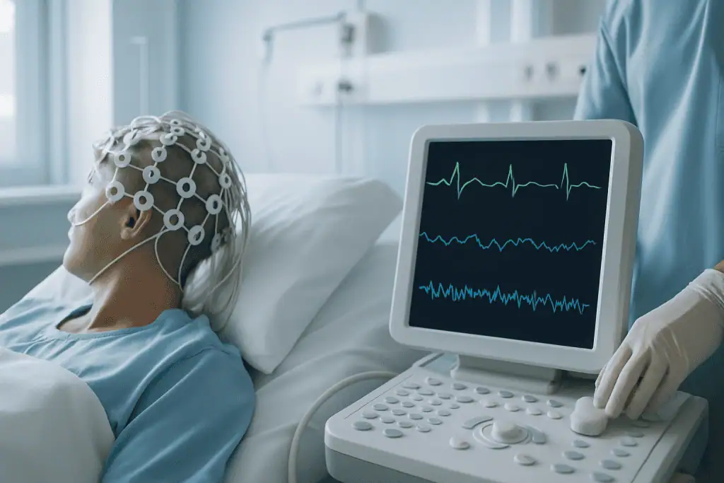
Quick answer
Neurocardiology is the medical field that evaluates how the nervous system and the heart influence each other. It is used to investigate fainting, near-fainting, palpitations with dizziness, and blood pressure regulation problems. Evaluation typically combines detailed history-taking, ECG, rhythm monitoring, echocardiography, tilt-table and autonomic testing, and neurological assessment where needed, so that treatment can target the actual mechanism behind the symptoms.
Neurocardiology: When Heart and Brain Symptoms Overlap
Neurocardiology is the medical field that examines how the nervous system and the heart influence each other — how the brain regulates heart rate, blood pressure and circulation, and how heart conditions can in turn affect the brain. It exists for patients whose symptoms sit between two specialties: fainting, near-fainting, sudden palpitations, dizziness on standing, unexplained fatigue, or episodes of almost blacking out. If your heart tests came back normal but the symptoms continue, or a neurological work-up did not fully explain why you briefly lost consciousness, neurocardiology is the discipline that looks at the connection itself rather than at each organ in isolation.
These symptoms are unsettling largely because they feel unpredictable. One patient faints in a queue or a hot shower. Another feels the heart race and the vision narrow while recovering from an illness. A third has episodes triggered by pain, fear, dehydration or prolonged standing — and a fourth has episodes while sitting or lying down, which raises different and sometimes more urgent questions. The same visible event, a brief loss of consciousness, can come from a heart rhythm problem, a blood pressure regulation problem, a disorder of the autonomic nerves, a seizure, a medication effect, or a combination. Neurocardiology exists to separate these possibilities in a structured way.
The stakes are practical as much as medical. Recurrent episodes affect work, travel, sport, driving and confidence. They also raise questions that deserve straight answers: Is this dangerous? Could it be an arrhythmia? Is it a seizure? Do I need medication, a device, lifestyle changes, or simply better information about my own physiology? A careful neurocardiology evaluation is designed to answer those questions, and to distinguish conditions that are benign but disruptive from disorders that need urgent or long-term treatment.
At Acibadem, neurocardiology is approached through coordinated assessment by physicians experienced in the interaction between the cardiovascular and nervous systems. Depending on the pattern of symptoms, the evaluation may involve cardiology, neurology, electrophysiology, internal medicine and rehabilitation specialists working from the same clinical question. The emphasis is on identifying the true cause of symptoms, reducing recurrence, and helping patients return to daily activity with a practical, evidence-based plan.
What Neurocardiology Is and How the System Works
What is neurocardiology?
Neurocardiology is the study and clinical management of the two-way relationship between the brain, the autonomic nervous system and the heart. The brain continuously regulates heart rate, blood pressure, blood vessel tone, and the body’s responses to posture, emotion, exercise, hydration and stress. Most of this regulation runs through the autonomic nervous system — the involuntary network made up of the sympathetic branch, which speeds the heart and tightens blood vessels, and the parasympathetic branch, which slows the heart and promotes rest and recovery.
When this system works well, the adjustments are invisible. Stand up, and your blood vessels tighten while your heart rate rises slightly so that blood keeps reaching your brain. Fall asleep, and heart rate and blood pressure drift down. Exercise or face a stressful moment, and the heart pumps faster while circulation redistributes. Symptoms appear when these responses become exaggerated, delayed or poorly coordinated — when the blood vessels fail to tighten on standing, when the heart rate overshoots, or when a reflex misfires and drops heart rate and blood pressure at the same time.
Much of the moment-to-moment control runs through the baroreflex: pressure sensors in the neck arteries and the aorta report blood pressure to the brainstem many times a minute, and the brainstem answers by adjusting heart rate and vessel tone. Tilt-table and autonomic testing are, in essence, ways of watching this reflex work — or fail — under controlled conditions, which is why they occupy such a central place in the evaluation.
It is important to understand what neurocardiology is not. It is not a single test, a single procedure or a single diagnosis. It is a diagnostic and treatment pathway. Depending on the patient, it may include detailed history-taking, physical examination, electrocardiography, ambulatory rhythm monitoring, blood pressure analysis in different postures, echocardiography, tilt-table testing, autonomic function testing, neurological evaluation and targeted laboratory tests. In selected patients, more advanced cardiac or neurological testing follows. The sequence is chosen to answer a specific question, not to run every available test.
The purpose of the pathway is to clarify the mechanism: is the symptom driven by a heart rhythm disorder, a blood pressure regulation problem, autonomic dysfunction, structural heart disease, a neurological event such as a seizure, a medication effect, dehydration, a metabolic condition — or several of these together? Once the mechanism is understood, treatment can be personalised. Some patients need education and lifestyle measures and nothing more. Others need medication review, rhythm treatment, pacemaker evaluation, rehabilitation or neurological management. The honest answer differs from patient to patient, which is exactly why the structured evaluation exists.
The Heart Nervous System: More Than a Pump
The heart nervous system is a real anatomical network, not a figure of speech. Alongside the electrical conduction system that spreads each heartbeat, the heart contains its own web of nerve cells and ganglia — clusters of nerve tissue embedded in and around the heart — that process information locally and communicate constantly with the brain through the vagus nerve and the sympathetic chains. Understanding the relationship between the nervous system and the heart is the foundation on which the whole specialty rests.
Does the heart have neurons?
Yes — the heart has neurons of its own. This intrinsic cardiac nervous system is sometimes informally described as the heart’s “little brain”, because it can integrate signals and adjust cardiac function locally, moment to moment, rather than waiting for every instruction to travel from the head. The neurons in the heart sit mainly in ganglia clustered on the heart’s surface, and they help fine-tune heart rate, the strength of contraction and the coordination between chambers. Signals also flow upward: the heart sends a continuous stream of information back to the brainstem about pressure, stretch and chemistry, and the brain uses that feedback to adjust circulation across the whole body.
This two-way traffic explains phenomena patients notice every day. Emotion, pain and fear can change heart rate within a beat or two. Slow breathing can calm a racing pulse. A strong vagal reflex — triggered by pain, the sight of blood, heat or standing too long — can drop heart rate and blood pressure abruptly enough to cause fainting. Conversely, disease or injury affecting the heart neurons and their connections can alter rhythm stability and blood pressure control. When physicians investigate a nervous system in heart tissue that is misfiring, over-reacting or under-reacting, they are working squarely inside neurocardiology, and this is why the specialty cannot be reduced to either cardiology or neurology alone.
How can brain events affect the heart?
Brain events can affect the heart directly, because the traffic between the two organs runs in both directions. Severe emotional or physical stress can trigger stress cardiomyopathy — sometimes called takotsubo or “broken heart” syndrome — in which a surge of stress signalling temporarily weakens part of the heart muscle and produces symptoms that closely resemble a heart attack. Acute neurological events such as stroke, subarachnoid haemorrhage and traumatic brain injury can provoke electrocardiogram changes, rhythm disturbances, blood pressure instability and, occasionally, injury to heart muscle, because the brain regions and pathways that regulate the circulation are themselves affected. Epileptic seizures can be accompanied by marked heart rate changes during and after the event. These interactions matter clinically: patients recovering from a neurological event may need cardiac monitoring, and an unexplained cardiac finding sometimes points back to a neurological cause. Recognising both directions of the relationship is part of what makes the field distinct.
Who May Need a Neurocardiology Evaluation
A neurocardiology assessment is usually considered when symptoms involve both circulation and nervous system responses, particularly when routine testing has not produced a clear explanation. The most common trigger for referral is syncope — the medical term for fainting — but many patients are evaluated before complete loss of consciousness ever occurs. Near-fainting that keeps recurring deserves the same structured attention as fainting itself.
Patients describe the episodes in recognisable ways: a sudden sense that they are about to go down, tunnel vision, sweating, nausea, weakness, ringing in the ears, palpitations, chest discomfort, tremor, shortness of breath, or confusion after an episode. Some notice symptoms in specific situations — standing in a queue, taking a hot shower, exercising, recovering from illness, skipping meals, travelling, or under emotional stress. Others have episodes while sitting or lying down, a pattern that points away from simple reflex fainting and typically prompts a more urgent cardiac evaluation.
The evaluation is also valuable when palpitations and dizziness occur together, because the same sensation can carry very different levels of risk. Palpitations can come from benign extra beats, anxiety-related adrenaline surges, supraventricular tachycardia, atrial fibrillation, ventricular arrhythmias, thyroid disease, anaemia, stimulant use or autonomic disorders. Which of these applies depends on the patient’s age, medical history, heart structure, family history and — decisively — the rhythm actually recorded while symptoms are happening. Conditions in this group overlap heavily with the broader field of heart rhythm disorders, and the two pathways often run together. Some presentations also differ between men and women, which is one reason women’s heart health is assessed with its own attention to symptom patterns.
Diagnosis begins with the story, and this is not a formality. A detailed history often carries the most important clues: what the patient was doing before the event, whether there were warning signs, how long the episode lasted, whether there were abnormal movements, whether the patient was confused afterwards, and whether injury occurred. Physicians also ask about medications, hydration, sleep, infection, recent weight loss, pregnancy, diabetes, neurological disease, existing heart disease, and any family history of sudden cardiac death or inherited rhythm disorders. A witness account — or, where safely available, a short video of an episode — can be worth more than an additional scan.
Why would I be referred to an electrophysiologist?
A referral to an electrophysiologist usually means your physicians suspect that the heart’s electrical system itself may be causing or contributing to your symptoms. Electrophysiologists are cardiologists who specialise in heart rhythm: they interpret ambulatory monitoring, decide when longer or implantable monitoring is justified, perform invasive electrical studies when the question demands it, and deliver rhythm treatments such as catheter ablation and device therapy. Within a neurocardiology pathway, the electrophysiologist’s role is to confirm or exclude an arrhythmic mechanism — because fainting caused by a heart that beats too fast, too slow or irregularly is managed very differently from fainting caused by a reflex or by low blood pressure on standing. Referral does not mean a procedure is planned; often it means the team wants a rhythm specialist’s judgement before committing to any long-term treatment.
Conditions Neurocardiology Can Address
What are the common neurological cardiac issues?
The common neurological cardiac issues are conditions in which heart rhythm, blood pressure regulation, brain perfusion and autonomic control interact — most frequently reflex fainting, orthostatic blood pressure problems, postural tachycardia, and arrhythmias that reduce blood flow to the brain. The evaluation is deliberately broad, because its job is to separate similar-looking conditions and identify the safest management plan for each.
- Vasovagal syncope: the most common cause of fainting, in which a nervous system reflex triggers a sudden drop in heart rate, blood pressure or both. It can be provoked by pain, fear, heat, dehydration, prolonged standing or medical procedures. It is rarely dangerous in itself, but it is disruptive and can cause injury.
- Orthostatic hypotension: a significant fall in blood pressure after standing, causing dizziness, weakness, blurred vision or fainting. It may relate to dehydration, medications, ageing, diabetes, Parkinson’s disease or autonomic failure.
- Postural orthostatic tachycardia syndrome (POTS): an excessive heart rate rise on standing, with palpitations, dizziness, fatigue, brain fog, exercise intolerance, and sometimes nausea or headache.
- Unexplained palpitations with dizziness: a combination that often needs rhythm monitoring and autonomic assessment to distinguish arrhythmia from non-arrhythmic causes.
- Arrhythmia-related fainting: episodes that occur when the heart beats too fast, too slow or irregularly, reducing blood flow to the brain. These situations can require prompt cardiology or electrophysiology care.
- Seizure versus syncope: some fainting episodes include brief jerking movements or confusion that can resemble seizures. Careful history, cardiac testing and neurological assessment separate the two.
- Autonomic dysfunction: disorders of the autonomic nerves that alter blood pressure, heart rate, sweating, temperature regulation, digestion and urinary function.
- Medication-related dizziness or fainting: blood pressure drugs, diuretics, antidepressants, sedatives, prostate medications and other treatments can contribute in susceptible patients.
- Cardiac conditions with neurological symptoms: structural heart disease, valve disease, cardiomyopathy, congenital heart disease and certain rhythm disorders can affect brain perfusion and need integrated evaluation. Reduced pumping function, as seen in heart failure, can also change how the circulation tolerates posture and exertion.
A word of honesty here: not every patient with dizziness needs a neurocardiology work-up. Inner-ear disorders, migraine, anxiety disorders, anaemia, infection, low blood sugar and many other conditions produce similar sensations and are managed elsewhere. The value of neurocardiology is its ability to organise the evaluation when symptoms involve circulation, consciousness, rhythm and neurological function at the same time — and to say plainly when the problem lies outside its remit.
How the Evaluation and Treatment Are Performed
Preparation Before the Visit
Preparation begins before the first appointment. Patients are typically asked to gather prior medical records: electrocardiograms, echocardiography reports, Holter or event monitor results, blood tests, hospital discharge summaries, current medication lists and any neurological test results. If fainting has occurred, a witness description of the event carries real diagnostic weight — how the patient looked, how long the episode lasted, whether there were movements, how quickly awareness returned. A short video recorded during an episode, when one exists, can be similarly useful.
Patients are usually encouraged to keep a simple symptom diary before the visit: when symptoms occur, what seems to trigger them, how long they last, what helps, and whether warning signs come first. Details about hydration, caffeine, alcohol, sleep, recent illness, exercise habits, menstrual cycle and medication timing can all be relevant. For certain tests, the team provides specific instructions about food, fluids or caffeine beforehand. Any change to medication belongs to the treating doctor; the preparation stage is about information, not adjustment.
The Clinical Assessment
The first step is a detailed consultation. The physician works through the history, then examines the patient with attention to both cardiovascular and neurological clues. Orthostatic measurements — blood pressure and heart rate taken lying, sitting and standing — show how the circulation responds to posture in real time. A standard electrocardiogram can reveal rhythm and conduction patterns, evidence of prior heart injury, or features suggesting inherited electrical disorders.
Blood tests assess anaemia, electrolytes, thyroid function, kidney function, glucose, inflammation, vitamin deficiencies and other contributors. Echocardiography may be recommended to evaluate heart structure, valve function, pumping strength, and congenital or acquired abnormalities. If arrhythmia is suspected, ambulatory monitoring is arranged for a day, several days or longer, depending on how often symptoms occur — the goal is always to catch the rhythm during a symptom, because that single correlation often settles the diagnosis.
Tilt-Table and Autonomic Testing
For patients with fainting, near-fainting, orthostatic symptoms or suspected autonomic dysfunction, a tilt-table test can be decisive. The patient lies on a special table while heart rate and blood pressure are monitored continuously; the table is then tilted upright to reproduce the effect of standing, in a controlled setting with the team present. The point is to see whether the patient’s usual symptoms appear and, if they do, exactly what the heart rate and blood pressure were doing at that moment. Reproducing the episode under observation is uncomfortable but informative — it converts a mysterious event into a measured one.
Autonomic testing extends this picture. It may include heart rate variability during deep breathing, the response to the Valsalva manoeuvre, blood pressure responses to posture, and other non-invasive assessments of nervous system control. Together these tests indicate whether symptoms are driven by abnormal autonomic reflexes, blood vessel response, or heart rate regulation — three mechanisms that feel identical to the patient but are treated differently.
Rhythm Evaluation and Cardiac Testing
If the pattern points towards arrhythmia, the pathway extends into longer rhythm monitoring, exercise testing, electrophysiology consultation or additional cardiac imaging. Ambulatory monitors record the rhythm during ordinary daily life. When events are rare but concerning — for example, infrequent faints without warning — an implantable monitor may be considered, because it can wait months for the next episode without the patient doing anything.
Exercise testing is used when symptoms occur during exertion, or when the team needs to assess heart rhythm, blood pressure response and exercise capacity under load. Cardiac imaging follows when structural disease is suspected. The technology is chosen to fit the clinical question, not the other way round: some patients need only focused testing, while others require the full staged pathway. A normal result at one level does not automatically trigger the next test; it triggers a decision about whether the question has been answered.
Neurological Evaluation
When episodes include prolonged confusion, tongue biting, unusual movements, loss of bladder control, headache, focal weakness, or events without typical fainting triggers, neurological evaluation joins the pathway. This may involve neurological examination, brain imaging, electroencephalography or other tests chosen from the history. The purpose is not to over-test — it is to avoid missing seizure disorders, cerebrovascular conditions or neurological diseases that can convincingly mimic syncope. Getting this distinction right early prevents years of treating the wrong condition.
Treatment Planning
Treatment follows the diagnosis, not the symptom. For vasovagal syncope, the plan may include education about triggers, hydration, salt intake when medically appropriate, physical counter-pressure manoeuvres, exercise conditioning, and medication in selected cases. For orthostatic hypotension, management may involve a medication review by the treating doctors, increased fluid intake, compression garments, gradual position changes, and targeted drugs when needed. For POTS, the plan often combines structured exercise, hydration strategies, sleep optimisation, symptom-directed medication and support for associated conditions.
If an arrhythmia is identified, treatment may include medication, catheter-based rhythm procedures, device therapy or ongoing electrophysiology follow-up. If a neurological condition is found, neurologists direct that arm of care. When medication side effects are part of the picture, physicians adjust timing, dosage or alternatives in coordination with the patient’s own doctors — never unilaterally, and never in a way that leaves the patient’s home team out of the loop.
How long does all of this take? It varies, and it is fair to say so plainly. Some patients complete the core testing within a short visit; others need staged monitoring precisely because their symptoms are intermittent. Recovery from the testing itself is usually brief — a patient may feel tired after a tilt-table test, or symptomatic if their usual episode was reproduced, and observation continues until they are stable. The longer “recovery” in neurocardiology is really the process of symptom control: learning triggers, adjusting treatment as monitoring results come back, and gradually rebuilding confidence in normal activity.
Why Acting Early Matters
Many causes of fainting and dizziness are manageable, but repeated episodes are not something to normalise. The most immediate risk is injury. Falls cause fractures, head trauma, dental injury and accidents — particularly when episodes strike on stairs, in bathrooms, during sport, while driving or in public places. Even near-fainting is hazardous when it happens during an activity that demands full alertness.
The second reason is that a small but important group of patients have cardiac causes that need timely treatment. Physicians treat certain features as higher-priority findings: fainting during exertion, fainting while lying down, palpitations immediately before loss of consciousness, known heart disease, abnormal electrocardiogram findings, a family history of sudden cardiac death, or chest pain accompanying the episodes. These patterns move a patient up the diagnostic queue precisely because they can signal conditions where waiting carries real risk.
Delay also makes the problem socially heavier. Patients start avoiding travel, exercise, school, work or social situations for fear of the next episode. Deconditioning then worsens orthostatic symptoms, creating a cycle of reduced activity and increased sensitivity. Early diagnosis breaks this cycle by replacing fear with an explanation and a plan — even when the explanation is a benign one, knowing it is benign changes how a person lives.
There is one further argument for acting early: it prevents fragmented care. Patients who move between clinics without a unifying diagnosis tend to accumulate partial test results — a normal Holter here, an unremarkable brain scan there — without anyone reconciling them into a single picture. A coordinated assessment brings prior records together, clarifies what has already been ruled out, identifies what still needs attention, and produces a structured plan that any treating physician can pick up and continue without repeating everything from scratch.
Benefits of a Neurocardiology Evaluation
The central benefit is precision: a clearer understanding of symptoms that do not fit neatly into one specialty, and a treatment plan matched to the actual mechanism rather than the most visible symptom.
| Benefit | What It Means for You |
|---|---|
| More accurate diagnosis | Testing is selected to determine whether symptoms come from rhythm problems, blood pressure regulation, autonomic dysfunction, neurological events, medication effects, or a combination. |
| Risk clarification | Physicians assess the warning features that can indicate a higher-risk cardiac or neurological condition, separating urgent concerns from disruptive but less dangerous ones. |
| Personalised treatment plan | Care may include lifestyle measures, medication review, targeted drug therapy, rehabilitation, rhythm treatment, neurological management or specialist follow-up — chosen by mechanism, not by habit. |
| Reduced recurrence and injury risk | When triggers and physiological patterns are understood, patients can take practical steps to reduce episodes and avoid situations where fainting could cause harm. |
| Coordination between specialties | Cardiology and neurology perspectives are brought together — especially valuable when previous evaluations have been incomplete or have reached conflicting conclusions. |
Evaluation and Follow-Up Timeline
Because neurocardiology is an evaluation and management pathway rather than a single operation, the timeline below describes testing, treatment adjustment and the return to normal activity rather than surgical recovery.
| Time Period | What Patients Can Expect |
|---|---|
| Day 1 | Consultation, examination, review of records, electrocardiogram, orthostatic measurements, and planning of any further tests. Some patients complete selected diagnostic tests the same day. |
| First week | Additional testing where needed: rhythm monitoring, echocardiography, tilt-table testing, autonomic assessment, exercise testing or neurological evaluation. Initial treatment recommendations are usually discussed. |
| First month | The treatment plan is put into practice. Medication adjustments, hydration strategies, exercise conditioning, compression use or rhythm management are refined according to symptom response. |
| Longer term | Follow-up focuses on reducing recurrence, improving tolerance of daily activity, reviewing monitoring results, and coordinating ongoing care with the patient’s own physician where needed. |
What Influences Outcomes and a Good Result
A good result in neurocardiology means more than a normal test report. It means the patient understands what is happening, serious causes have been properly considered, and the treatment plan fits real daily life. Outcomes vary — that is worth stating plainly — because symptoms arise from different mechanisms, and some of the underlying conditions are chronic or fluctuate over time.
The single most important factor is the accuracy of the initial diagnosis. Fainting from vasovagal syncope is managed completely differently from fainting caused by heart block, ventricular arrhythmia, seizure or medication-induced low blood pressure. When the mechanism is clearly identified, treatment can be focused and unnecessary interventions can usually be avoided; when it is guessed at, patients accumulate treatments that do not work.
Symptom frequency and documentation matter almost as much. Frequent episodes are easier to capture on monitoring; rare ones need longer or implantable monitoring and more patience. Patients who keep a detailed symptom diary, bring their prior test results and describe triggers carefully genuinely change the quality of their own evaluation — this is one area of medicine where the patient’s notes are part of the diagnostic toolkit.
Underlying health conditions shape both the treatment options and the response to them. Diabetes, Parkinson’s disease, autoimmune disorders, connective tissue conditions, heart failure, congenital heart disease, valve disease, thyroid disease, anaemia, sleep disorders and anxiety can all influence symptoms. Medication burden is another major factor, particularly in older adults or anyone taking drugs that affect blood pressure, heart rhythm or alertness — reviewing that burden is a task for the treating doctors, and it often yields more improvement than any new prescription.
Patient participation is central to the result. Many neurocardiology treatment plans rest on daily habits: hydration, deliberate posture changes, salt intake when medically appropriate, exercise reconditioning, compression garments, regular sleep and avoiding known triggers. These measures sound modest. Matched to the correct diagnosis and practised consistently, they are often the difference between recurrent episodes and a stable, ordinary life.
Can heart disease be reversed with diet and exercise?
Partly, and honestly only partly. Diet and exercise can substantially improve the factors that drive many heart conditions — blood pressure, cholesterol, weight, blood sugar, fitness and autonomic balance — and in some patients this slows or stabilises disease and reduces symptoms meaningfully. Deconditioning-related orthostatic symptoms, in particular, often improve with structured exercise. What lifestyle change generally cannot do is undo established structural damage: scarred heart muscle does not regrow, and a badly narrowed valve does not reopen through diet. That is why lifestyle measures are treated as a foundation of care rather than a replacement for it, and why conditions with a structural or electrical basis still need their own targeted treatment alongside the healthier routine.
For arrhythmia-related conditions specifically, outcomes depend on the type of rhythm disorder, the heart’s structure, associated disease, and the response to medication, ablation or device-based therapy where these are indicated. For autonomic disorders, improvement is often gradual and requires ongoing adjustment. The best care plans are flexible by design: they evolve as monitoring results return and as the patient’s symptoms change.
Neurocardiology at Acibadem
Patients often reach a neurocardiology pathway after symptoms have proved difficult to explain, when previous tests have not matched the severity of what they experience, or when they want a second opinion before committing to long-term medication, a rhythm procedure or device therapy. Neurocardiology rewards organised collaboration, and Acibadem’s hospital network is structured to bring the relevant specialists — cardiologists, neurologists, electrophysiologists, imaging specialists, internal medicine physicians and rehabilitation professionals — together around a single clinical question rather than passing the patient between departments.
In complex cases, multidisciplinary discussion aligns the diagnostic pathway and avoids isolated decision-making. This matters most when a symptom such as fainting has several plausible causes: the cardiologist’s, neurologist’s and electrophysiologist’s readings of the same episode need to be reconciled before treatment is chosen, not after. The diagnostic infrastructure supports this: electrocardiography, echocardiography, ambulatory rhythm monitoring, exercise testing, tilt-table evaluation, autonomic function assessment, advanced cardiac imaging, neurological testing and laboratory evaluation are available within the same system. The purpose of the technology is practical — to connect symptoms with measurable changes in rhythm, blood pressure, brain function or heart structure, so that treatment decisions rest on evidence rather than inference.
Physicians work from international, evidence-based treatment protocols while adapting recommendations to each patient’s history, risk profile and daily circumstances. For some patients the right plan is conservative and education-focused; for others the findings lead to medication, rhythm intervention, device evaluation, neurological treatment or a coordinated long-term management programme. Continuity after the visit is planned rather than assumed: patients receive medical reports, test results and treatment recommendations prepared so that their own physicians can continue monitoring, medication adjustment, rehabilitation or specialist review without starting over.
Living With the Diagnosis
Symptoms that involve the heart, the brain and the autonomic nervous system are frightening precisely because they are sudden and hard to predict. A neurocardiology evaluation replaces that unpredictability with a mechanism: a named condition, a measured pattern, and a plan built around it. For many patients the diagnosis turns out to be benign but disruptive, and the treatment is knowledge — recognising the warning signs, managing triggers, and trusting the body again in queues, showers, journeys and gyms. For a smaller group, the evaluation finds a condition that needs specific cardiac or neurological treatment, and finding it early is exactly what the pathway is for.
Either way, the measure of success is the same: fewer episodes, less fear around the episodes that remain, a plan the patient actually understands, and a set of records that any physician, anywhere, can pick up and continue. That is what a well-run neurocardiology pathway is designed to deliver — not certainty about everything, which medicine cannot honestly promise, but clarity about what is happening, what has been ruled out, and what to do next.
Our Specialists Explain
 Vasovagal Syncope: Diagnosis and Treatment at Acibadem
Preparation
- Patients should bring previous ECGs, Holter reports, neurological evaluations, imaging, and a full medication list. Depending on planned autonomic or rhythm testing, the doctor may advise avoiding caffeine, nicotine, or certain medications before the appointment.
Aftercare
- Most patients can resume normal activities immediately after evaluation. Follow-up may include cardiac monitoring, neurological tests, medication adjustment, lifestyle guidance, or referral to cardiology and neurology specialists for coordinated care.
Turkey vs UK, Germany & USA
Neurocardiology cost comparisons depend on whether symptoms require mainly cardiac testing, neurological testing, autonomic assessment, or inpatient monitoring. The points below outline common cost and experience factors for international patients considering evaluation abroad.
For symptoms such as fainting, palpitations, dizziness, or autonomic complaints, the final cost is usually driven by the depth of diagnostic testing and whether several specialties are involved.
| Factor | Turkey | UK | Germany | USA |
|---|---|---|---|---|
| Price drivers | Private hospital evaluation, cardiology and neurology input, rhythm monitoring, autonomic testing, imaging, and laboratory work shape the package. | Private care costs vary by consultant, hospital, diagnostic tests, and whether additional specialist referrals are needed. | Costs are influenced by hospital type, specialist consultations, advanced diagnostics, and whether inpatient observation is required. | Costs may vary widely by hospital network, physician fees, facility charges, diagnostics, and insurance arrangements. |
| Hospital and specialist factors | International hospitals may coordinate cardiology, neurology, electrophysiology, and imaging in the same care pathway. | Access may be through private consultants or hospital clinics, with referrals between specialties as needed. | Specialist centres may offer structured cardiac and neurological diagnostics, often with strong subspecialty pathways. | Large academic and private centres may provide highly specialised testing, with billing often separated by provider and facility. |
| Accreditation and quality | Some hospitals serving international patients hold JCI accreditation; patients should confirm the accreditation status of the selected facility. | Quality oversight is based on national regulation and hospital governance systems. | Quality is supported by national healthcare regulation and institutional quality programmes. | Hospitals may hold national or international accreditations; coverage and billing rules should be checked carefully. |
| Waiting times | Private international patient departments may help arrange appointments and diagnostics with relatively coordinated scheduling. | Private care may offer faster access than public pathways, depending on consultant and test availability. | Scheduling depends on the centre, specialist availability, and the complexity of required investigations. | Access can be rapid in private settings, but authorisations and network rules may affect timing. |
| Travel and language logistics | International patient teams may support translation, appointment planning, airport transfers, and hotel coordination. | English language access is straightforward; overseas patients may need to organise travel and accommodation separately. | Interpreter support may be available in larger centres, but should be confirmed before travel. | English language access is standard; travel distance, accommodation, and insurance administration can add complexity. |
| Typical package scope | Packages may combine consultation, selected tests, care coordination, translation, and follow-up planning. | Fees are often arranged by consultation and test, with packages varying by provider. | Packages may be available for international patients, but detailed inclusions should be reviewed in advance. | Billing is often itemised across consultations, tests, facility use, and professional fees. |
What affects your final cost
- The main symptom pattern, such as fainting, palpitations, dizziness, or suspected autonomic dysfunction.
- The need for cardiology, neurology, electrophysiology, or other specialist input.
- The type and duration of rhythm monitoring, autonomic testing, imaging, and laboratory investigations.
- Whether evaluation is outpatient or requires hospital observation.
- Interpreter support, airport transfers, accommodation, and other international patient services.
- Whether a written treatment plan, medication review, or follow-up consultation is included.
Compare your options
Neurocardiology assessment is tailored to the suspected cause of symptoms. Suitability for each option is decided by a specialist after reviewing the patient’s history, examination findings, and prior test results.
| Option | What it is | Typical use | Key considerations |
|---|---|---|---|
| Specialist neurocardiology consultation | A coordinated review of cardiac, neurological, autonomic, medication, and lifestyle factors. | Often used when symptoms may have mixed heart, brain, or nervous system causes. | The consultation guides which tests are necessary and helps avoid unnecessary investigations. |
| ECG and ambulatory rhythm monitoring | Tests that record heart rhythm during rest or daily activities. | Used for palpitations, fainting, near-fainting, or suspected rhythm disturbances. | Monitoring choice depends on symptom frequency and clinical suspicion. |
| Tilt table and autonomic testing | Assessment of blood pressure, heart rate, and autonomic responses to posture and controlled stimuli. | Commonly considered for unexplained fainting, orthostatic intolerance, or suspected autonomic dysfunction. | Results must be interpreted alongside symptoms, medications, hydration status, and other conditions. |
| Echocardiography and cardiac imaging | Imaging tests that evaluate heart structure and function. | Used when structural heart disease, valve disease, or heart muscle problems may contribute to symptoms. | The need for imaging depends on examination findings, previous records, and risk profile. |
| Neurological testing | May include neurological examination, EEG, brain imaging, or other targeted assessments. | Considered when seizure, migraine, neuropathy, or central nervous system causes are possible. | Testing is selected according to symptom description, triggers, recovery pattern, and prior results. |
| Electrophysiology review | A detailed rhythm specialist assessment, with further invasive testing only if indicated. | Used when significant arrhythmia is suspected or non-invasive tests suggest a rhythm cause. | Benefits, risks, and alternatives should be discussed carefully before any invasive procedure. |
General information only — not medical or financial advice. Final costs depend on the factors above and your individual case; request a free, personalised quote.
Frequently Asked Questions
What affects the cost of neurocardiology evaluation?
Cost depends on the complexity of symptoms, the specialists involved, the tests selected, whether monitoring or observation is needed, and whether international patient services such as translation and transfers are included. A personalised quote can be prepared after a medical review.
How can I get a personalised quote from Acibadem?
You can request a free consultation by sharing your symptoms, previous test results, medication list, and relevant medical history. The clinical team can then suggest an appropriate evaluation plan and provide a tailored quote.
Is neurocardiology usually a single test or a combined assessment?
It is usually a combined assessment rather than a single test. The care plan may include cardiology, neurology, rhythm monitoring, autonomic testing, imaging, or laboratory work depending on specialist assessment.
Will my package include translation and travel support?
International patient packages may include care coordination, interpreter support, appointment planning, and assistance with transfers or accommodation. Exact inclusions should be confirmed before travel.
Do I need to travel if I already have test results?
Existing ECG reports, Holter results, imaging, discharge summaries, and neurological tests may help the specialist plan your visit more efficiently. However, whether further in-person testing is needed can only be decided after medical review.
Medically reviewed by the Acıbadem International Medical Board — August 30, 2026
See our medical review board →
Update history
- PublishedJune 8, 2026
- Medical review approvedAugust 30, 2026
- Last content updateAugust 30, 2026
Trusted care for international patients
Doctors Performing This Treatment

Prof. Dr. Altay Bedük
Neurosurgery
Prof. Dr. Cahide Soydaş Çınar
Cardiology
Prof. Dr. Elif Ilgaz Aydınlar
Neurology
Prof. Dr. Sertaç İşlekel
Neurosurgery
Prof. Dr. Ayşe Sağduyu Kocaman
Neurology
Prof. Dr. Elif Eroğlu Büyüköner
Cardiology
Prof. Dr. Ahmet Akyol
Cardiology
Prof. Dr. Ahmet Karabulut
Cardiology
Prof. Dr. Dilaver Kaya
Neurology
Prof. Dr. Ahmet Kaya Bilge
Cardiology
Prof. Dr. Duhan Fatih Bayrak
Cardiology
Prof. Dr. Barış Kılıçaslan
Cardiology
Prof. Dr. Hüseyin Hayrı Kertmen
Neurosurgery
Prof. Dr. Melih Bozkurt
Neurosurgery
Prof. Dr. Çağın Şentürk
Interventional Neuroradiology
Prof. Dr. Akın Sabancı
Neurosurgery
Prof. Dr. Aleks Değirmencioğlu
Cardiology
Prof. Dr. Alper Özkan
Cardiology
Prof. Dr. Erkin Sönmez
Neurosurgery
Prof. Dr. Mert İlker Hayıroğlu
Cardiology
Prof. Dr. Ali Aydınlar
Cardiology
Prof. Dr. Ender Semiz
Cardiology
Prof. Dr. Ercüment Yılmaz
Cardiology
Prof. Dr. Bekir Sıtkı Cebeci
CardiologyMedical Units
Available at These Hospitals