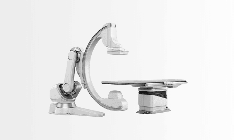
Renal Angio
Renal angio is an imaging-guided procedure used to visualize the kidney arteries and assess narrowing, blockage, aneurysm, or abnormal blood flow. It may guide further treatment such as angioplasty or stenting.

Quick answer
A renal angio, or renal angiogram, is an imaging procedure that shows the arteries supplying the kidneys. A thin catheter is guided from the groin or wrist to the renal arteries, contrast dye is injected, and X-ray images map the blood flow. It is used to diagnose narrowing, aneurysms and abnormal vessels, and in selected cases treatment such as angioplasty or stenting can follow in the same session.
Renal Angio: Seeing the Kidney Arteries Clearly Before Important Decisions
Renal angio, also called renal angiography, renal arteriography or a renal angiogram, is an imaging procedure that shows the arteries carrying blood from the aorta to your kidneys. A thin, flexible catheter is guided into an artery, usually at the groin or wrist, contrast dye is injected near the kidney arteries, and X-ray images record exactly how blood flows through them. Doctors recommend it when they need a precise vascular map: to confirm or rule out a narrowing, to assess an aneurysm, or to understand abnormal blood flow before deciding on treatment.
If a doctor has recommended a renal angio for you, it is usually because something important needs to be clarified about the blood supply to your kidneys. You may have high blood pressure that resists several medications, unexplained changes in kidney function, a suspected narrowing in a renal artery, or an aneurysm that needs closer evaluation. The recommendation tends to raise the same practical questions in most patients. Is this only a diagnostic test, or could treatment happen at the same time? Will it hurt? Could it affect kidney function? How long does recovery take? This page answers those questions in order, as plainly as the evidence allows.
The kidneys depend on a steady blood supply to filter waste, regulate fluid balance and help control blood pressure. When blood flow to one or both kidneys is reduced, the effects are often subtle at first. Some patients feel entirely well but have worsening blood pressure readings or changing kidney blood test results. Others notice fluid retention, sudden shifts in heart failure symptoms, or a decline in kidney function after starting certain medications. A renal angio helps clarify whether the kidney arteries themselves are part of the problem, or whether the explanation lies elsewhere.
It helps to understand from the start what this procedure is and is not. Renal angio is not a routine screening test, and it is not ordered on suspicion alone. It sits near the end of a diagnostic pathway, after simpler tests have raised a question that only detailed vascular imaging can settle. It is a decision-making tool: the pictures it produces are only as useful as the clinical judgement applied to them.
The procedure is usually one step within a wider evaluation involving nephrology, cardiology, urology, vascular surgery or interventional radiology, depending on the reason for the test. The practical questions, how the study is planned, what happens on the day, and what follows once the diagnostic images have been reviewed, deserve the same clear answers as the medical ones, and this page addresses those too.
What Is Renal Angio?
Renal angio is a catheter-based imaging procedure used to examine the renal arteries, the vessels that carry oxygen-rich blood from the aorta to each kidney. In most cases it is performed by inserting a thin catheter into an artery at the groin (the femoral artery) or the wrist (the radial artery) and guiding it under continuous X-ray imaging to the point where the kidney arteries branch off the aorta. Contrast dye is then injected through the catheter while a rapid sequence of images captures the flow of blood in real time. Most units use digital subtraction angiography, a technique that removes bone and soft tissue from the image so the vessels stand out sharply against a blank background.
This level of detail matters because the renal arteries are relatively small, and their disease patterns can be subtle. A renal angio can show the exact location and severity of a narrowing, whether blood flow is delayed or reduced, whether an aneurysm is present and how it relates to branch vessels, and whether abnormal connections exist between arteries and veins. It can resolve questions that renal ultrasonography, CT or MRI have raised but not fully answered. In some patients the angiogram follows other imaging; in others it is used when symptoms and blood test findings strongly suggest a vascular cause but the diagnosis remains uncertain.
What is a renal angiogram for?
A renal angiogram is used to diagnose problems in the blood vessels supplying the kidneys and, where appropriate, to guide their treatment. The most common reasons include:
- Confirming and grading a suspected narrowing (renal artery stenosis) found or suspected on other imaging
- Investigating high blood pressure that behaves as if it has a vascular cause
- Mapping a renal artery aneurysm before deciding whether it needs treatment or observation
- Identifying abnormal connections between arteries and veins, including those that can follow a kidney biopsy or trauma
- Locating active bleeding from a kidney or its vessels, sometimes with treatment in the same session
- Providing detailed vascular mapping before selected operations or transplant-related decisions when non-invasive imaging is not clear enough
Is renal angio a diagnostic test or a treatment?
It can be both, and that is one of its main practical advantages. Renal angiography is primarily diagnostic, but the same catheter access that produces the images can also deliver treatment. If a significant narrowing is found and your condition supports intervention, the physician may perform angioplasty, widening the vessel with a small balloon, place a stent to hold it open, or carry out embolisation to close an abnormal vessel or stop bleeding. Whether treatment happens in the same session depends on the angiographic findings, your kidney function, your blood pressure history, your age and other medical conditions, and on what was agreed and consented to before the procedure began.
In modern practice, not every renal artery narrowing requires a procedure. Many patients do well with carefully managed medication and follow-up imaging, and large clinical studies have made specialists more selective about who genuinely benefits from stenting. The value of renal angio is precisely that it helps distinguish findings that can be safely observed from those that may warrant intervention. It is not simply a picture-taking exercise; it is the point at which a treatment decision becomes properly informed.
Who May Need Renal Angio?
A renal angio may be recommended when a physician suspects that reduced or abnormal blood flow to the kidneys is contributing to symptoms, abnormal test results, or a potentially serious vascular finding. The most common concern is renal artery stenosis, a narrowing of one or both kidney arteries. This usually develops through atherosclerosis, plaque building up in the artery wall, typically in older patients with other cardiovascular risk factors, or through fibromuscular dysplasia, a non-atherosclerotic condition of the artery wall that tends to affect younger and middle-aged patients, particularly women.
Some patients are referred because of high blood pressure that resists control despite several medications taken correctly. Others have a sudden worsening of previously stable blood pressure, or develop hypertension at an age when essential hypertension is unusual, very young or comparatively late in life. A renal vascular cause may also be considered when blood pressure deteriorates quickly, when imaging shows a difference in kidney size between the two sides, or when kidney function declines noticeably after starting medicines that act on the renin-angiotensin system. These patterns do not prove a vascular cause, but they raise the question clearly enough to justify detailed imaging.
Renal angio may also be considered when kidney function worsens without a clear explanation, particularly if other imaging suggests poor blood supply. Patients with widespread vascular disease, coronary artery disease, peripheral artery disease, a long smoking history, carry a higher likelihood of renal artery narrowing, because atherosclerosis rarely confines itself to one vascular territory. In some people, recurrent episodes of sudden fluid overload or pulmonary oedema prompt evaluation of the renal arteries, because severe bilateral narrowing can destabilise both blood pressure and fluid regulation.
Before a renal angio is recommended, patients normally pass through a structured diagnostic pathway. This may include blood and urine tests to assess kidney function, ambulatory blood pressure monitoring, Doppler ultrasound of the renal arteries, CT angiography or MR angiography. The care team also reviews current medications, previous procedures, allergies, cardiovascular history and any prior scans. Catheter angiography is typically chosen only when the expected diagnostic value is meaningful and when the answer could realistically change management: a principle that protects patients from unnecessary invasive testing.
Wherever the evaluation takes place, the same principle applies. Previous imaging, laboratory results, medication lists and physician notes allow the team to judge whether renal angio is genuinely appropriate, whether additional non-invasive imaging should come first, and how to plan the procedure safely around kidney function. A thorough record review often shortens the time between referral and a clear answer.
Conditions and Indications a Renal Angio Can Address
Renal angio is used across several vascular conditions involving the kidneys. The indication differs from patient to patient, and the same finding can lead to different plans depending on context, which is why the conditions below are described individually.
Renal artery stenosis
Renal artery stenosis is the most common indication for renal angio. The narrowing reduces blood flow to the kidney, which responds by activating hormonal systems that raise blood pressure throughout the body. In advanced cases, sustained low flow can contribute to loss of kidney tissue and function. The angiogram defines the severity, location and anatomy of the narrowing, whether it sits at the origin of the artery, along its trunk or in the branches, which directly shapes the choice between medical therapy, angioplasty and stenting. In selected cases, pressure measurements across the narrowing during the same procedure help establish whether it is functionally significant rather than merely visible.
Fibromuscular dysplasia
Fibromuscular dysplasia produces a characteristic beaded pattern of alternating narrowing and widening in the renal arteries, quite different from the focal plaque of atherosclerosis. Patients often present with high blood pressure at a younger age or with hypertension that resists standard treatment. Renal angio can confirm the diagnosis when non-invasive imaging is equivocal, and in carefully selected patients it guides balloon angioplasty, which is the intervention most often considered for this condition. Stents are used less often here than in atherosclerotic disease, another example of why the underlying diagnosis matters as much as the image.
Renal artery aneurysm
A renal artery aneurysm is an abnormal widening of the artery wall. Many are found incidentally on scans done for other reasons and simply need measurement and periodic follow-up. Some require detailed assessment because of their size, documented growth, symptoms, pregnancy considerations or concern about rupture. Renal angio maps the aneurysm precisely: its neck, its relationship to branch vessels, and the kidney tissue those branches supply, which is essential information if endovascular or surgical treatment is being weighed.
Arteriovenous malformations and fistulas
Arteriovenous malformations and fistulas are abnormal direct connections between arteries and veins. They may be congenital, follow trauma, or develop after procedures such as a renal biopsy. Because blood short-circuits from artery to vein at high pressure, these lesions can cause blood in the urine, strain on the heart or, occasionally, high blood pressure. Renal angio identifies the feeding vessels and the drainage pattern, and it can guide embolisation, deliberately closing the abnormal connection from inside the vessel, when treatment is needed.
Vascular injury, bleeding and trauma
Injury to the kidney or its vessels, from accidents, procedures or spontaneous rupture of an abnormal vessel, may require urgent angiographic evaluation. The angiogram shows where blood is escaping the circulation. If active bleeding is identified, minimally invasive embolisation can often be performed during the same session, controlling the bleeding while preserving as much functioning kidney tissue as possible. This is one of the situations where the diagnostic and therapeutic roles of renal angio merge completely.
Preoperative and transplant-related vascular assessment
Detailed vascular mapping is sometimes needed before kidney surgery or in transplant-related evaluation. Most of this work is now done with CT or MR angiography, which are non-invasive. Catheter angiography is reserved for situations where those tests leave genuine uncertainty, for example, complex branch anatomy, prior vascular procedures, or findings that would change the surgical plan if confirmed. A substantial minority of people also have accessory renal arteries, additional vessels supplying part of a kidney, and confirming their presence and course can matter considerably when surgery is planned.
How Renal Angio Is Performed
Renal angio is performed in an angiography suite by an interventional radiologist, interventional cardiologist or vascular specialist experienced in image-guided arterial procedures. The process is planned to balance diagnostic quality against patient safety: a balance that gets particular attention in people who already have reduced kidney function or significant cardiovascular disease.
What is the protocol for a renal angiogram?
The protocol for a renal angiogram begins well before the procedure day, with a structured pre-assessment. Your physician reviews the indication and your medical history, then checks kidney function, blood clotting status, blood count, allergies and any previous reactions to contrast dye. Diabetes medication and blood thinners receive specific attention, because both can interact with the procedure; decisions about continuing, pausing or adjusting any medicine rest entirely with your treating doctor, who will give you explicit written instructions. If you have chronic kidney disease, the team may plan a lower contrast dose, arrange hydration before and after the procedure, and consider whether an alternative imaging method should be tried first.
You will usually be asked not to eat or drink for several hours beforehand. On the day itself, the team confirms your identity, the indication, your consent, your allergies and your latest laboratory results: a deliberate checklist, repeated for every patient. An intravenous line is placed. Most patients receive local anaesthetic at the access site plus mild sedation, enough to stay relaxed while remaining awake and able to respond to instructions. Deeper sedation or anaesthesia is reserved for specific situations, decided case by case.
Step by step: what happens during the procedure
- The access site, groin or wrist, is cleaned, draped and numbed with local anaesthetic. Ultrasound guidance is often used to place the needle accurately.
- A small puncture is made in the artery and a short sheath is inserted, through which the catheter passes.
- The catheter is guided under continuous X-ray imaging through the arterial system to the point near where the renal arteries leave the aorta. You do not feel it moving; arteries have no sensation of touch from the inside.
- Contrast dye is injected while rapid X-ray images are recorded. You may feel a brief warm flush as the contrast circulates, normal and short-lived.
- Images are taken from several angles so the physician can judge each narrowing, aneurysm or abnormal vessel in three dimensions rather than one projection.
- In selected cases, a pressure wire measures the pressure drop across a narrowing, clarifying whether it is functionally significant.
- If the study is purely diagnostic, the catheter and sheath are removed. If a treatable problem is found and prior consent and planning support it, angioplasty, stenting or embolisation may follow immediately.
Modern angiography combines high-resolution fluoroscopy, digital image processing and careful contrast delivery to visualise small vessels while limiting exposure. Closure devices may seal the puncture at the end of the procedure where the anatomy suits them, shortening the period of enforced rest afterwards. None of this changes the fundamental logic of the study: get clear images, interpret them in context, decide with the patient.
Is a renal angiogram painful?
Most patients describe discomfort rather than pain, though experiences vary. The local anaesthetic injection at the groin or wrist stings briefly; after that, the commonest sensations are pressure at the puncture site, the short warm flush when contrast is injected, and the stiffness of lying still on the procedure table. The catheter itself is not felt as it moves through the arteries. Sedation takes the edge off anxiety for most people. Afterwards, the access site is typically sore and bruised for a few days, comparable to a deep bruise, and this is managed with simple measures your team will explain. If an intervention such as stenting is added, some patients notice a dull ache in the flank for a short period.
How long does a renal angiogram take?
The imaging itself often takes less than an hour in a straightforward diagnostic case. Your total time in the department is longer, commonly several hours, because it includes preparation, positioning, post-procedure monitoring and the initial recovery period. Complex anatomy, additional pressure measurements or treatment performed in the same session all extend the procedure. Many patients go home the same day; an overnight stay may be advised after an intervention, when kidney function needs rechecking, or when other medical conditions call for closer observation. It is sensible to keep the rest of the day free: sedation, monitoring and the first discussion of the images all take time even when the imaging itself is quick.
After the procedure
Once the catheter is removed, firm pressure is applied to the puncture site, or a closure device is used. You are monitored while the sedation wears off: nurses check blood pressure, pulse, the puncture site and urine output at intervals. If the groin was used, you will lie flat for a period to protect the artery; if the wrist was used, you can usually sit up sooner, though the arm needs protecting for a short time. Before discharge, the team explains which sensations are expected, which signs should be reported, and when your follow-up tests are scheduled.
Most patients return to light activities quickly, depending on the access site, the sedation used, kidney function and whether treatment was performed. Typical advice includes drinking fluids if your condition allows, avoiding heavy lifting for several days, and keeping the puncture site clean and dry. Follow-up usually involves repeat kidney function blood tests and blood pressure review, timed according to your individual situation.
Risks, Contrast Safety and Kidney Protection
Renal angio is an invasive procedure, and an honest account includes its risks. The most frequent issues are minor: bruising or tenderness at the access site, and short-lived effects of sedation. Less common problems include bleeding at the puncture, damage to the accessed vessel, allergic-type reactions to contrast dye, and a temporary effect of contrast on kidney function: a particular consideration in this procedure, since the patients who need it often have kidneys that are already vulnerable. Serious complications are uncommon in experienced hands, but they exist, which is why the pre-assessment is thorough and why the procedure is only offered when the expected benefit justifies it.
Kidney protection is planned rather than assumed. Depending on your kidney function, the team may limit the volume of contrast, choose imaging angles that answer the question with fewer injections, arrange intravenous hydration before and after the study, and review any medicines that interact with contrast, with all medication decisions made by your treating doctor. In some patients, alternative contrast agents or a different imaging strategy altogether may be more appropriate, and a good team will say so plainly rather than proceed by default. In selected patients whose kidney function makes standard iodinated contrast a concern, some centres can perform parts of the study using carbon dioxide as the contrast medium, a gas the body clears through the lungs, accepting somewhat different image characteristics in exchange for sparing the kidneys.
Radiation exposure is managed the same way: modern angiography systems use dose-reduction technology, and operators keep screening time and image counts to what the clinical question requires. The exposure from a diagnostic renal angio is a recognised part of the risk-benefit discussion, not an afterthought, and it is one more reason the procedure follows, rather than replaces, non-invasive tests.
How Renal Angio Compares With Other Vascular Imaging
Renal angio sits at the top of a stepwise imaging pathway, not at the start of it. Doppler ultrasound of the renal arteries is usually the first test, because it is non-invasive, uses no contrast and can measure flow velocities that hint at narrowing; its limitation is that image quality depends on body habitus and operator experience. CT angiography and MR angiography produce detailed three-dimensional pictures of the renal arteries without catheterisation and answer the question outright in many patients. Catheter angiography is reserved for cases where those tests disagree, where fine branch detail matters, where pressure measurements are needed, or where treatment may follow in the same sitting: the situations in which its invasiveness earns its keep.
The catheter technique itself is shared across vascular territories. If you have had, or been offered, coronary angiography for the heart arteries or a carotid angio for the neck vessels, the renal version will feel familiar: the same type of access, the same contrast injections, similar monitoring afterwards. What differs is the anatomy being studied and the decisions that hang on the result. Patients with disease in one territory not infrequently have it in another, which is why findings from a renal angio are read alongside the rest of your cardiovascular picture rather than in isolation.
Why Acting Early Matters
Kidney artery disease can progress quietly. You may feel no kidney pain and no obvious symptoms even when blood flow is significantly reduced. Often the earliest signs are indirect: blood pressure creeping upward, more medications being needed to hold it, changing kidney blood tests, or new fluid retention. Delaying evaluation allows avoidable strain on the kidneys, heart and blood vessels to continue in the background.
When renal artery narrowing contributes to uncontrolled hypertension, the heart works harder over time. That raises the long-term risk of heart enlargement, heart failure, stroke and further vascular complications, and it can feed the two-way strain between heart and kidneys seen in cardiorenal syndrome. If kidney function is already declining, identifying, or excluding, a vascular cause changes medication choices, the monitoring plan and whether intervention should even be on the table.
For aneurysms, vascular malformations or suspected bleeding, timely diagnosis matters for a different reason: some findings can be safely observed for years, while others need planned intervention or urgent treatment, and the two can look similar on a routine scan. Renal angio provides the detailed vascular map that lets physicians judge how serious a finding is and which treatment route is safest.
Acting early does not mean rushing to intervene. It means obtaining the right diagnosis before the condition causes further harm or before options narrow. A careful evaluation may end in medication adjustment, surveillance, endovascular therapy or a surgical opinion, and any of those outcomes is a good one if it is the right one. Decisions are simply better when they rest on precise information.
Potential Benefits of Renal Angio
What renal angio delivers depends on why it is performed, but in each scenario the procedure produces information that directly shapes the next step in care. The table below summarises the main benefits in practical terms.
| Benefit | What It Means for You |
|---|---|
| Detailed visualisation of kidney arteries | Physicians see the exact location and pattern of narrowing, blockage, aneurysm or abnormal blood flow, not an estimate, a map. |
| More confident treatment planning | The results help determine whether medication, monitoring, angioplasty, stenting, embolisation or surgery should be considered. |
| Possibility of same-session treatment | In selected cases, a treatable vascular problem can be addressed during the angiography procedure itself, if safe and pre-agreed. |
| Clarification after inconclusive imaging | When ultrasound, CT or MRI leave the question open, renal angio can settle it, including with pressure measurements where needed. |
| Support for blood pressure and kidney care decisions | Understanding renal blood flow guides medication strategy, monitoring intervals and follow-up with nephrology or vascular specialists. |
Recovery After Renal Angio
Recovery from a diagnostic renal angio is usually straightforward. The timeline stretches somewhat if an intervention was performed in the same session or if other medical conditions need attention, but for most patients it follows the pattern below.
| Time Period | What Patients Can Expect |
|---|---|
| Day 1 | Monitoring after the procedure, mild soreness at the access site, hydration if appropriate, and clear instructions on activity and medications. |
| First week | Return to light daily activities for most patients, avoidance of heavy lifting, and routine care of the puncture site as it heals. |
| First month | Follow-up blood pressure review, kidney function testing where indicated, and a structured discussion of the angiography results and plan. |
| Longer term | Ongoing care may include medication optimisation, repeat imaging, lifestyle risk reduction, or follow-up after angioplasty, stenting or embolisation. |
Two variables shape your individual recovery more than any other: the access site and whether treatment was added. Wrist access generally allows earlier mobility than groin access, and a purely diagnostic study needs less observation than one that included stenting or embolisation. Your team tailors the discharge plan to those two facts plus your kidney function and any other medical conditions that need watching.
Factors That Influence Outcomes
The result of a renal angio is shaped by both the quality of the imaging and the clinical context in which it is read. A technically successful angiogram gives clear pictures of the arteries; the real goal is using those pictures to make the right decision for the person they belong to.
The underlying condition matters first. Renal artery narrowing caused by atherosclerosis behaves differently from fibromuscular dysplasia. Atherosclerotic disease tends to occur alongside diabetes, smoking history, high cholesterol or coronary artery disease, and those companions affect both procedural risk and long-term prospects. Fibromuscular dysplasia may respond differently to angioplasty in carefully selected patients, but selection is the operative word, and each case is assessed individually.
The degree and location of narrowing matter next. A mild narrowing may not be responsible for the symptoms at all and may not warrant intervention. A severe narrowing restricting flow to a still-functioning kidney is a different proposition. Branch vessel involvement, kidney size, how long the blood pressure has been elevated, and how much kidney function remains all influence whether restoring blood flow is likely to help, or whether the kidney downstream is already too damaged to benefit.
Kidney function before the procedure is a key consideration in its own right. Patients with reduced function need specific precautions around contrast, hydration and post-procedure monitoring, and the team weighs diagnostic benefit against those risks before proceeding, adjusting the imaging strategy where sensible.
Blood pressure history also sets expectations. Recent-onset or rapidly worsening hypertension raises different possibilities from hypertension that has been present for decades. When vascular disease has existed for a long time, blood pressure may not normalise even after a technically successful intervention, the vessels and kidneys have adapted around the problem, though treatment can still be worthwhile in selected situations. An honest team says this before the procedure, not after.
Coordination among specialists carries real weight. Nephrologists assess kidney function and medical therapy. Cardiologists and hypertension specialists evaluate cardiovascular risk and blood pressure management. Interventional radiologists and vascular specialists perform the angiographic study and any image-guided treatment. Surgeons join when anatomy or disease characteristics point to an open approach. In complex cases, multidisciplinary review ensures the angiogram is interpreted as one input to a complete care plan rather than as a verdict in isolation.
Finally, your own participation matters. Following instructions on hydration, activity restriction and follow-up testing reduces avoidable complications. Long-term vascular health usually asks for more: stopping smoking, controlling cholesterol and diabetes, managing weight, and monitoring blood pressure consistently. The angiogram identifies the vascular problem; keeping the benefit depends on the medical care and habits that follow it.
Questions Worth Discussing Before a Renal Angio
Whoever performs your procedure, the conversation beforehand should leave you clear on the essentials. Reasonable questions to raise with your treating team include:
- What question is this angiogram meant to answer, and could a non-invasive test answer it instead?
- If a significant narrowing is found, will treatment happen in the same session, and what exactly am I consenting to in advance?
- How will my kidney function be protected around the contrast injection?
- Which of my medicines will be reviewed before the procedure, and who decides any changes?
- Which access site is planned, groin or wrist, and what does that mean for my recovery?
- When and how will I receive the results, and who coordinates the follow-up plan?
How Renal Angio Is Approached at Acibadem
At Acibadem, renal angio is treated as one component of a complete vascular, nephrology, cardiology, urology or interventional radiology evaluation, depending on the reason for the test. Cases that need broader input can be discussed across specialties, particularly when the findings may lead to angioplasty, stenting, embolisation, surgery or complex medical management, where the choice between options benefits from more than one perspective.
The diagnostic pathway is deliberately selective. Not every patient with a suspected renal artery narrowing needs an angiogram, and not every angiographic finding needs a stent. The team reviews prior imaging, kidney function, blood pressure patterns, medication response and overall vascular risk before recommending the invasive study. This review matters twice over: it avoids unnecessary procedures, and it ensures that significant disease is not overlooked when it could still be treated on favourable terms.
Technically, the approach draws on high-resolution fluoroscopic imaging, digital vascular imaging, ultrasound-guided arterial access, contrast-sparing strategies where kidney function requires them, and intraprocedural pressure measurements in selected cases. The purpose of the technology is practical rather than decorative: safer access, clearer diagnosis, and treatment decisions made on measurement rather than impression.
Personalised planning is especially relevant here because patient goals differ. One person needs a definitive diagnosis before their blood pressure treatment is restructured. Another is being evaluated for an incidentally discovered renal artery aneurysm. A third has declining kidney function and a folder of inconclusive tests. The plan should reflect the individual’s condition, risk profile and preferences, after a clear discussion of benefits, limitations and alternatives, including the alternative of not intervening at all.
Moving Forward With Clarity
A renal angio is worth considering when kidney artery disease is suspected or when previous imaging has raised a question that needs a definitive answer. Its value lies not only in what it shows but in what it makes possible: a clearer diagnosis, a more precise treatment plan, and a better understanding of how kidney blood flow is affecting your overall health.
For anyone facing a possible renal angiography, or weighing the options around renal artery stenosis, a renal artery aneurysm, abnormal kidney blood flow or hard-to-control blood pressure: the most useful preparation is informational. Existing imaging studies, laboratory results, the current medication list and the wider medical history all belong in the evaluation, and a careful team reviews them before confirming that the procedure is appropriate at all. The best decisions in this field are made when patient, family and medical team share the same understanding of the diagnosis, the realistic options, and what recovery involves. A renal angio, used at the right moment for the right reason, is what makes that shared understanding possible.
Preparation
- Before renal angio, patients usually have blood tests to check kidney function and clotting status. Your doctor may ask you to stop blood thinners or adjust diabetes medications. Fasting for several hours before the procedure is commonly required, and contrast allergy or kidney disease should be discussed in advance.
Aftercare
- After the procedure, the puncture site is monitored and patients are advised to rest for several hours. Drinking fluids helps clear contrast material unless restricted by your doctor. Avoid heavy lifting and strenuous activity for a few days, and report severe pain, swelling, fever, or bleeding promptly.
Frequently Asked Questions
What affects the cost of renal angio?
The final cost depends on whether the procedure is diagnostic only or combined with treatment, the need for CT, MRI, ultrasound or laboratory tests, the specialist team, hospital setting, anaesthesia or sedation, devices such as stents or coils, and follow-up needs. A personalised quote is prepared after medical records and imaging are reviewed.
How can I get a personalised quote from Acibadem?
You can request a free consultation by sharing your medical history, current medications, kidney function tests, previous imaging reports, and the reason renal angio has been recommended. The international patient team can then coordinate specialist review and provide a tailored treatment plan and quote.
Is renal angio usually priced differently from renal artery stenting?
Yes. Diagnostic renal angiography and therapeutic procedures such as angioplasty, stenting, or embolisation involve different equipment, devices, procedure time, and recovery planning. Your quote should clearly state what is included and whether additional treatment is anticipated.
What is typically included in an international patient package?
Depending on the hospital plan, a package may include specialist consultation, procedure preparation, catheter laboratory use, standard medications, nursing care, basic follow-up coordination, interpreter support, and assistance with travel logistics. Inclusions should always be confirmed before travel.
Will I need to stay in hospital after renal angio?
Some patients can be monitored and discharged after recovery, while others may need observation depending on the access site, kidney function, procedure complexity, and whether an intervention was performed. The specialist team decides the safest plan after evaluation.
Is this information medical or financial advice?
No. This is general educational information about renal angio cost factors and treatment options. Medical suitability, risks, benefits, and financial details should be discussed during a free consultation and confirmed in a personalised quote.
Medically reviewed by the Acıbadem International Medical Board August 30, 2026
See our medical review board →
Update history
- PublishedJune 8, 2026
- Medical review approvedAugust 30, 2026
- Last content updateAugust 30, 2026
References1
- Renal Angiography ncbi.nlm.nih.gov
Trusted care for international patients
From the Acibadem Blog

After a Renal Angiogram: Puncture Site Rest, Fluids and When Normal Activity Resumes
Recovery after a renal angiogram is usually short. Most people lie flat for several hours so the artery puncture can seal, drink fluids to…

What Happens During a Renal Angiogram: Contrast, Imaging and the Option to Treat Narrowing
During renal angiography, a doctor threads a thin catheter through an artery in the wrist or groin up to the kidney arteries, injects iodine…