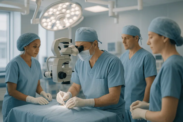
Traumatic Brain Injury Treatment
Traumatic brain injury treatment focuses on rapid stabilization, brain imaging, pressure control, neurosurgery when needed, and rehabilitation to support neurological recovery after head trauma.

Quick answer
A traumatic brain injury (TBI) is damage to the brain caused by an external force such as a fall, road accident, sports impact, assault or blast. Treatment ranges from monitored rest and a graded return to activity after concussion, through intensive care and pressure control, to emergency neurosurgery for bleeding or swelling, followed by rehabilitation for movement, speech, memory and mood.
What Is a Traumatic Brain Injury (TBI)?
A traumatic brain injury (TBI) is damage to the brain caused by an external force: a blow, a jolt, a penetrating object, or the violent acceleration and deceleration of the head. It disrupts how the brain works, sometimes briefly, sometimes lastingly, and it sits on a wide spectrum. At one end is a concussion that settles over days or weeks with guided rest and a careful return to activity. At the other is a severe injury that demands emergency neurosurgery, intensive care and months of rehabilitation. Treatment exists for every point on that spectrum, and choosing the right treatment begins with working out where a particular injury falls.
A TBI can change the course of a day, a journey or a life within seconds. It may follow a car accident, a fall, a sports collision, an assault or a blast-related event. Sometimes the injury announces itself: loss of consciousness, severe headache, vomiting, confusion, weakness, a seizure, bleeding from the scalp. In other cases the person initially appears fine and then becomes drowsy, disoriented or difficult to wake as bleeding or swelling develops inside the skull. This gap between how an injury looks from the outside and what it is doing on the inside is one of the defining problems of TBI, and it shapes almost everything about how doctors assess and treat it.
Treatment for traumatic brain injury is not a single procedure. It is a sequenced pathway: emergency stabilisation, accurate imaging, control of pressure inside the skull, timely surgery when indicated, prevention of complications, and rehabilitation tailored to the patient’s neurological, physical, cognitive and emotional needs. Depending on severity, that pathway may involve emergency physicians, neurosurgeons, neurologists, anaesthesiologists, intensive care specialists, rehabilitation physicians, radiologists, nurses, physiotherapists, speech and language therapists and neuropsychologists working together. The goal is not only survival, but preservation of brain function and the best neurological recovery the injury allows.
What does TBI mean?
TBI means traumatic brain injury: an injury to the brain caused by an outside physical force rather than by an internal disease process. The word “traumatic” refers to mechanical trauma, not emotional trauma, although the two can coexist after a serious accident. The distinction in the TBI meaning matters clinically, because brain damage from internal causes: a stroke, an infection, or brain tumoursis classified as acquired, non-traumatic brain injury and follows different treatment pathways. When you see TBI written in a medical report, it tells you two things at once: the mechanism was external force, and the clinical team will be working from trauma-based protocols that prioritise identifying bleeding, swelling and pressure early.
Is a concussion a traumatic brain injury?
Yes. A concussion is a form of mild traumatic brain injury: the mildest and by far the most common form. The force involved is enough to disturb brain function temporarily, typically causing headache, dizziness, confusion, nausea or brief memory loss, usually without visible damage on a standard CT scan. “Mild” describes the initial clinical grading, not necessarily the experience of recovery: even a mild TBI should be taken seriously if symptoms persist or worsen, and repeated concussions before the brain has recovered carry particular risk. Most people improve with appropriate guidance, but a minority develop symptoms that last weeks or months and need structured follow-up.
Head injury and traumatic brain injury: what is the difference?
A head injury and a traumatic brain injury are related terms, but they are not the same thing. “Head injury” covers any injury to the scalp, skull or brain. A scalp laceration can bleed dramatically without disturbing brain function at all; conversely, the brain can be significantly injured with no external mark whatsoever. TBI specifically means the force reached the brain and disrupted its function or structure. Clinicians therefore approach every significant head injury with one central question, has the brain been affected?, and use examination and imaging to answer it, rather than judging by the appearance of the wound.
When a Head Injury Becomes a Medical Emergency
For patients and families, uncertainty is one of the most frightening parts of TBI. The brain is delicate, and symptoms in the first minutes or hours do not always reflect the full extent of injury. Some intracranial bleeds enlarge over time: a person may speak normally after an accident and deteriorate hours later as pressure builds inside the skull: a pattern clinicians call a lucid interval. This is why medical assessment after a significant head injury matters even when symptoms seem to improve at first, particularly after high-energy trauma, loss of consciousness, repeated vomiting, severe headache, blood thinner use or any neurological change.
Rapid assessment matters for a second reason: some complications are most treatable before they cause secondary damage. The priorities in the emergency phase are fixed and universal, protect breathing and circulation, assess neurological function, identify bleeding or swelling on imaging, control pressure inside the skull, and decide whether neurosurgery or intensive care is needed. Each of these steps buys the injured brain time.
Warning features that raise clinical concern include worsening headache, repeated vomiting, confusion or unusual behaviour, seizure, weakness or numbness on one side of the body, slurred speech, unequal pupils, clear fluid or blood from the ears or nose, increasing drowsiness, agitation, poor coordination, and inability to remember the event itself. None of these findings proves that dangerous bleeding is present, but each one lowers the threshold for urgent imaging and close observation.
Types of Traumatic Brain Injury
TBI is an umbrella term. The treatment plan depends on the specific type of injury, where it is located, how quickly it is evolving and how it affects brain function. Several injury types can occur together in the same patient, which is one reason two people with the “same” diagnosis can follow very different pathways.
Concussion and mild traumatic brain injury may cause headache, dizziness, nausea, sensitivity to light, fatigue, poor concentration, memory problems, sleep disturbance or mood changes. Most patients improve with guided rest, careful monitoring and a gradual, symptom-limited return to school, work, travel and physical activity. Persistent symptoms warrant neurological assessment and, in some cases, targeted rehabilitation.
Brain contusion is bruising of brain tissue. Contusions matter because they are not static: they can swell or bleed further in the hours and days after injury, so observation and repeat imaging are often necessary. Treatment ranges from monitoring and medication to surgery if swelling or bleeding creates dangerous pressure.
Epidural haematoma is bleeding between the skull and the outer covering of the brain, often linked to a skull fracture tearing an artery. It can expand rapidly and may require urgent neurosurgical evacuation, especially when it causes neurological decline or visible pressure on the brain. This is the classic injury behind the lucid interval described above.
Subdural haematoma is bleeding between the layers covering the brain. Acute subdural haematomas after significant trauma can be life-threatening and may need emergency surgery. Chronic subdural haematomas, more common in older adults and in patients taking blood thinners, can develop gradually over weeks after a seemingly minor knock, causing headache, confusion, weakness or balance problems that families sometimes mistake for ageing.
Traumatic subarachnoid haemorrhage involves bleeding into the fluid-filled space around the brain. Patients typically need close monitoring, symptom management and evaluation for associated injuries, since this pattern often accompanies other forms of TBI.
Diffuse axonal injury occurs when rapid acceleration or deceleration stretches and damages nerve fibres throughout the brain. It is not always visible on early CT imaging, which can make the first scans look deceptively reassuring in a patient who remains unconscious. Treatment is supportive: intensive care where needed, prevention of secondary injury, and rehabilitation.
Skull fractures may be linear, depressed, open, or involve the skull base. Some need observation only. Others require surgery to lift depressed bone off the brain, clean contaminated wounds, address leakage of cerebrospinal fluid, or manage bleeding underneath the fracture.
Penetrating head injuriesfrom projectiles, sharp objects or bone fragments, require specialised trauma and neurosurgical care to manage bleeding, contamination, damaged tissue, retained foreign material, infection risk and, where appropriate, reconstruction.
Secondary brain injury is the damage that develops after the initial trauma because of low oxygen, low blood pressure, swelling, fever, seizures, infection or rising pressure inside the skull. It is worth understanding this concept clearly, because much of modern TBI care is built around a single idea: the first injury cannot be undone, but the second one can often be limited.
Symptoms of Traumatic Brain Injury
Symptoms depend on the severity and location of the injury, and they can appear immediately or emerge over hours to days. They also cross several domains at once, physical, cognitive and emotional, which is why a TBI can affect work, relationships and mood as much as it affects headache or balance.
What are 5 symptoms of TBI?
Five of the most common symptoms of TBI are headache, confusion, nausea or vomiting, dizziness, and memory problems around the time of the injury. These five appear across the whole severity spectrum, from concussion to severe injury; what changes with severity is their intensity, their persistence and what accompanies them. A fuller picture includes:
- Physical symptoms: headache, dizziness, nausea and vomiting, fatigue, sensitivity to light or noise, blurred vision, ringing in the ears, loss of balance or coordination, weakness or numbness, slurred speech, seizures, loss of consciousness.
- Cognitive symptoms: confusion, disorientation, inability to remember the event, poor concentration, slowed thinking, difficulty finding words, problems with planning and decision-making.
- Emotional and behavioural symptoms: irritability, agitation, anxiety, low mood, impulsivity, personality change, sleep disturbance, sleeping much more or much less than usual.
In children, symptoms can look different again: persistent crying, unusual irritability, sleepiness, refusal to eat, vomiting, loss of interest in favourite toys or activities, or changes in behaviour that parents recognise as abnormal even when they cannot name them. Because infants and young children cannot describe what they feel, their assessment relies more heavily on examination, observation and, where indicated, imaging.
Who Needs Assessment After a Head Injury?
Any person who has experienced a blow, jolt, penetrating injury, acceleration-deceleration force or blast exposure involving the head may need evaluation for traumatic brain injury. The need becomes more urgent when symptoms suggest the brain may be bleeding, swelling or under pressure, but the absence of dramatic symptoms is not, by itself, reassurance, particularly in the groups below.
Some patients are at higher risk of complications even when symptoms seem modest at first. Older adults, people taking anticoagulant or antiplatelet medications, patients with clotting disorders, individuals with previous brain surgery, and people affected by alcohol or other substances at the time of injury all warrant a lower threshold for imaging and observation. In older adults on blood thinners, even a low-energy fall can produce slow bleeding that declares itself days or weeks later. Alcohol adds a second problem: it can mask or mimic the confusion and drowsiness that clinicians rely on to detect deterioration.
Infants and young children also need careful assessment, both because they cannot report symptoms and because their injury patterns differ from adults’. At the other end of the spectrum, athletes returning to contact sport after concussion need structured clearance, because a second impact on an unrecovered brain is more dangerous than the first.
How Traumatic Brain Injury Is Diagnosed
Diagnosis begins with a medical history and a neurological examination. Clinicians ask how the injury happened, whether there was loss of consciousness, how long any confusion lasted, whether symptoms are stable or changing, and what medications the patient takes. The examination covers level of alertness, pupil size and reaction, limb strength, sensation, coordination, speech and memory. In severe trauma, this assessment happens alongside life-saving stabilisation rather than as a separate step, and the cervical spine is protected until neck injury has been excluded.
How do doctors grade the severity of a TBI?
Doctors grade TBI severity primarily with the Glasgow Coma Scale (GCS), which scores eye opening, verbal response and movement to produce a single number. Broadly, a GCS of 13–15 is described as mild, 9–12 as moderate, and 3–8 as severe. The score is combined with the duration of any loss of consciousness, the length of memory loss around the event, and imaging findings. Crucially, the GCS is repeated over time: serial examinations are often more informative than any single assessment, because traumatic brain injury can evolve, and a falling score is one of the clearest signals that something inside the skull is changing.
What scans are used after a head injury?
Computed tomography (CT) is the main emergency imaging tool after a head injury because it is fast and effective at detecting acute bleeding, bruising, swelling, skull fractures and shifting of brain structures. Repeat CT may be needed if the patient deteriorates or if the first scan shows a lesion that could progress. CT angiography is added in selected cases where injury to blood vessels is suspected. Magnetic resonance imaging (MRI) is usually reserved for later: it is more sensitive to injuries that CT can miss, such as diffuse axonal injury or small areas of damage, and it becomes valuable when symptoms persist despite an unremarkable CT, or when detailed assessment of brain tissue is needed for prognosis and rehabilitation planning. Imaging of the spine, chest, abdomen or pelvis may run in parallel when the mechanism of injury suggests additional trauma. Laboratory tests round out the picture: haemoglobin, electrolytes, clotting function, kidney function, blood sugar and medication effects all influence both bleeding risk and treatment decisions.
Based on all of this, patients may be observed in hospital, admitted to an intensive care unit, taken to surgery, or, for genuinely mild injuries without risk features, assessed and monitored without admission. The pathway is individualised, because two patients with similar scans can differ substantially in age, medication use, medical risks and neurological examination.
How Traumatic Brain Injury Treatment Is Performed
Treatment begins before all the questions are answered. In significant trauma, the medical team first stabilises the patient using an organised emergency approach. The airway may need protection if the patient cannot breathe safely, has a reduced level of consciousness or is at risk of aspiration. Oxygen levels and blood pressure are actively supported, because the injured brain is exquisitely sensitive to reduced oxygen and poor blood flow. If the patient takes blood thinners, the team weighs reversal strategies against the specific medication, the imaging findings and the bleeding risk: a decision that belongs entirely to the treating clinicians and changes case by case.
Treatment for concussion and mild TBI
For mild traumatic brain injury without dangerous findings, treatment centres on observation, symptom control and a structured return to normal life. The care team typically addresses pain and nausea, gives guidance on sleep and on avoiding alcohol and high-risk activities, and sets out clearly what changes should prompt reassessment. Patients are usually advised to rest briefly and then resume mental and physical activity gradually, guided by symptoms. Prolonged complete inactivity is not generally recommended: recovery after concussion is typically best supported by a stepwise, symptom-limited return to normal routines rather than by weeks in a dark room.
Intensive care for moderate and severe TBI
For moderate or severe traumatic brain injury, admission is usually required, often to an intensive care unit where neurological status, oxygenation, blood pressure, temperature, fluid balance and laboratory results can be monitored continuously. Some patients need mechanical ventilation, sedation, or medication to control agitation and protect the brain. Seizure prevention may be used in higher-risk injury patterns. Fever, low sodium, high blood sugar, infection and anaemia are each managed deliberately, because every one of these factors can worsen the condition of injured brain tissue. This is unglamorous, detail-heavy medicine, and it is where much of the difference in severe TBI care is made.
How is pressure inside the skull controlled?
Pressure control is central to severe TBI care because the skull is rigid: swelling or bleeding has nowhere to expand, so pressure rises and blood flow to the brain falls. In selected patients, a monitor is placed to measure intracranial pressure directly. Treatment builds in steps, head elevation, sedation, controlled ventilation strategies, drainage of cerebrospinal fluid in some cases, osmotic medications that draw fluid out of swollen tissue, and surgical decompression when other measures cannot hold the pressure down. The goal throughout is the same: maintain enough blood flow and oxygen delivery to keep vulnerable brain tissue alive while the injury stabilises.
When is surgery needed for a TBI?
Surgery is needed when imaging or clinical findings show a problem that can be relieved mechanically. Common procedures include removing an epidural or subdural haematoma, evacuating a blood clot within the brain itself, repairing a depressed skull fracture, treating a penetrating injury, placing a drain, or performing a decompressive craniectomy. In a decompressive craniectomy, a section of skull is temporarily removed and stored or preserved so that a swollen brain has room to expand without crushing itself; it is generally reserved for selected severe cases where pressure cannot be controlled any other way, and the bone is replaced later once swelling has resolved. The specific approach depends on the location and size of the bleeding, the patient’s neurological condition, the degree of swelling and any associated injuries. Surgeons work from detailed imaging, using magnification, navigation and monitoring tools where appropriate. The purpose of the technology is entirely practical: define the injury accurately, plan the safest route, remove pressure from the brain, protect critical structures, and track changes during and after the operation.
Timeframes vary enormously. Emergency imaging and stabilisation may take minutes to hours depending on the patient’s condition and associated injuries. Some neurosurgical procedures are relatively focused; complex trauma surgery takes longer. Intensive care can last days or weeks in severe cases, while a genuinely mild TBI may need no admission at all, but only after a proper medical evaluation has excluded dangerous features.
Rehabilitation After a Traumatic Brain Injury
Rehabilitation is part of the treatment, not an afterthought, and it begins early, often while the patient is still in intensive care, once they are stable. Physiotherapy starts with positioning, range of motion and prevention of complications, then progresses to mobility, balance and strength. Occupational therapy addresses daily activities, coordination and independence. Speech and language therapy covers swallowing as well as speech, communication and aspects of cognitive function. Neuropsychological assessment maps memory, attention, mood, behaviour and executive function, and shapes the plan as recovery unfolds. Nutrition, sleep, pain control and family education run alongside all of it.
For patients whose main problem is persistent symptoms after a milder injury, rehabilitation looks different: management of ongoing post-traumatic headache, sometimes with input from headache medicine specialists, vestibular therapy for dizziness, vision therapy, cognitive strategies for concentration and memory, and support for mood and sleep. The emotional aftermath deserves attention in its own right; some patients develop anxiety or post-traumatic stress disorder after the accident that caused their injury, and treating these alongside the physical symptoms improves the whole recovery.
Discharge planning is individualised. Some patients go home with instructions and outpatient follow-up. Others need inpatient rehabilitation or continued nursing care before they can safely leave hospital. A thorough discharge plan typically covers medication planning by the treating team, wound care, seizure precautions where relevant, a written rehabilitation summary, and clear communication with the physicians who will take over ongoing care, so that recovery continues without a gap.
Why Early Treatment Matters
The first hours after a significant head injury can influence the entire course of recovery. The initial trauma cannot be undone, but secondary injury can often be reduced when problems are recognised and treated promptly. Low oxygen, low blood pressure, expanding bleeding, uncontrolled swelling, seizures, fever and delayed treatment of associated injuries can each add damage on top of the original injury, and each is, to a meaningful degree, preventable or treatable.
Delay also complicates the quieter injuries. Untreated dizziness, vision problems, headaches, sleep disturbance, cognitive changes or mood symptoms after concussion can interfere with work, study, travel and family life for far longer than necessary. Early recognition lets clinicians guide activity levels, identify red flags, prevent repeat injury and refer to rehabilitation when symptoms persist rather than letting them settle into a pattern.
For moderate and severe injuries, early access to neurosurgical judgement and intensive care resources matters most of all. Not every patient needs an operation, but patients who do benefit from timely decision-making, and patients who do not still require careful monitoring to catch changes before they become dangerous. The value of an experienced team lies as much in knowing when not to operate as in operating well.
Benefits of Traumatic Brain Injury Treatment
The benefits of treatment depend on the injury type and severity, but the overall aim is constant: protect the brain, treat complications, and support recovery in a structured way.
| Benefit | What It Means for You |
|---|---|
| Rapid identification of bleeding or swelling | Emergency imaging and neurological assessment help doctors decide whether observation, intensive care, medication or surgery is needed. |
| Protection from secondary brain injury | Careful control of oxygen, blood pressure, temperature, seizures and intracranial pressure may reduce additional stress on injured brain tissue. |
| Timely neurosurgical intervention when needed | Removing a blood clot, repairing a fracture or relieving pressure can be critical in selected patients with dangerous imaging or neurological changes. |
| Coordinated intensive care | Continuous monitoring allows the team to respond quickly to changes in consciousness, breathing, circulation, pressure, infection risk or metabolic status. |
| Early rehabilitation planning | Therapies for movement, speech, swallowing, cognition and daily function can begin as soon as medically appropriate and continue after discharge. |
| Clear guidance for return to daily life | Patients and families receive recommendations about activity, travel, medications, warning symptoms, follow-up imaging and rehabilitation needs. |
Recovery Timeline After Traumatic Brain Injury
Recovery varies widely, but the following timeline gives a general view of what many patients and families can expect after evaluation and treatment.
| Time Period | What Patients Can Expect |
|---|---|
| Day 1 | Emergency stabilisation, neurological examination, brain imaging, treatment of associated injuries, and a decision about observation, intensive care, surgery, or discharge with strict instructions. |
| First Week | Close monitoring for worsening swelling, bleeding, seizures, infection or changes in consciousness. Rehabilitation may begin if the patient is stable. Mild injuries focus on symptom control and gradual activity. |
| First Month | Follow-up appointments, medication review by the treating team, wound care if surgery was performed, and assessment of headaches, dizziness, sleep, concentration, mood, strength, balance, speech or swallowing. |
| Longer Term | Ongoing rehabilitation may be needed for moderate or severe injuries. Some patients return to previous activities gradually, while others require longer support for cognitive, physical, emotional or occupational recovery. |
Can you ever fully recover from a TBI?
Some people do return to their previous level of function, particularly after mild injuries, and others live with lasting changes. No honest clinician promises a specific outcome in the early phase, because recovery depends on the injury type, its severity, the patient’s age and health, how quickly secondary injury was prevented, and how consistently rehabilitation is pursued. What can be said with confidence is that recovery after TBI often continues for far longer than people expect, improvement can carry on over many months, and that recovery is rarely a straight line. Plateaus, setbacks and uneven progress across different abilities are part of the normal pattern, not signs of failure.
Is TBI a permanent disability?
Not automatically. TBI can result in permanent disability, particularly after severe injuries involving extensive damage, but many people, especially those with mild or moderate injuries, return to work, study, driving and sport. Between those two ends sits a large group who recover most functions while retaining specific difficulties: fatigue, slower processing, memory gaps, mood changes or headaches. Whether a given injury is formally recognised as a disability also depends on the legal and insurance framework of the patient’s home country, which is a separate question from the medical one. Medically, the honest answer is that permanence cannot be judged early; it emerges from how the injury evolves and how rehabilitation progresses.
Factors That Influence Outcomes
Outcomes after traumatic brain injury are shaped by many factors, and no responsible medical team predicts recovery with certainty in the early phase. The severity of the initial injury matters, but it is not the only variable. Age, overall health, oxygen levels, blood pressure, associated injuries, medication use, timing of treatment, imaging findings and the evolving neurological examination all contribute.
The type of injury matters. A concussion may resolve with appropriate guidance, while diffuse axonal injury or severe swelling can require prolonged intensive care and rehabilitation. A surgically removable blood clot may improve markedly once pressure is relieved, but recovery still depends on how much damage occurred before treatment and whether complications develop afterwards.
Early neurological status is one of the most important clinical indicators. Level of consciousness, pupil response, limb movement and the trend over time help clinicians judge urgency and prognosis. Imaging findings guide expectations too: the size and location of bleeding, the degree of swelling, whether the brain’s midline structures have shifted, whether there are multiple contusions, and whether the brainstem is involved. Repeat scans show whether the injury is stable or progressing.
Prevention of secondary injury influences recovery. Maintaining oxygen delivery, avoiding low blood pressure, treating fever, controlling seizures, correcting metabolic problems and managing intracranial pressure are all part of evidence-based care. These interventions do not erase the original trauma, but they help protect the brain tissue that remains at risk.
Finally, rehabilitation intensity and continuity matter. Recovery is often uneven: a patient may regain strength before memory, speech before attention, mobility before emotional regulation. Families may notice fatigue, irritability, impulsivity, low mood, anxiety, sleep disruption or personality change. These are medical consequences of the injury, not character flaws, and structured rehabilitation helps patients and families understand what is happening and adapt the plan as recovery unfolds.
Can a traumatic brain injury cause bipolar disorder?
A TBI can contribute to significant mood disturbance, and research has explored links between brain injury and later psychiatric conditions, including mood disorders with features resembling bipolar disorder. What is well established is that TBI can cause depression, anxiety, irritability, emotional volatility and impulsivity, particularly when the frontal and temporal regions are injured. Whether an individual patient’s mood disorder was caused by the injury, unmasked by it, or unrelated to it is a question for careful psychiatric and neurological assessment: the timing of symptoms, family history and injury location all inform the answer. The practical point for families is simpler: marked mood or personality change after a TBI deserves the same medical attention as a physical symptom.
Long-Term Effects of Traumatic Brain Injury
Long-term effects of TBI vary from none at all to substantial changes in daily life. The most common lasting complaints after significant injuries are fatigue, headaches, difficulty concentrating, memory problems, slowed thinking, dizziness, sleep disturbance and changes in mood or behaviour. Physical effects such as weakness, coordination problems or speech and swallowing difficulties are more typical of severe injuries and are the main targets of longer-term rehabilitation.
Safety after discharge shapes the long term as much as anything done in hospital. Preventing a repeat head injury, following the treating team’s guidance on medications, taking a gradual approach to work and sport, respecting seizure precautions where they apply, and attending follow-up appointments all support a safer recovery. Families play a larger role in TBI recovery than in most conditions, because the patient may not fully perceive their own changes in memory, judgement or behaviour, education and support for caregivers is a legitimate part of the treatment, not an optional extra.
Traumatic Brain Injury Care at Acibadem
Patients with traumatic brain injury need more than a hospital bed. They need rapid access to specialists, coordinated decision-making, reliable imaging, intensive care capability, neurosurgical expertise, rehabilitation planning and clear communication. At Acibadem, TBI care is organised around these practical needs, within the wider framework of traumatic neurology services.
Care is multidisciplinary by design. Neurosurgeons, neurologists, emergency medicine physicians, anaesthesiologists, intensive care specialists, radiologists, rehabilitation physicians, nurses and therapists collaborate according to the patient’s condition, and patients may move quickly between the emergency department, imaging unit, operating room, intensive care unit, inpatient ward and rehabilitation services. In complex cases, multidisciplinary discussions review the imaging, neurological status, surgical options, critical care priorities and rehabilitation needs together, which is particularly valuable when a case is not straightforward, when multiple injuries are present, or when a family is weighing a second opinion on whether surgery is necessary or what rehabilitation plan is realistic.
Diagnostic pathways are built to answer specific questions rather than to accumulate tests: Is there bleeding? Is pressure rising? Is surgery needed? Is the injury stable? What rehabilitation plan fits this patient? Emergency CT, advanced MRI in selected cases, vascular imaging, laboratory testing and serial neurological examinations are used in whatever combination the clinical question demands. When surgery is required, the neurosurgical team plans the approach from detailed imaging with intraoperative tools suited to the individual case, and intensive care monitoring continues afterwards for swelling, bleeding, infection, seizures and respiratory complications.
Treatment planning is personalised because no two brain injuries are identical. A young athlete with persistent concussion symptoms, an older adult with a chronic subdural haematoma, a traffic accident patient with multiple injuries and a critically ill patient with severe brain swelling require very different pathways, guided by evidence-based protocols, adapted to the individual’s condition, imaging, risk factors and recovery goals. The same protocols scale in both directions: a straightforward concussion is not over-treated, and a deteriorating patient is escalated without delay.
Moving Forward After a Traumatic Brain Injury
A traumatic brain injury brings urgent medical questions and deeply personal ones. Families want to know whether their loved one will wake up, whether surgery is necessary, whether symptoms will improve, and what life may look like after discharge. The honest answer is that recovery depends on the injury and the patient, but careful early treatment, disciplined prevention of secondary injury and structured rehabilitation each make a genuine difference to what recovery is possible.
Where care crosses hospitals or teams, continuity is what protects the patient: imaging studies, operative notes, medication lists, intensive care summaries and a clear account of the injury allow any receiving team to pick up the thread accurately. TBI recovery is measured in weeks and months rather than days, and the patients who tend to do best are those whose care, medical, rehabilitative and emotional, stays organised for the whole of that time, not just the dramatic first chapter.
Preparation
- Traumatic brain injury is usually managed as an emergency, so preparation is limited to rapid assessment and stabilization. Doctors may perform neurological examination, CT or MRI imaging, blood tests, and monitoring of breathing, blood pressure, and intracranial pressure. Family members should share details about the injury, medications, allergies, and previous medical conditions.
Aftercare
- Aftercare may include intensive care monitoring, medication, wound care if surgery was performed, and prevention of complications such as seizures or infections. Many patients need neurological rehabilitation, physical therapy, speech therapy, or cognitive support. Follow-up visits and repeat imaging help track recovery and guide return to daily activities.
Frequently Asked Questions
What affects the cost of traumatic brain injury treatment?
The main factors are injury severity, need for ICU care, imaging, neurosurgery, medications, monitoring, hospital stay, rehabilitation, and transfer logistics. A personalised quote requires review of medical records and current neurological status.
Can I get a quote before travelling to Turkey?
For stable patients or families seeking transfer, hospitals can review available scans, reports, discharge notes, and current medication lists. In emergency situations, the final plan may change after assessment by the medical team.
What is usually included in an international patient treatment plan?
Depending on the case, it may include specialist consultation, imaging review, hospital admission planning, ICU coordination, surgery if needed, rehabilitation assessment, interpreter support, and assistance with transfer arrangements.
Is traumatic brain injury treatment always surgical?
No. Some patients need observation and medical management, while others require urgent neurosurgery. The decision is based on symptoms, neurological examination, imaging findings, and overall trauma status.
How does rehabilitation influence the total cost?
Rehabilitation can be a significant part of recovery and may include physiotherapy, speech therapy, occupational therapy, cognitive support, and follow-up planning. Cost depends on intensity, setting, and duration recommended by the specialist.
How can Acibadem International help with planning?
Acibadem International can review medical information, coordinate specialist input, explain expected package elements, and provide a personalised quote after clinical evaluation. This information is general and is not a substitute for medical or financial advice.
Medically reviewed by the Acıbadem International Medical Board September 13, 2026
See our medical review board →
Update history
- PublishedJune 8, 2026
- Medical review approvedSeptember 13, 2026
- Last content updateSeptember 12, 2026
References3
- Traumatic Brain Injury medlineplus.gov
- Traumatic Brain Injury (TBI) my.clevelandclinic.org
- Traumatic Brain Injury and Concussion cdc.gov
Trusted care for international patients
From the Acibadem Blog

Can You Return to Work After a Traumatic Brain Injury? Pacing, Fatigue and Gradual Steps
Many people do return to work after a traumatic brain injury, but the path is usually gradual rather than a single return date. Clinicians…