

Carotid Angio
Carotid angio is a catheter-based imaging test that visualizes the carotid arteries supplying the brain. It helps detect narrowing, blockage, or aneurysm and guides stroke-prevention planning.
Quick answer
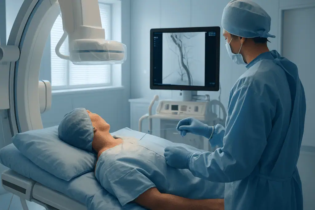
Carotid angio is a catheter-based imaging procedure used to show the carotid arteries that carry blood to the brain and identify problems such as narrowing, blockage, or aneurysm. At Acibadem in Turkey, it is performed by advancing a thin catheter through a blood vessel, injecting contrast dye, and taking detailed X-ray images to support diagnosis and stroke-prevention treatment planning.
When Carotid Artery Disease Raises Questions About Stroke Risk
Learning that there may be narrowing, blockage, or an abnormal bulge in the arteries that supply blood to the brain can be unsettling. Many patients first hear about carotid artery disease after a warning symptom, such as a transient ischemic attack, temporary weakness, brief vision loss, or dizziness. Others are told that an ultrasound has found a significant narrowing in the neck arteries, even though they feel completely well. In either situation, the central concern is often the same: What is my risk of stroke, and what should be done to reduce it?
Carotid angiography, often called carotid angio, is a specialized catheter-based imaging test that helps answer that question with a high level of anatomical detail. It allows physicians to see the carotid arteries and related blood vessels in real time, assess the severity and shape of a narrowing, identify blockages or aneurysms, and plan the safest next step. For some patients, this means confirming that medication and careful monitoring are appropriate. For others, carotid angio helps guide treatment such as carotid stenting, carotid endarterectomy planning, or further neurovascular care.
Because the carotid arteries carry oxygen-rich blood to the brain, decisions about diagnosis and treatment require precision. The goal is not simply to “find a narrowing,” but to understand whether that narrowing is likely to cause symptoms, whether blood flow to the brain is affected, and which treatment pathway offers the most appropriate balance of benefit and risk for the individual patient. This is especially important for international patients who may be comparing medical opinions, seeking confirmation before a procedure, or looking for coordinated care in another country.
At an experienced center, carotid angio is performed within a broader diagnostic and clinical framework. The angiogram is not interpreted in isolation. It is considered together with neurological symptoms, vascular risk factors, ultrasound findings, CT or MRI results, heart rhythm evaluation, and the patient’s overall health. This careful approach supports more confident stroke-prevention planning and helps avoid both undertreatment and unnecessary intervention.
What Is Carotid Angio?
Carotid angio is a minimally invasive imaging procedure used to visualize the carotid arteries, which run along both sides of the neck and supply blood to the brain, face, and head. During the test, a thin flexible tube called a catheter is inserted into an artery, most commonly through the groin or sometimes the wrist. The catheter is guided through the blood vessels to the area being examined. A contrast dye is then injected, and specialized X-ray imaging records the flow of blood through the carotid arteries and, when needed, the arteries inside the brain.
The medical term often used for this test is catheter-based cerebral or carotid angiography, depending on the extent of the imaging. It differs from noninvasive imaging tests such as carotid ultrasound, CT angiography, or MR angiography because it involves placing a catheter inside the blood vessels. This allows detailed, dynamic visualization of blood flow and vessel anatomy. In selected situations, catheter angiography may provide information that is difficult to obtain from external imaging alone.
Carotid angio can show the degree of narrowing, the length and location of a plaque, irregular vessel surfaces, complete occlusions, collateral circulation, aneurysms, dissections, and other vascular abnormalities. It can also help physicians evaluate whether a narrowing is technically suitable for stenting or whether surgical treatment may be more appropriate. In emergency or complex neurovascular cases, angiography may be part of a pathway that includes immediate intervention, although diagnostic carotid angio is often performed as a planned procedure.
The test is typically carried out by specialists trained in vascular imaging and endovascular techniques, such as interventional radiologists, interventional neurologists, neurointerventional specialists, vascular surgeons, or cardiologists with relevant expertise. The patient is monitored throughout the procedure, and imaging is reviewed in the context of clinical findings. For many patients, carotid angio is a decision-making tool: it helps define the problem clearly so the care team can recommend the most appropriate strategy to lower stroke risk.
Who May Need Carotid Angio?
Carotid angio may be recommended when physicians need more detailed information about the arteries supplying the brain than can be obtained from standard tests. Many patients are first evaluated with carotid Doppler ultrasound because it is noninvasive and widely available. If the ultrasound suggests moderate or severe narrowing, if results are unclear, or if treatment is being considered, additional imaging may be needed. CT angiography or MR angiography is often used before catheter angiography. Carotid angio is usually reserved for situations where precise vascular mapping is important or where other tests do not provide enough clarity.
Some patients are referred after symptoms that suggest a transient ischemic attack, often called a TIA or “mini-stroke.” These symptoms may last only minutes and then resolve, but they should always be taken seriously. Temporary weakness or numbness on one side of the body, difficulty speaking, sudden loss of vision in one eye, facial drooping, imbalance, or sudden confusion may indicate that blood flow to part of the brain has been interrupted. If carotid disease is suspected as the cause, angiographic assessment may help guide stroke-prevention treatment.
Other patients have no symptoms at all. They may have carotid narrowing found during evaluation for a heart condition, screening after detection of a bruit in the neck, or imaging performed for another reason. In asymptomatic patients, the decision to perform carotid angio is individualized. The care team considers the severity of narrowing, plaque appearance, age, medical conditions, kidney function, previous stroke or TIA history, and whether any procedure is being contemplated.
Diagnosis begins with a careful medical history and physical examination, including a neurological assessment. Physicians review vascular risk factors such as high blood pressure, diabetes, high cholesterol, smoking history, coronary artery disease, peripheral artery disease, sleep apnea, and family history of vascular disease. Blood tests may be ordered to assess kidney function, blood count, clotting profile, and other safety factors before contrast dye is used. Noninvasive imaging is usually reviewed before angiography is scheduled, so the procedure can be focused and efficient.
Patients who travel internationally for evaluation may bring previous imaging and reports from their home country. When possible, original images are more useful than written reports alone. A specialist team can compare prior ultrasound, CT, MRI, and clinical records to determine whether carotid angio is necessary or whether another diagnostic or treatment pathway is more suitable.
Conditions and Indications Carotid Angio Can Address
Carotid angio is most commonly used to evaluate carotid artery stenosis, which means narrowing of the carotid artery. This is often caused by atherosclerosis, a buildup of fatty plaque within the artery wall. Plaques may gradually restrict blood flow, but stroke risk is also related to plaque instability and the possibility that small clots or debris can travel to the brain. Angiography helps characterize the vessel lumen, the open channel through which blood flows, and can show how blood moves beyond the narrowed segment.
Another indication is suspected or known carotid artery occlusion, where the artery is completely blocked. In these cases, angiography can help determine whether the blockage is truly complete, how far it extends, and whether other vessels are supplying the brain through collateral circulation. This information may influence medical management and, in selected cases, interventional planning.
Carotid angio may also be used to evaluate carotid artery dissection, a tear in the inner lining of the artery that can occur after trauma, sudden neck movement, or sometimes without a clear cause. Dissection can narrow the artery, form a pseudoaneurysm, or create a risk of clot formation. Detailed imaging can support treatment decisions, especially if symptoms are present or noninvasive imaging is uncertain.
In selected patients, carotid angio is used to assess aneurysms or abnormal vessel dilations involving the carotid circulation. It may help define the size, shape, neck, branch involvement, and relationship to surrounding vessels. These details are important when evaluating the need for monitoring, endovascular treatment, or surgery.
The procedure can also contribute to the evaluation of complex neurovascular conditions, including arteriovenous malformations, vascular tumors, unusual collateral pathways, and recurrent symptoms after prior carotid surgery or stenting. For patients who have already had a carotid stent or endarterectomy, angiography may be used in selected circumstances to assess restenosis, stent patency, or anatomy before a repeat intervention.
Importantly, carotid angio is not required for every patient with carotid disease. Many patients can be managed using ultrasound, CT angiography, MR angiography, and clinical evaluation. The decision to proceed with catheter angiography is made when the expected diagnostic value is meaningful and when the benefits outweigh the procedural risks.
How Carotid Angio Is Performed
Preparation Before the Procedure
Preparation begins with a consultation and review of medical records. Your physician will ask about previous stroke or TIA symptoms, allergies, kidney disease, bleeding disorders, medications, and any history of reactions to contrast dye. It is especially important to discuss blood thinners, antiplatelet medications, diabetes medications, and supplements that may affect bleeding. Some medications may need to be adjusted, while others should be continued to protect against clotting; the plan is individualized.
Blood tests are commonly performed before the procedure to evaluate kidney function, hemoglobin level, platelet count, and clotting status. If you have kidney disease, diabetes, dehydration risk, or previous contrast reactions, additional precautions may be taken. These may include hydration, alternative imaging considerations, or medication to reduce the chance of an allergic-type reaction when appropriate.
You may be asked not to eat or drink for several hours before the angiogram. The care team will explain whether you can take your usual morning medications with a small sip of water. On arrival, nurses check vital signs, review consent, place an intravenous line, and prepare the access site. For international patients, interpreters or multilingual patient coordinators can help ensure that instructions, consent details, and aftercare recommendations are clearly understood.
During the Procedure
Carotid angio is performed in an angiography suite equipped for high-resolution vascular imaging and continuous monitoring. Most patients receive local anesthesia at the access site and light sedation to help them feel relaxed while remaining able to respond if needed. In some complex cases, different anesthesia arrangements may be considered.
The physician inserts a small sheath into an artery, commonly in the groin through the femoral artery or in the wrist through the radial artery. A thin catheter is then guided through the arterial system under live X-ray guidance. Patients generally do not feel the catheter moving inside the blood vessels, though they may feel pressure at the access site. Once the catheter is positioned near the arteries being studied, contrast dye is injected while images are taken.
During contrast injection, some patients notice a brief warm sensation, a metallic taste, or a feeling of flushing. These sensations usually pass quickly. The imaging equipment captures the contrast as it travels through the carotid arteries and, when necessary, into the intracranial circulation. The physician may take images from several angles to understand the three-dimensional shape of a narrowing, aneurysm, or abnormal vessel segment.
Modern angiography uses digital imaging techniques that enhance visualization of blood vessels while reducing background structures. Road-mapping tools can help guide catheter movement with precision. Dose-management protocols are used to keep radiation exposure as low as reasonably achievable while obtaining the necessary diagnostic information. In some cases, rotational or three-dimensional angiographic views may be used to clarify complex anatomy. The choice of imaging method depends on the clinical question and the patient’s condition.
A purely diagnostic carotid angio often takes less than a few hours from preparation through completion, with the imaging portion itself usually shorter. The exact duration depends on vascular anatomy, whether one or both sides are examined, whether brain circulation is included, and whether any additional measurements or views are needed. If an intervention is planned at the same session, such as carotid stenting in selected patients, the procedure will take longer and require a separate treatment discussion and consent.
Immediately After the Procedure
When the angiogram is complete, the catheter and sheath are removed. The access site is closed either with manual pressure or a vascular closure method, depending on the artery used, anatomy, and physician preference. You will be monitored in a recovery area while nurses check your pulse, blood pressure, neurological status, and access site. You may need to lie flat for a period of time if the groin was used.
Most patients can eat and drink after the procedure once fully awake, unless another medical reason prevents this. Drinking fluids helps the body clear contrast dye. The team will ask you to report new headache, weakness, numbness, speech difficulty, vision change, chest discomfort, increasing access-site pain, or swelling. These events are uncommon, but rapid recognition is important.
Some patients go home the same day, while others may stay overnight depending on their overall health, the results, the access route, distance from the hospital, and whether further treatment is planned. International patients should receive clear written instructions about activity restrictions, medication use, wound care, warning signs, and follow-up appointments before returning to their accommodation or traveling onward.
Why Acting Early Matters
Carotid artery disease is important because it can be associated with stroke, one of the leading causes of long-term disability worldwide. A stroke can affect movement, speech, memory, vision, swallowing, independence, and quality of life. Not every carotid narrowing leads to stroke, and not every stroke is caused by the carotid arteries. However, when carotid disease is significant or symptomatic, timely evaluation can change the prevention strategy.
Symptoms such as temporary weakness, loss of speech, or brief blindness in one eye may disappear quickly, but they can be warning signs of a future stroke. Delaying evaluation after these symptoms may leave an important cause untreated. Early assessment allows physicians to identify whether the carotid artery is the source, start or optimize medications, address blood pressure and cholesterol, and determine whether a procedure may reduce future risk in selected patients.
For patients with severe narrowing but no symptoms, timing is more nuanced. Some may benefit most from intensive medical therapy and monitoring, while others may be considered for intervention depending on anatomy, plaque features, age, surgical risk, and overall life expectancy. Accurate imaging helps avoid decisions based on incomplete information. If prior tests disagree, or if a treatment decision depends on precise measurements, carotid angio can help clarify the situation.
Delay can also matter in aneurysms, dissections, or unusual vascular abnormalities. Some require urgent care, while others can be monitored. The key is obtaining the right diagnosis and risk assessment before a preventable event occurs. Acting early does not always mean having a procedure immediately; it means getting an expert evaluation in time to make an informed, medically appropriate decision.
Potential Benefits of Carotid Angio
The main value of carotid angio is that it provides detailed information that can guide safer and more individualized stroke-prevention planning.
| Benefit | What It Means for You |
|---|---|
| Detailed visualization of the carotid arteries | Physicians can see the location, severity, length, and shape of a narrowing or blockage more clearly when noninvasive tests are inconclusive or treatment planning requires precision. |
| Real-time assessment of blood flow | The test shows how blood moves through the neck and brain circulation, including collateral pathways that may help supply the brain if one artery is narrowed or blocked. |
| Improved treatment planning | Angiographic findings can help determine whether medication, carotid stenting, surgery, or further monitoring is the most appropriate next step. |
| Clarification after conflicting test results | If ultrasound, CT, or MRI findings do not match, catheter angiography can provide additional detail to support a more confident diagnosis. |
| Evaluation of complex vascular conditions | Dissections, aneurysms, restenosis after prior treatment, and unusual anatomy can be assessed in a way that supports specialized neurovascular decision-making. |
Recovery Timeline After Carotid Angio
Recovery is usually short after a diagnostic carotid angiogram, although individual instructions depend on the access site, medical condition, and whether any additional treatment was performed.
| Time Period | What Patients Can Expect |
|---|---|
| Day 1 | You will be monitored after the procedure for vital signs, neurological status, and bleeding or swelling at the access site. Mild tenderness or bruising may occur. Some patients return to their hotel or home the same day; others stay overnight. |
| First Week | Most patients resume light daily activities within a short time. Heavy lifting, strenuous exercise, and long periods of standing may be limited for several days, especially after groin access. Follow wound-care instructions carefully. |
| First Month | Your physician reviews results and confirms the treatment plan. This may include medication adjustment, lifestyle recommendations, follow-up imaging, or referral for carotid stenting, surgery, or neurological care if needed. |
| Longer Term | Ongoing stroke prevention usually focuses on controlling blood pressure, cholesterol, diabetes, smoking exposure, heart rhythm problems, and other vascular risks. Surveillance imaging may be scheduled depending on the diagnosis. |
What Influences the Results and a Good Outcome?
A good outcome after carotid angio begins with selecting the right patients for the test. Because catheter angiography is invasive, physicians weigh the expected diagnostic benefit against possible risks such as bleeding, contrast reaction, kidney strain, vessel injury, or stroke. Serious complications are uncommon in experienced hands, but they are possible, which is why careful preparation and monitoring matter.
The quality of the result also depends on the completeness of the clinical picture. Symptoms, timing, prior imaging, medications, and vascular risk factors all affect interpretation. A narrowing that appears severe on imaging may have different implications depending on whether the patient has had recent neurological symptoms, whether the opposite carotid artery is healthy, and whether the brain has adequate collateral blood flow. For this reason, the angiogram should be assessed by physicians who understand both vascular anatomy and stroke mechanisms.
Patient-related factors influence procedural safety. Kidney function is important because contrast dye is cleared through the kidneys. Allergies, blood-thinning medications, clotting disorders, uncontrolled blood pressure, active infection, and severe vascular disease can affect preparation and technique. The access route may be chosen based on anatomy, prior procedures, body habitus, and the physician’s judgment. Some patients benefit from additional hydration, medication adjustment, or observation after the test.
Technical expertise is another important factor. Catheter manipulation within arteries requires training, judgment, and careful attention to minimizing plaque disruption, clot formation, radiation exposure, and contrast volume. Imaging quality must be high enough to answer the clinical question without unnecessary views. The team should also be prepared to recognize and manage complications promptly if they occur.
Finally, the value of carotid angio depends on what happens afterward. A detailed diagnosis is useful only if it leads to an appropriate prevention plan. Many patients require optimized medical therapy, including antiplatelet medication when indicated, cholesterol-lowering treatment, blood pressure control, diabetes management, smoking cessation, nutrition guidance, and physical activity recommendations. Others may require a procedural solution. The best result is a plan that is medically sound, clearly explained, and realistic for the patient’s life after returning home.
Why International Patients Choose Acibadem for Carotid Angio
International patients considering carotid angio often want more than a single test. They want a careful second opinion, a clear explanation of stroke risk, access to experienced specialists, and a coordinated plan that can be completed efficiently while they are away from home. At Acibadem, carotid angiography is approached as part of a comprehensive vascular and neurological evaluation rather than as an isolated procedure.
Care is delivered within JCI-accredited hospitals, where safety processes, infection control standards, imaging protocols, and patient monitoring systems are designed to support complex diagnostic and interventional care. Patients with carotid disease may be evaluated by neurologists, vascular surgeons, interventional radiology or neurointerventional teams, cardiologists, anesthesiology specialists, and rehabilitation professionals when needed. In complex cases, multidisciplinary boards or specialist case discussions help align imaging findings with the most appropriate treatment pathway.
Modern diagnostic pathways are especially important in carotid artery disease because treatment decisions can be finely balanced. Acibadem physicians may review carotid ultrasound, CT angiography, MR angiography, brain MRI, cardiac evaluation, laboratory results, and catheter angiography findings together. This integrated review helps determine whether symptoms are likely related to the carotid arteries, whether intervention is advisable, and which option is safest for the patient’s anatomy and medical profile.
The angiography suites used for carotid angio are equipped with advanced imaging systems that allow real-time visualization of blood vessels, digital subtraction techniques, dose-conscious imaging protocols, and detailed views of complex vascular anatomy. These technologies help physicians guide catheters accurately and obtain images that are useful for decision-making. The emphasis is on using technology appropriately: enough detail to support a safe clinical plan, while avoiding unnecessary exposure or testing.
Physician experience is also central. Carotid angio may appear to be a diagnostic test, but it requires the same discipline as other endovascular procedures: careful patient selection, precise catheter technique, awareness of stroke mechanisms, and readiness to respond to unexpected findings. For patients who may need carotid stenting, surgery, or further neurovascular treatment, having diagnostic and therapeutic expertise within the same healthcare group can make planning more coherent.
For international patients, communication and logistics can significantly affect the care experience. Acibadem International provides dedicated support in more than 20 languages, including assistance with appointment coordination, medical record transfer, translation, hospital admission, accommodation guidance, and communication between the patient, family, and clinical teams. This support is particularly valuable when patients are traveling after a recent TIA, managing multiple medications, or seeking a second opinion before deciding on an intervention.
Personalized treatment planning is a key part of the approach. Some patients arrive expecting they will need a procedure and learn that intensive medical management is the better first step. Others come with uncertain or conflicting reports and benefit from a more definitive vascular assessment. Some require urgent coordination between neurology and vascular specialists. The aim is to match the level of care to the patient’s actual risk, not to apply a single pathway to everyone.
Acibadem’s position in Turkey can also be practical for patients traveling from Europe, the Middle East, Central Asia, Africa, and North America. However, medical travel for carotid disease should be planned carefully. Patients with recent neurological symptoms may need timely evaluation and may not be suitable for long flights without medical advice. The international patient team can help coordinate pre-arrival review so that the receiving physicians understand the urgency and can recommend the safest timing for travel and assessment.
Taking the Next Step
If you have been told that you may have carotid artery narrowing, blockage, aneurysm, or another vascular abnormality, it is reasonable to seek a clear explanation before making treatment decisions. Carotid angio can provide important information in selected patients, particularly when stroke-prevention planning depends on precise vascular detail. It is not the right test for everyone, but when used appropriately, it can help physicians define the problem and recommend a plan based on your anatomy, symptoms, and overall health.
For patients and families, the most helpful consultation often begins with specific questions: How severe is the narrowing? Did it cause my symptoms? What is my risk if I do nothing beyond medication? Would stenting or surgery be considered? Are there safer alternatives to catheter angiography in my case? What should I do before traveling? A specialist review can help answer these questions in a structured way.
Acibadem offers evaluation for international patients seeking carotid angio, stroke-risk assessment, or a second opinion on carotid artery disease. By sharing your medical history, imaging, medication list, and symptom timeline, you can help the clinical team advise whether catheter angiography is appropriate and what the next step may involve.
This information is general and is not a substitute for professional medical advice. Diagnosis and treatment decisions should always be made with a qualified physician who can evaluate your individual medical condition.
Preparation
- Before carotid angio, your doctor reviews medical history, medications, allergies, kidney function, and blood tests. You may be asked to stop blood thinners temporarily and avoid eating or drinking for several hours. Inform the team if you are pregnant or have contrast dye allergy.
Aftercare
- After the procedure, you will rest while the catheter entry site is monitored for bleeding or swelling. Drink fluids as advised to help clear contrast dye, and avoid heavy lifting for 24 to 48 hours. Seek medical help for severe headache, weakness, chest pain, fever, or increasing groin or arm pain.
Turkey vs UK, Germany & USA
Carotid angio is usually planned after a specialist reviews symptoms, prior imaging, stroke risk factors, and kidney function. Costs and patient experience vary by country, hospital setting, imaging technology, and whether the angiogram is diagnostic only or linked to a treatment plan.
The comparison below focuses on practical factors that commonly influence the cost and experience of having carotid angio in different healthcare systems.
| Factor | Turkey | UK | Germany | USA |
|---|---|---|---|---|
| Care pathway | Often arranged through private hospitals with international patient coordination and bundled planning. | May be public or private; access depends on referral route, urgency, and insurance or self-pay status. | Often organised through specialist vascular, neurology, or interventional radiology centres; referrals are structured. | Usually insurance-led or self-pay; hospital network status and prior authorisation can strongly affect cost. |
| Hospital and accreditation factors | Private hospitals may offer internationally recognised accreditation such as JCI, multidisciplinary teams, and dedicated coordinators. | Quality standards are regulated nationally; private hospital amenities and consultant choice may affect fees. | Strong specialist infrastructure; university and private centres may differ in facilities, billing, and scheduling. | Wide variation between community hospitals, academic centres, and specialist vascular or neurointerventional units. |
| Specialist factors | Cost may vary by the interventional radiologist, vascular specialist, neurologist, or neurospecialist involved. | Consultant fees and facility charges may be billed separately in private care. | Physician and hospital fees may depend on the centre type and complexity of evaluation. | Professional fees, facility fees, anaesthesia or sedation services, and imaging interpretation may be billed separately. |
| Waiting times | International self-pay pathways may allow coordinated scheduling, depending on medical urgency and preparation needs. | Public pathways are triaged by clinical need; private care may offer more flexible scheduling. | Scheduling depends on referral urgency, centre capacity, and insurer or self-pay arrangements. | Timing is influenced by insurance approval, hospital availability, and specialist scheduling. |
| Package and logistics | Packages may include consultations, angiography, standard pre-procedure tests, hospital coordination, translation, and airport or hotel support. | Packages vary; travel, accommodation, and translation are usually separate for international patients. | International offices may assist, but inclusions vary by hospital and region. | Care is often itemised; travel, accommodation, and coordination services are usually separate. |
| Language support | International hospitals commonly provide interpreter and patient liaison services. | Interpreter support availability depends on the provider and setting. | Interpreter services may be available at larger centres, especially for international patients. | Language services are commonly available in major hospitals, but arrangements may vary. |
- What affects your final cost
- Whether the procedure is diagnostic only or part of a broader stroke-prevention plan.
- The need for pre-procedure tests such as blood tests, kidney function assessment, ultrasound, CT, or MRI review.
- The type of catheter angiography, contrast material, sedation, monitoring, and recovery requirements.
- The expertise of the interventional team and whether additional specialists are involved.
- Hospital category, accreditation status, room choice, and length of observation.
- Travel, accommodation, interpreter support, companion services, and follow-up planning.
Compare your options
Several imaging options may be considered when assessing the carotid arteries. Suitability is decided by a specialist based on symptoms, previous imaging, stroke risk, kidney function, allergies, and overall medical condition.
| Option | What it is | Typical use | Key considerations |
|---|---|---|---|
| Carotid Doppler ultrasound | A non-invasive ultrasound test that evaluates blood flow in the carotid arteries. | Often used as an initial screening or follow-up test for suspected narrowing. | No catheter is needed; results may depend on anatomy, image quality, and operator experience. |
| CT angiography | A CT scan performed with contrast to show the carotid and related blood vessels. | Commonly used to map narrowing, blockage, calcification, or vessel anatomy before treatment planning. | Uses contrast and radiation; kidney function and contrast allergy history are important. |
| MR angiography | An MRI-based technique that images blood vessels, sometimes with contrast. | May be used when detailed vessel assessment is needed or when CT contrast is less suitable. | Not suitable for all implants or patients with severe claustrophobia; contrast choice depends on kidney function and clinical need. |
| Catheter-based carotid angiography | A minimally invasive test in which a catheter is guided through a blood vessel and contrast is injected to visualise the carotid arteries in detail. | Used when precise vessel imaging is required, when non-invasive tests are unclear, or when planning intervention. | Involves arterial access, contrast, and monitoring; risks such as bleeding, contrast reaction, kidney strain, or neurological complications are reviewed by the specialist. |
| Angiography with possible endovascular planning | Diagnostic angiography performed in a setting where an interventional team can assess whether treatment such as carotid stenting may be appropriate. | Considered for selected patients with significant narrowing or complex anatomy after multidisciplinary assessment. | Treatment is not automatic; decisions depend on symptoms, degree and location of narrowing, surgical risk, and overall stroke-prevention strategy. |
Trusted care for international patients
General information only — not medical or financial advice. Final costs depend on the factors above and your individual case; request a free, personalised quote.
Doctors Performing This Treatment

Prof. Dr. Ahmet Akyol
Cardiology
Prof. Dr. Ahmet Karabulut
Cardiology
Prof. Dr. Ahmet Kaya Bilge
Cardiology
Prof. Dr. Ahmet Oytun Baykan
Cardiology
Prof. Dr. Aleks Değirmencioğlu
Cardiology
Prof. Dr. Ali Aydinlar
Cardiology
Prof. Dr. Alpay Turan Sezgin
Cardiology
Prof. Dr. Alper Özkan
Cardiology
Prof. Dr. Barış Kılıçaslan
Cardiology
Prof. Dr. Bekir Sıtki Cebeci
Cardiology
Prof. Dr. Burak Pamukçu
Cardiology
Prof. Dr. Cahide Soydaş Çınar
Cardiology
Prof. Dr. Duhan Fatih Bayrak
Cardiology
Prof. Dr. Elif Eroğlu Büyüköner
Cardiology
Prof. Dr. Ender Semiz
Cardiology
Prof. Dr. Ercüment Yılmaz
Cardiology
Prof. Dr. Ergün Seyfeli
Cardiology
Prof. Dr. Ertuğrul Zencirci
Cardiology
Prof. Dr. Ethem Kumbay
Cardiology
Prof. Dr. Gültekin Karakuş
Cardiology
Prof. Dr. Haldun Akgöz
Cardiology
Prof. Dr. Mert İlker Hayıroğlu
Cardiology
Prof. Dr. Metin Gürsürer
Cardiology
Prof. Dr. Murat Turfan
CardiologyMedical Units
Available at These Hospitals












Frequently Asked Questions
What affects the cost of carotid angio?
The final cost depends on the hospital, specialist team, pre-procedure tests, contrast and sedation needs, recovery monitoring, whether extra imaging is required, and whether the angiogram is linked to a wider treatment plan. Travel, accommodation, translation, and follow-up arrangements may also affect the overall budget.
How can I get a personalised quote for carotid angio in Turkey?
You can request a free consultation and share your medical reports, previous ultrasound, CT or MRI images, medication list, and any history of stroke, transient neurological symptoms, kidney disease, or contrast allergy. A specialist review is needed before a personalised quote and care plan can be prepared.
Is carotid angio always necessary if ultrasound shows narrowing?
Not always. Some patients can be assessed with ultrasound, CT angiography, or MR angiography, while others need catheter-based angiography for more detailed evaluation. The decision is made by a vascular, neurology, interventional radiology, or related specialist according to clinical risk and imaging findings.
What is typically included in an international patient package?
Packages vary by hospital, but may include specialist consultation, standard pre-procedure assessment, the angiography procedure, routine monitoring, medical reporting, interpreter assistance, and care coordination. Items such as additional tests, longer observation, treatment procedures, flights, and hotel stays may be separate.
Can carotid angio and treatment be done during the same visit?
In selected cases, diagnostic angiography may be part of a pathway that evaluates whether an endovascular treatment such as stenting is appropriate. This is not suitable for every patient and depends on symptoms, vessel anatomy, imaging results, and multidisciplinary specialist assessment.
Is this information medical or financial advice?
No. This is general educational information only. A specialist consultation is required to decide suitability, risks, alternatives, and the most appropriate care plan, and a personalised quote is needed to understand the expected cost.