Coronary Angiography vs CT Coronary Angiogram: Which Test Shows More?

A CT coronary angiogram is noninvasive and is often used to look for or rule out coronary artery narrowing. Coronary angiography is an invasive catheter procedure and remains the reference test when treatment may be needed immediately.
Key Takeaways
- A CT coronary angiogram is noninvasive and is often used to look for or rule out coronary artery narrowing.
- Coronary angiography is an invasive catheter procedure and remains the reference test when treatment may be needed immediately.
- Neither test is universally better; the right choice depends on symptoms, risk level, kidney function, heart rhythm, and the clinical question.
- Both tests usually involve contrast dye and some radiation exposure, though the amount can vary.
- Doctors often choose CT for evaluation and coronary angiography when severe disease is suspected or an intervention is likely.
Coronary angiography and CT coronary angiogram both help doctors see the coronary arteries, but they are used in different ways. One is a minimally invasive catheter-based test that can diagnose and treat blockages in the same session, while the other is a noninvasive CT scan that is often used to assess the likelihood of coronary artery disease.
Overview: How the Two Tests Differ
Coronary angiography and CT coronary angiogram are both imaging tests used to examine the arteries that supply blood to the heart. They are often discussed together because each can help identify narrowing or blockages related to coronary artery disease. Even so, they are not interchangeable in every situation, and each answers slightly different clinical questions.
A CT coronary angiogram uses a computed tomography scanner and intravenous contrast dye to create detailed pictures of the coronary arteries from outside the body. It does not require a catheter to be threaded into the heart arteries. For many people with chest pain and a low-to-intermediate likelihood of significant disease, it can be a very helpful first test.
Conventional coronary angiography, also called invasive coronary angiography or cardiac catheter angiography, is performed by passing a thin catheter through a blood vessel in the wrist or groin to the coronary arteries. Contrast dye is then injected directly into the arteries while X-ray images are taken. This test is more invasive, but it has an important advantage: if a severe blockage is found, treatment such as angioplasty or stenting may sometimes be done during the same procedure.
When patients ask which test shows more, the answer depends on what “more” means. CT often shows the artery wall, calcium, and patterns of plaque very well. Invasive coronary angiography shows the inside of the artery lumen in real time with excellent detail and is the preferred test when a doctor needs the most direct view before treatment.
What Each Test Can Show
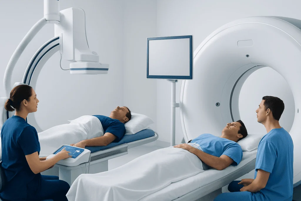
A CT coronary angiogram is especially useful for detecting plaque buildup and estimating whether the coronary arteries are narrowed. It can help identify both calcified and non-calcified plaque and may be combined with a coronary calcium score in some settings. Because it provides three-dimensional images, it can give a broad overview of the coronary anatomy and may help rule out significant disease in selected patients.
Invasive coronary angiography is very good at showing the degree and location of narrowing inside the artery channel itself. It is often considered the reference standard for defining blockages that may be causing symptoms such as chest pain or reduced blood flow to the heart. It is commonly used when there is concern for acute coronary syndrome, significant ischemia on another test, or symptoms suggesting a higher likelihood of a serious blockage.
Still, each test has limitations. CT image quality can be affected by a fast or irregular heart rhythm, heavy calcification, body habitus, or difficulty holding the breath. Coronary angiography provides less information about the vessel wall and plaque composition than CT, unless additional intravascular tools are used. It also does not always show whether a narrowing is functionally significant without further measurements.
In practice, doctors may use these tests as complementary rather than competing options. A CT scan can help clarify whether symptoms are likely related to coronary disease, while invasive angiography is often reserved for cases where the chance of important blockage is higher or where treatment planning is needed.
When Doctors Prefer CT Coronary Angiogram

CT coronary angiogram is often chosen for people who have chest discomfort or shortness of breath but are medically stable and not in the middle of a heart attack. It can be especially helpful when the diagnosis is uncertain and the doctor wants a reliable, noninvasive way to look at the coronary arteries. In many cases, a normal or near-normal CT scan can provide reassurance and reduce the need for an invasive procedure.
This test may be useful for patients with a low-to-intermediate pretest probability of coronary artery disease, including some people with stable angina symptoms. It can also be considered when stress test results are unclear or when an anatomic picture of the coronary arteries would help guide next steps. Because it is noninvasive, it is often easier for patients to accept and generally involves a shorter recovery time.
However, CT is not ideal for everyone. It may be less suitable in people with severe coronary calcification, certain arrhythmias, inability to slow the heart rate, contrast dye allergy, or significantly reduced kidney function. It is also not the preferred choice when a patient has symptoms or test findings that strongly suggest a high-risk blockage needing urgent treatment.
In some centers, CT findings may lead to additional evaluation with other heart tests such as echocardiography or stress imaging, depending on the symptoms and the degree of narrowing seen. The goal is to match the test to the clinical question while avoiding unnecessary procedures.
When Doctors Prefer Invasive Coronary Angiography
Invasive coronary angiography is usually preferred when symptoms, blood tests, ECG findings, or prior imaging suggest a high likelihood of significant coronary blockage. It is commonly used in urgent or emergency settings, including suspected heart attack or unstable symptoms. In those situations, doctors need fast, precise information and the ability to move directly to treatment if needed.
For example, a patient with severe chest pain, elevated cardiac enzymes, or evidence of myocardial infarction may go directly to coronary angiography because time matters. The same applies to some people with worsening chest pain at rest or features concerning for high-risk unstable angina. In these cases, CT generally does not replace invasive angiography.
The main advantage of coronary angiography is that it is both diagnostic and potentially therapeutic. If a critical narrowing is found, doctors may proceed with balloon angioplasty and stenting during the same session. If the artery disease is more extensive or complex, the results can also help plan procedures such as coronary artery bypass surgery or input from a cardiothoracic surgery team.
Because it is invasive, coronary angiography carries a small risk of complications such as bleeding at the access site, blood vessel injury, heart rhythm problems, contrast-related kidney issues, or, rarely, stroke or heart attack. Even so, in the right clinical setting, the benefits clearly outweigh the risks, and the test remains central to modern cardiac care.
How the Tests Are Performed and What Patients Can Expect
For a CT coronary angiogram, a patient usually lies on a CT scanner table while ECG leads monitor the heartbeat. An intravenous line is placed so contrast dye can be injected. In many cases, medication is given to slow the heart rate and improve image quality, and sometimes nitroglycerin is used to widen the coronary arteries temporarily. The scan itself is quick, often completed within minutes, though preparation may take longer.
For invasive coronary angiography, the patient is taken to a catheterization laboratory. After numbing the skin, the doctor inserts a catheter into an artery in the wrist or groin and guides it to the heart under X-ray imaging. Contrast dye is injected into the coronary arteries while a series of images is recorded. Patients are usually awake but may receive medicine to help them relax.
Recovery is generally simpler after CT. Most people can return to normal activities soon unless their doctor advises otherwise. After invasive angiography, patients may need monitoring for several hours, especially if treatment was performed or if the groin was used as the access site. Mild bruising or soreness can occur at the puncture site.
Before either test, patients should tell their care team about kidney disease, pregnancy, previous reactions to contrast dye, asthma, bleeding disorders, diabetes medications, and any symptoms of arrhythmia such as heart palpitations. This helps the team choose the safest approach and prepare appropriately.
Risks, Accuracy, and Important Limitations
Both tests can be highly informative, but no test is perfect. CT coronary angiogram is very good at ruling out significant coronary disease in appropriately selected patients. Its strength is often in its negative predictive value, meaning a normal result can be very reassuring. However, CT may sometimes overestimate the severity of blockage, especially when there is a lot of calcium in the artery walls.
Invasive coronary angiography is extremely accurate for showing the size of the artery opening and the location of a narrowing. It is less affected by motion or calcium than CT and provides real-time images during a procedure. Yet even invasive angiography does not always tell the full story about plaque characteristics or whether every narrowing is definitely reducing blood flow enough to cause symptoms.
Radiation exposure and contrast dye are relevant with both tests, although modern equipment and protocols aim to minimize these risks. Kidney function is an important consideration because contrast can stress the kidneys in susceptible patients. A history of allergy to contrast dye also matters, and premedication or alternative planning may be needed.
Accuracy is highest when the right test is chosen for the right person. Doctors consider age, symptoms, ECG findings, blood tests, previous heart disease, kidney function, heart rhythm, and the urgency of the situation. In some patients, additional studies such as stress testing or nuclear cardiology may be used to understand whether a narrowing seen on imaging is actually limiting blood flow.
Choosing the Right Test and What Happens Next
The choice between coronary angiography and CT coronary angiogram is individualized. For a stable patient with uncertain chest pain, CT may be the preferred first step because it is noninvasive and informative. For a patient with high-risk symptoms, abnormal cardiac blood tests, or strong evidence of active ischemia, invasive coronary angiography is often the better option because treatment can begin right away if necessary.
Results from either test guide next steps. If there is little or no narrowing, treatment may focus on risk reduction, symptom management, and controlling contributing conditions such as hypertension. If there is moderate disease, doctors may recommend medications, lifestyle changes, and sometimes more testing. If severe or complex blockages are found, procedures or surgery may be discussed.
Many people benefit from a broader plan that includes cholesterol management, smoking cessation, exercise guidance, blood pressure control, diabetes care, and follow-up with a cardiologist. After treatment for coronary disease, some patients are referred for cardiac rehabilitation to support recovery and reduce future risk.
Patients should seek urgent medical attention for new, severe, or persistent chest pain, chest pressure with sweating or nausea, fainting, or shortness of breath that comes on suddenly. For planned evaluation, it is best to discuss symptoms and medical history with a qualified doctor who can recommend the safest and most useful test. Acibadem International’s multidisciplinary heart specialists at JCI-accredited hospitals diagnose and treat coronary conditions for international patients when advanced evaluation is needed.
Frequently asked questions
Is a CT coronary angiogram the same as coronary angiography?
No. A CT coronary angiogram is a noninvasive scan performed with a CT machine and an IV contrast injection. Coronary angiography is an invasive catheter-based procedure done in a cardiac catheterization laboratory.
Which test is more accurate for blocked arteries?
The answer depends on the situation. Invasive coronary angiography is usually considered the reference test for defining the inside of the artery and planning treatment, while CT coronary angiogram is very effective for detecting or ruling out significant disease in selected stable patients.
Can a CT coronary angiogram replace invasive angiography?
Sometimes, but not always. In stable patients with lower or intermediate risk, CT may provide enough information to avoid an invasive test. If symptoms are high risk or immediate treatment may be needed, invasive angiography is usually still preferred.
Do both tests use contrast dye and radiation?
Yes, both usually involve contrast dye and radiation exposure. The amount varies depending on the equipment, the protocol, and whether treatment is performed during invasive angiography.
Is one test safer than the other?
CT coronary angiogram is generally less invasive and therefore has fewer procedure-related risks. Invasive coronary angiography has a higher risk profile because it involves catheter insertion, but it is very safe when performed by experienced teams and can provide immediate treatment if needed.
How does a doctor decide which test I need?
Doctors look at symptoms, ECG findings, blood test results, age, risk factors, kidney function, heart rhythm, and whether the situation is urgent. The best test is the one that answers the clinical question clearly and safely.
References
- American Heart Association
- American College of Cardiology
- European Society of Cardiology
- National Heart, Lung, and Blood Institute
- Radiological Society of North America
This article is for general information only and is not a substitute for professional medical advice. Please consult a qualified doctor about your individual situation.
Coronary Angiography in Turkey — costs, top hospitals & a free quote
JCI-accredited · board-certified surgeons · reply within 24h
Add us as a Preferred Source to see more of our trusted health content across Google Search, AI Overviews and Discover.
Check your numbers in seconds
BMI, calories, due date, blood pressure and 30+ more clinical calculators — free, instant, doctor-reviewed ranges.

Cardiology Department
Diagnosis and treatment of heart and vascular conditions, from prevention to advanced interventional procedures.
91 specialists in this unit