How Neurological Conditions Are Diagnosed: Tests You May Need

Neurological diagnosis begins with symptoms, medical history, and a focused neurological examination. Tests are chosen based on the person’s symptoms and may include blood tests, imaging, EEG, EMG, or lumbar puncture.
Key Takeaways
- Neurological diagnosis begins with symptoms, medical history, and a focused neurological examination.
- Tests are chosen based on the person’s symptoms and may include blood tests, imaging, EEG, EMG, or lumbar puncture.
- Not every patient needs every test; a neurologist tailors the evaluation to the most likely causes.
- Many neurological symptoms are treatable, and an accurate diagnosis helps guide the safest next steps.
- Urgent symptoms such as sudden weakness, speech trouble, seizures, or severe sudden headache need immediate medical attention.
Neurological diagnosis usually starts with a careful medical history and neurological exam, followed by targeted tests based on symptoms. The goal is to identify the cause clearly while avoiding unnecessary procedures whenever possible.
Overview: How neurological diagnosis works
Neurological conditions affect the brain, spinal cord, nerves, and muscles. Because these systems control movement, sensation, balance, speech, memory, vision, and many automatic body functions, neurological symptoms can vary widely from person to person. A diagnosis is often made step by step, starting with the simplest and most informative parts of the evaluation.
In many cases, the most important tools are a detailed conversation and a physical examination. A neurologist will ask when symptoms began, whether they started suddenly or gradually, what makes them better or worse, and whether there is a family history of similar problems. The pattern of symptoms often provides strong clues about whether the issue may involve the brain, spinal cord, peripheral nerves, muscles, or blood flow.
Tests are then used to confirm or rule out possible causes. These may include blood tests, imaging scans such as MRI or CT, electrical studies of the brain or nerves, and sometimes spinal fluid analysis. The process may take one visit or several, depending on how complex the symptoms are and whether they are ongoing, changing, or urgent.
The first step: medical history and neurological exam

The medical history helps place symptoms into context. Doctors usually ask about headaches, weakness, numbness, dizziness, balance problems, fainting, sleep changes, memory concerns, tremor, pain, vision changes, and episodes such as seizures or brief loss of awareness. Information about medications, alcohol use, recent infections, injuries, and long-term conditions such as diabetes or high blood pressure can also be important.
The neurological exam is a structured assessment of how the nervous system is working. It commonly includes testing strength, reflexes, coordination, walking, sensation, eye movements, speech, and mental status. Even small findings on the exam can help identify where a problem may be located.
For example, one pattern of symptoms may suggest a migraine or cluster headache, while another may point to a spinal cord problem, nerve compression, or a movement disorder such as essential tremor. If symptoms started suddenly, the evaluation may focus first on urgent causes such as stroke or bleeding in the brain.
- Sudden onset symptoms often need urgent evaluation.
- Gradual or fluctuating symptoms may suggest inflammatory, degenerative, or metabolic causes.
- Family history can be especially relevant in inherited neurological disorders.
Imaging tests: seeing the brain, spine, and blood vessels
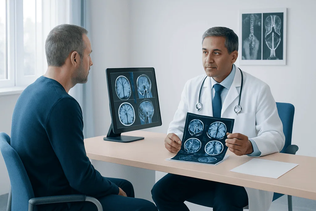
Imaging tests help doctors look for structural changes in the nervous system. A CT scan is often used quickly in emergencies because it can detect bleeding, large strokes, fractures, and some masses. It is widely available and can be especially helpful when someone has a sudden severe headache, head injury, or sudden weakness.
MRI provides more detailed images of the brain and spinal cord. It is often preferred for conditions involving multiple sclerosis, tumors, inflammation, spinal problems, or small strokes that may not be seen clearly on CT. Sometimes contrast material is used to highlight inflammation, blood vessels, or abnormal tissue.
When blood vessels are a concern, doctors may request CT angiography, MR angiography, ultrasound of the neck vessels, or specialized studies through neuroradiology or interventional neuroradiology. These tests can help evaluate narrowing, aneurysms, malformations, or reduced blood flow. The choice depends on the symptoms, how urgent the situation is, and the part of the nervous system being examined.
Imaging does not always provide a complete answer on its own. A normal scan can still occur in some neurological conditions, especially early in the course of disease or when symptoms are caused by electrical, chemical, or functional changes rather than structural damage.
Electrical and functional tests: EEG, EMG, and related studies
Some neurological disorders are better understood by measuring how the brain, nerves, or muscles function. An electroencephalogram, or EEG, records the brain’s electrical activity using small sensors placed on the scalp. It is commonly used when seizures are suspected, but it may also help assess unexplained episodes of staring, confusion, loss of awareness, or certain sleep-related events.
Nerve conduction studies and electromyography, often called EMG, evaluate peripheral nerves and muscles. These tests may be recommended for numbness, tingling, weakness, muscle wasting, or suspected nerve injury. They can help distinguish whether symptoms are coming from a nerve, the junction between nerve and muscle, or the muscle itself.
Other specialized assessments may include evoked potentials, autonomic testing, sleep studies, vestibular tests, or formal cognitive evaluation. Depending on symptoms, care may involve services such as neurophysiology, neurological sleep medicine, or neuropsychology. These tests can be particularly helpful when symptoms involve memory, attention, daytime sleepiness, fainting, dizziness, or subtle changes that do not appear on routine scans.
Although these studies can sound technical, they are usually well tolerated. A doctor or technician will explain what to expect, how long the test takes, and whether any preparation is needed, such as adjusting caffeine intake, sleep schedule, or certain medications.
Laboratory tests and lumbar puncture
Blood and urine tests are often used to look for medical causes that can affect the nervous system. Examples include vitamin deficiencies, thyroid disease, infections, autoimmune conditions, inflammation, electrolyte imbalances, liver or kidney problems, and medication-related effects. These tests can also help rule out non-neurological conditions that may mimic neurological disease.
In some cases, a lumbar puncture, also called a spinal tap, is recommended. During this procedure, a small amount of cerebrospinal fluid is collected from the lower back and examined in a laboratory. It can help diagnose meningitis, encephalitis, multiple sclerosis, bleeding around the brain, autoimmune disorders, and some cancers affecting the nervous system.
A lumbar puncture is usually considered when the expected information is likely to change treatment decisions. Imaging may be performed first in some patients to make sure the procedure can be done safely. Many people worry about the test, but the medical team takes steps to improve comfort and reduce risks such as headache afterward.
Specialized diagnosis for different neurological symptoms
Neurological diagnosis is not the same for every symptom. A person with recurrent headaches may need a different workup from someone with tremor, memory decline, facial weakness, or numbness in the feet. For example, patients with frequent headaches may be assessed for triggers, associated symptoms, medication use, and warning signs, and may be referred for headache medicine evaluation if needed. Some headaches are primary disorders such as tension-type headache, while others require investigation for an underlying cause.
People with dizziness or balance problems may need hearing and vestibular testing in addition to neurological assessment, especially if the concern is a condition such as vestibular neuritis. Those with tremor, stiffness, slowed movement, or abnormal postures may need a movement disorder assessment. Memory or behavior changes may lead to cognitive screening, brain imaging, and a review of mood, sleep, medications, and general medical health.
Some diagnoses require longer follow-up because symptoms evolve over time. This can happen in autoimmune, neurodegenerative, genetic, or neuromuscular conditions. In those situations, repeat examination, repeat imaging, or additional testing may be needed before the diagnosis becomes clear. The process can feel uncertain, but careful follow-up often prevents misdiagnosis and helps treatment stay appropriately targeted.
What patients can expect before, during, and after testing
Preparation depends on the test. Before imaging, a patient may be asked about metal implants, kidney function, pregnancy, or allergies to contrast agents. Before EEG, it may help to arrive with clean hair and avoid styling products. Before EMG or nerve conduction studies, the doctor may ask about blood thinners, skin conditions, or implanted electrical devices.
Most neurological tests are outpatient procedures. Some are very quick, while others take longer or involve a short observation period. It is often helpful to bring a list of symptoms, medications, prior test results, and key medical events. If symptoms involve memory, language, or episodes of confusion, having a family member or caregiver present can make the history more accurate.
After testing, results may be available immediately or may take several days, especially if images or samples require expert review. A diagnosis is sometimes made from one test, but often the final answer comes from combining history, examination, and multiple findings. If a result is unclear, the doctor may recommend follow-up rather than jumping to conclusions.
Near the end of the diagnostic journey, some people benefit from coordinated care across specialties. Acibadem International’s multidisciplinary specialists and JCI-accredited hospitals diagnose and treat neurological conditions for international patients when advanced evaluation is needed.
When to seek urgent medical care
Some neurological symptoms should never be ignored. Sudden weakness or numbness on one side of the body, trouble speaking, facial drooping, new confusion, sudden vision loss, a seizure, or a severe sudden headache can signal a medical emergency. Immediate assessment is important because early treatment can reduce complications in time-sensitive conditions.
Urgent evaluation is also needed after a significant head injury, when there is persistent loss of consciousness, or when a neurological symptom is accompanied by fever, stiff neck, or rapidly worsening weakness. Progressive difficulty walking, new bladder or bowel problems with back symptoms, or severe eye pain with vision changes also deserves prompt medical attention.
Even when symptoms are not an emergency, persistent or unexplained neurological complaints should be discussed with a qualified doctor. Early evaluation can provide reassurance, identify treatable causes, and help people understand what tests are truly necessary for their situation.
Frequently asked questions
What is usually the first test for neurological symptoms?
The first step is usually not a scan, but a detailed medical history and neurological examination. These help the doctor decide which tests, if any, are most likely to be useful. The exact next step depends on the symptom pattern and whether the situation is urgent.
Will everyone with neurological symptoms need an MRI?
No. MRI is very helpful for many conditions, but it is not necessary for every patient. Some people may need blood tests, an EEG, nerve studies, or no immediate testing at all if the diagnosis is clear from the history and examination.
What is the difference between CT and MRI in neurology?
CT is often used quickly in emergencies because it can detect bleeding, major stroke, and fractures rapidly. MRI usually provides more detailed images of the brain and spinal cord and is often preferred for inflammation, tumors, small strokes, and many chronic neurological conditions.
Are EEG and EMG painful?
An EEG is generally painless because it records brain activity from sensors placed on the scalp. EMG and nerve conduction studies can cause temporary discomfort because they involve mild electrical stimulation and, in EMG, a small needle in certain muscles. Most people tolerate these tests well.
Why might a doctor order a lumbar puncture?
A lumbar puncture can provide important information about infections, inflammation, autoimmune disease, bleeding around the brain, or certain cancers affecting the nervous system. It is usually recommended only when the expected information could meaningfully help confirm a diagnosis or guide treatment.
How long does it take to get a neurological diagnosis?
Some diagnoses are made quickly, especially in emergencies or when findings are straightforward. Others take more time because symptoms can overlap between conditions or evolve gradually. Follow-up visits and repeat testing may be necessary to reach the most accurate conclusion.
References
- World Health Organization
- National Institute of Neurological Disorders and Stroke
- American Academy of Neurology
- NHS
- Mayo Clinic
This article is for general information only and is not a substitute for professional medical advice. Please consult a qualified doctor about your individual situation.
Joint and spine care in Turkey — expert assessment & treatment
JCI-accredited · board-certified surgeons · reply within 24h
Add us as a Preferred Source to see more of our trusted health content across Google Search, AI Overviews and Discover.
Check your numbers in seconds
BMI, calories, due date, blood pressure and 30+ more clinical calculators — free, instant, doctor-reviewed ranges.

Neurology
Diagnosis and treatment of disorders of the brain, spinal cord, nerves and muscles.
51 specialists in this unit