

Uterine Polyps Before IVF: Diagnosis, Removal, and Implantation Planning

Uterine polyps before IVF are common and often benign, but they may interfere with implantation in some patients. Transvaginal ultrasound, saline infusion sonography, and hysteroscopy are commonly used to assess the uterine cavity.
Key Takeaways
- Uterine polyps before IVF are common and often benign, but they may interfere with implantation in some patients.
- Transvaginal ultrasound, saline infusion sonography, and hysteroscopy are commonly used to assess the uterine cavity.
- Hysteroscopic polypectomy is the standard method for removing polyps when treatment is recommended.
- Embryo transfer timing is planned after healing, pathology review, and the fertility specialist’s assessment.
- Small, symptom-free polyps may sometimes be monitored, but decisions should be individualized before IVF.
- Patients should seek medical advice for abnormal bleeding, recurrent implantation failure, or polyps found during fertility treatment.
Uterine polyps are usually benign growths inside the womb that can sometimes affect embryo implantation, especially when they distort the uterine cavity. Before IVF, careful diagnosis and individualized planning help decide whether removal is needed and when embryo transfer should take place.
Overview
Uterine polyps, also called endometrial polyps, are soft overgrowths of the lining of the uterus. They can be small and flat, or they may grow on a thin stalk inside the uterine cavity. Most are benign, but they are important in fertility care because the embryo must implant into a healthy, receptive endometrium.
When uterine polyps are found before in vitro fertilization, the fertility team considers their size, number, location, symptoms, and the patient’s reproductive history. A polyp near the area where an embryo may implant can change the shape of the uterine cavity or create local inflammation. For this reason, some polyps are removed before embryo transfer, while very small and symptom-free polyps may sometimes be observed.
The goal is not simply to remove every finding, but to create the best possible environment for implantation while avoiding unnecessary procedures. Patients planning IVF treatment should discuss whether a polyp is likely to affect their individual treatment plan, especially if they have had previous failed embryo transfers or abnormal uterine bleeding.
Symptoms and How Polyps Are Found
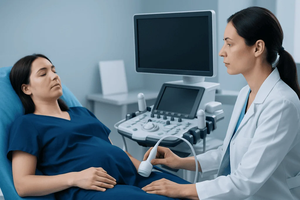
Many uterine polyps cause no symptoms and are discovered during routine fertility evaluation. Others may be associated with irregular menstrual bleeding, spotting between periods, bleeding after intercourse, or unusually heavy periods. In some patients, a polyp is suspected because the endometrium appears uneven or thickened on ultrasound.
Symptoms do not always reflect the size or importance of a polyp. A small polyp may cause spotting, while a larger one may be silent. In fertility care, even silent polyps are considered carefully because the uterine cavity is central to implantation and early pregnancy development.
Common situations in which polyps are detected include:
- Baseline ultrasound before ovarian stimulation or embryo transfer
- Evaluation for abnormal bleeding
- Investigation after miscarriage or recurrent implantation failure
- Assessment before frozen embryo transfer
- Follow-up of an unclear endometrial finding on ultrasound
Because symptoms can overlap with other gynecologic conditions, such as fibroids, hormonal imbalance, or endometrial thickening, imaging and sometimes hysteroscopy are used to confirm the diagnosis.
Causes and Risk Factors
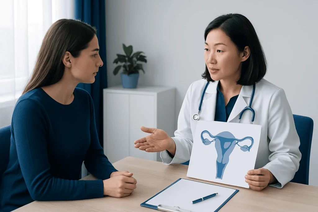
Uterine polyps develop from the endometrial tissue that responds to hormones during the menstrual cycle. Their exact cause is not always clear, but estrogen stimulation appears to play a role. Polyps may grow when local areas of the endometrium respond differently to hormonal signals than the surrounding lining.
Risk factors can include increasing reproductive age, obesity, high blood pressure, anovulation, and the use of certain medications such as tamoxifen. Conditions that affect ovulation and hormone balance, including polycystic ovary syndrome, may also be associated with endometrial changes. Patients being evaluated for female infertility often have a broad assessment to identify both uterine and hormonal factors.
In IVF cycles, polyps may occasionally be noticed during ovarian stimulation, when estrogen levels rise. This does not mean the stimulation caused a dangerous problem; rather, the higher hormone environment can make an existing polyp more visible or contribute to endometrial thickening. The fertility team then decides whether to proceed, freeze embryos for later transfer, or adjust the plan.
Diagnosis Before IVF
Diagnosis usually begins with transvaginal ultrasound. This test uses a small ultrasound probe placed in the vagina to view the uterus and ovaries. It is often most informative shortly after menstruation, when the uterine lining is thin and focal abnormalities are easier to see.
If the ultrasound is unclear, saline infusion sonography may be recommended. During this test, sterile fluid is gently placed into the uterine cavity while ultrasound images are taken. The fluid outlines the cavity and helps distinguish polyps from other findings, such as fibroids that bulge into the cavity or folds of thickened endometrium.
Hysteroscopy is considered the most direct way to evaluate the inside of the uterus. A thin camera is passed through the cervix, allowing the doctor to see the uterine cavity and often remove the polyp during the same procedure. If tissue is removed, it is sent to a pathology laboratory to confirm the diagnosis and rule out uncommon precancerous or cancerous changes.
The choice of test depends on the patient’s symptoms, previous imaging results, treatment timeline, and clinic protocols. In fertility care, the diagnostic plan should be coordinated with ovarian stimulation, embryo freezing, or embryo transfer scheduling.
When Removal Is Recommended
Removal may be recommended when a polyp is large, multiple, causing bleeding, distorting the uterine cavity, or located where implantation is likely to occur. It is also commonly considered before embryo transfer in patients with previous unsuccessful transfers, recurrent pregnancy loss, or unexplained infertility. Evidence suggests that an abnormal uterine cavity can reduce the chance of implantation, although the effect of very small polyps is less certain.
The standard treatment is hysteroscopic polypectomy. This procedure allows the doctor to see the polyp and remove it precisely, usually with small instruments passed through the hysteroscope. Blind dilation and curettage alone is generally less preferred for polyps because it may miss focal lesions and does not provide the same direct visualization.
Hysteroscopic removal is commonly performed as an outpatient or short-stay procedure. Patients may experience mild cramping and light bleeding afterward. Serious complications are uncommon, but as with any procedure, possible risks include infection, bleeding, uterine perforation, or intrauterine adhesions. The doctor explains these risks in relation to the patient’s health and reproductive plan.
After removal, pathology results help confirm that the polyp was benign. If unexpected findings are reported, the fertility plan may need to pause while the patient receives appropriate gynecologic care.
Implantation and IVF Planning After Polyp Removal
Implantation planning depends on healing, menstrual cycle timing, pathology results, and the type of IVF treatment being used. Many fertility teams wait until the next menstrual cycle before embryo transfer, especially if the polyp was removed close to the planned transfer date. This allows the endometrium to regenerate and gives time to review pathology.
If a polyp is discovered during ovarian stimulation, the clinic may recommend completing egg retrieval and freezing embryos, then removing the polyp before a later frozen embryo transfer. This approach can avoid canceling the entire cycle while still protecting the uterine environment for implantation. In some cases, if a very small polyp is found late in the cycle and does not appear to distort the cavity, the team may discuss the risks and benefits of proceeding.
Planning is especially important when patients are also using advanced laboratory fertilization methods such as ICSI or when embryos are limited. A careful uterine assessment helps ensure that the embryo transfer is timed for a cavity that appears normal and receptive.
Patients should ask their fertility specialist how polyp removal affects medication schedules, embryo freezing decisions, progesterone timing, and the planned transfer date. A clear written plan can reduce stress during treatment and help patients understand each step.
Prevention, Self-Care, and Follow-Up
There is no guaranteed way to prevent uterine polyps. However, general reproductive health measures may support a healthier endometrial environment. These include managing body weight, treating hormonal disorders, controlling blood pressure, and seeking care for irregular bleeding rather than assuming it is normal.
Patients with irregular cycles, anovulation, or conditions such as polycystic ovary syndrome may benefit from individualized hormonal and metabolic evaluation. Broader infertility assessment can identify additional factors that may influence IVF planning, including ovulation, sperm quality, ovarian reserve, and tubal or uterine conditions.
After polypectomy, follow-up is usually based on symptoms and fertility plans. Some patients do not need repeated hysteroscopy if symptoms resolve and imaging is reassuring. Others may need repeat imaging before embryo transfer, especially if they have recurrent polyps, persistent bleeding, or previous implantation problems.
For international patients, Acibadem International’s multidisciplinary specialists and JCI-accredited hospitals can evaluate uterine polyps and coordinate fertility treatment planning. Any decision about diagnosis, removal, or embryo transfer timing should be made with a qualified gynecologist or reproductive medicine specialist who knows the patient’s full medical history.
When to See a Doctor
A doctor should be consulted if there is bleeding between periods, unusually heavy menstrual bleeding, bleeding after intercourse, or bleeding after menopause. Patients preparing for IVF should also seek review if an ultrasound report mentions a suspected polyp, focal endometrial thickening, or an irregular uterine cavity.
Medical advice is particularly important before embryo transfer, because timing can affect treatment decisions. If a polyp is found after fertility medications have started, patients should not stop medications on their own. The fertility team can explain whether the cycle should continue, be adjusted, or be converted to a freeze-all plan.
Patients should also contact their doctor after polyp removal if they develop fever, worsening pelvic pain, heavy bleeding, foul-smelling discharge, or symptoms that feel unusual for their recovery. Most recoveries are straightforward, but prompt guidance helps address concerns safely.
Frequently asked questions
Can uterine polyps reduce IVF success?
Uterine polyps may reduce the chance of implantation if they distort the uterine cavity or affect the endometrial environment. The impact is less clear for very small polyps that do not change the cavity shape. A fertility specialist can interpret the finding in relation to embryo quality, prior transfers, and the planned treatment.
Do all uterine polyps need to be removed before IVF?
Not all polyps automatically require removal. Many clinics recommend removing larger, symptomatic, multiple, or cavity-distorting polyps before embryo transfer. Small, symptom-free polyps may sometimes be monitored, depending on their location and the patient’s treatment history.
What is the best test for diagnosing a uterine polyp?
Transvaginal ultrasound is often the first test, especially early in the menstrual cycle. Saline infusion sonography can provide a clearer outline of the uterine cavity. Hysteroscopy is the most direct test because it allows the doctor to see the polyp and often remove it at the same time.
How long after polyp removal can embryo transfer be done?
Timing varies by clinic and patient situation, but many fertility teams wait until at least the next menstrual cycle. This allows the lining to heal and gives time for pathology results. The final decision depends on the size of the polyp, the procedure performed, and the embryo transfer plan.
Is hysteroscopic polypectomy painful?
Discomfort varies depending on whether the procedure is done in an office setting or operating room and what type of anesthesia or pain control is used. Many patients describe mild to moderate cramping afterward. The doctor will explain the expected experience and recovery instructions before the procedure.
Can polyps come back after removal?
Yes, uterine polyps can recur, although many patients do not have repeated problems. Recurrence risk may be higher when underlying hormonal factors remain present. Follow-up imaging may be recommended if symptoms return or before a future fertility treatment cycle.
What happens if a polyp is found during IVF stimulation?
The fertility team may continue stimulation and egg retrieval, then freeze embryos and remove the polyp before transfer. In selected cases, the team may consider proceeding if the polyp is very small and not cavity-distorting. Patients should follow their clinic’s guidance and avoid changing medications without medical advice.
This article is for general information only and is not a substitute for professional medical advice. Please consult a qualified doctor about your individual situation.
IVF & fertility treatment in Turkey — success rates and costs
JCI-accredited · board-certified surgeons · reply within 24h
Add us as a Preferred Source to see more of our trusted health content across Google Search, AI Overviews and Discover.
Check your numbers in seconds
BMI, calories, due date, blood pressure and 30+ more clinical calculators — free, instant, doctor-reviewed ranges.

Related Treatments
Related Conditions
More from the Health Library

IVF With Adenomyosis: Uterine Changes, Testing, and Treatment Planning

Adenomyosis and IVF: Diagnosis, Transfer Planning, and Pregnancy Outlook

Open vs Minimally Invasive IVF: What Changes for Patients?

IVF Injections: Side Effects, Injection Tips, and When to Call

Donor Sperm IVF: Candidates, Screening, and Treatment Steps

ICSI vs IVF: Which Fertility Treatment May Be Recommended?
Gynecology & Obstetrics Specialists at Acibadem

Dr. Edıs Kahraman
Gynecology & Obstetrics
Dr. Elif Avşaroğlu
Gynecology & Obstetrics
Dr. Elif Demir
Gynecology & Obstetrics
Assoc. Prof. Dr. Elif Ganime Aygün
Gynecology & Obstetrics