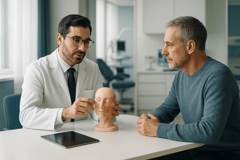
Auricular (Ear) Reconstruction
Auricular reconstruction rebuilds or reshapes the external ear after congenital absence, trauma, burns, or tumor removal using cartilage, implants, or tissue techniques for natural appearance.
Quick answer
Auricular (ear) reconstruction is surgery that rebuilds or reshapes the auricle, the visible outer ear. Depending on the defect, surgeons use the patient's own rib cartilage, a biocompatible implant framework, local flaps and grafts, or a custom prosthesis. It is performed for microtia, traumatic ear loss, burn deformity and defects left after tumour removal, often in planned stages.
Rebuilding the Auricle After Congenital Difference, Injury, Burn or Tumour Surgery
Auricular (ear) reconstruction is surgery that rebuilds or reshapes the auricle: the visible outer ear on the side of the head. It restores ear shape for children born with microtia, and for adults and children who have lost part or all of the ear through accidents, burns or the removal of a skin cancer or other tumour. The aim is an ear that is balanced with the opposite side, stable over time, and appropriate for your age, skin quality and overall health.
The auricle is a small structure, but it carries considerable personal meaning. It frames the face, supports eyeglasses and hearing devices, and is closely tied to identity and confidence. When part or all of the auricle is missing, underdeveloped, scarred or changed by surgery, the concern is rarely only cosmetic. Patients and families ask about appearance, social attention, the number of procedures ahead, scarring, hearing, safety during sports, and whether the reconstructed ear will look natural from the front and the side. These are reasonable questions, and honest answers depend on the specific defect and the tissue available to repair it.
Weighing up this surgery is rarely simple. You may be comparing surgical methods, wondering how many stages a reconstruction will require, and trying to work out whether rib cartilage, an implant-based framework, a local tissue repair or a prosthetic solution suits your case. You may be seeking a second opinion after a previous reconstruction that fell short of expectations. Careful evaluation matters here more than in many other operations, because auricular reconstruction is highly individualised. The best plan depends on anatomy, scar tissue, previous procedures, hearing needs and your own priorities, and a method that suits one patient may be a poor fit for another with an apparently similar ear.
At Acibadem, patients are assessed by physicians experienced in reconstructive and aesthetic facial surgery, paediatric and adult care, oncology-related reconstruction when needed, and imaging-based planning. The purpose of that assessment is to give you clear guidance, realistic expectations and a treatment plan that respects both function and appearance. The sections below explain the anatomy of the outer ear, who is a candidate for reconstruction, how each surgical option works, what recovery involves and which factors genuinely influence the result.
The Auricle: Ear Pinna Anatomy in Brief
Ear pinna anatomy is worth understanding before you compare surgical options, because the quality of a reconstruction is judged against the real thing. The outer ear is not a flat flap of skin. It is a three-dimensional framework of elastic cartilage covered by thin skin, with distinct curves, hollows and shadows that the eye recognises instantly, and notices instantly when they are missing.
What is the auricle in the ear?
The auricle of the ear is the visible, shell-shaped outer portion that sits on the side of the head, everything you can see and touch before the ear canal begins. Its cartilage framework creates the familiar landmarks: the helix, which is the curled outer rim; the antihelix, the raised inner fold running roughly parallel to it; the concha, the deep central bowl that funnels sound toward the ear canal; the tragus and antitragus, the two small projections guarding the canal entrance; and the lobule, or earlobe, which is the only major part of the auricle that contains no cartilage at all. The skin is tightly bound to the cartilage on the front surface and looser at the back, which is one reason the front of the ear shows every contour so clearly. Functionally, the auricle gathers sound waves and helps you work out where a sound is coming from; practically, it also supports glasses, masks and many hearing devices.
What is another name for the auricle?
The most common alternative name for the auricle is the pinna. The word auricle comes from the Latin auricula, meaning “little ear”, while pinna is Latin for a wing, feather or fin. Both terms appear throughout anatomy textbooks and clinical notes, and both refer to exactly the same structure. The plural of pinna is pinnae, in bilateral microtia, for example, both pinnae are underdeveloped rather than one.
What is the pinna vs auricle?
There is no difference between the pinna and the auricle: they are two names for one structure, the external ear. The pinna of the ear and the auricle both describe the cartilage-and-skin projection on the side of the head, as distinct from the ear canal, the eardrum and the middle and inner ear. In surgical writing you will more often see “auricle” and the adjective “auricular”; in general anatomy and audiology texts, “pinna” is common. When this page refers to reconstructing the ear pinna or the auricle, it means the same operation.
What does auricular mean?
Auricular simply means “relating to the ear”, and in surgical language it refers specifically to the auricle, the outer ear. Auricular cartilage is the elastic cartilage of the outer ear; auricular reconstruction is surgery to rebuild that structure. You may come across the same adjective in unrelated contexts, auricular acupuncture and auricular therapy, for instance, are ear-based complementary practices with no connection to reconstructive surgery, and in older cardiac texts “auricle” once described an appendage of the heart’s atrium. On this page, auricular always refers to the external ear.
Why does this anatomy matter for surgery? Because a reconstructed ear pinna is only convincing when the framework beneath the skin recreates the helix, antihelix and concha in correct proportion, at the correct height and angle relative to the eyebrow, nose and jawline, and with the correct projection away from the scalp. Even small differences in placement or angle are visible in daily life. Good reconstruction is therefore as much about planning and sculpture as it is about wound healing.
What Auricular Reconstruction Is
Auricular reconstruction is surgery to create, rebuild or refine the external ear. It concentrates on the visible cartilage-and-skin structure, the auricle, rather than the ear canal or middle ear, although hearing evaluation is often part of the overall plan for some patients, particularly children with congenital ear differences whose canal may be narrow or absent.
The reconstructed ear may be built from your own tissue, most commonly cartilage taken from the ribs and carved into an ear-shaped framework, or from a biocompatible implant framework covered by your own vascularised tissue and skin. In smaller defects, a missing rim after trauma or tumour removal, for example, reconstruction may instead use local skin flaps, cartilage grafts, skin grafts or staged tissue movement. In some patients, a custom external prosthetic ear is recommended instead of, or alongside, surgical reconstruction. None of these is universally “best”; each has a profile of advantages, limitations and demands on the patient.
It helps to think of the field in four broad categories. Autologous reconstruction uses the patient’s own tissue, usually rib cartilage, to sculpt a living framework. Implant-based reconstruction uses a synthetic porous framework that is shaped and then covered with well-vascularised tissue and skin. Local reconstruction addresses partial defects using nearby tissue, cartilage grafts and skin grafts. Revision reconstruction corrects contour, position, scarring or framework problems after previous surgery. The choice between them should rest on a surgeon’s assessment of safety, durability and expected appearance in your specific tissues, not on a standard pathway applied to everyone.
The surgical objective is never simply to create an ear-shaped object. A natural auricle has depth, shadow and asymmetry in subtle ways: the outer rim curls, the inner folds catch light, the central bowl sits at a particular depth, the earlobe hangs with a particular softness, and the whole structure projects from the scalp at a particular angle. Reconstruction has to account for these details while also working within the constraints of scars, blood supply, skin thickness and symmetry with the opposite ear. This is why surgeon experience with ear anatomy carries so much weight in this field.
Timing is a genuine planning question rather than an afterthought. In children born with microtia, some techniques are performed when the child is older and has enough rib cartilage for shaping; others may be considered earlier depending on anatomy, family goals and the technique selected. In adults, reconstruction can usually be planned after trauma, burns or tumour treatment once the tissues are healthy enough to support a reliable repair. The same principles that govern this operation also apply to neighbouring reconstructive fields: the planning logic overlaps considerably with nasal reconstruction and lip reconstruction, where thin skin must again be draped over a shaped framework in a highly visible location.
Who May Need Auricular Reconstruction
Auricular reconstruction may be appropriate for children, adolescents or adults who have absent, underdeveloped, damaged or surgically altered external ear tissue. The need may be present from birth or arise later after injury or disease. The first consultation usually concentrates on four questions: what exactly is missing, what tissue is available to rebuild it, how the ear difference affects daily life, and whether hearing or other medical issues need attention at the same time.
Microtia and congenital ear differences
Microtia is a condition in which the external ear is small, malformed or absent, and it is one of the most common congenital reasons for auricular reconstruction. It can occur on one side or both. It is sometimes associated with a narrow or absent ear canal, known as aural atresia, which can affect hearing. Children with microtia are typically evaluated by plastic or reconstructive surgeons, ear specialists, audiologists and, where appropriate, paediatric specialists working together, so that decisions about ear reconstruction, hearing support and developmental needs are coordinated rather than made in isolation. Anotia, complete absence of the external ear, sits at the severe end of the same spectrum and is planned along similar lines.
Traumatic ear loss
Adults and children may need reconstruction after partial or complete ear loss from road traffic accidents, sports injuries, bite injuries, workplace accidents or other lacerations. In acute injuries, the first priorities are preserving tissue, preventing infection and restoring blood supply where possible. In delayed cases, reconstruction is planned once swelling has settled, wounds have closed and scars have stabilised. The principles here belong to the wider discipline of reconstruction after trauma, where the surgeon must work with whatever tissue the injury has left behind rather than with the predictable anatomy of a congenital case.
Burn deformity
Burn injuries can distort the auricle badly because the skin over it is thin and the cartilage is vulnerable to exposure and infection. Burn-related reconstruction is technically demanding: scarring reduces skin mobility and blood supply, and the surrounding scalp and hairline may be involved as well, sometimes requiring coordinated planning with scalp reconstruction. These patients often need staged procedures, scar release, grafting, flap coverage and framework reconstruction, sequenced according to the extent of the damage.
Reconstruction after tumour removal
Patients may require auricular reconstruction after removal of a skin cancer or another tumour affecting the ear. The ear is a common site for sun-related skin cancers, particularly on the rim and upper portions. After tumour excision, the reconstructive plan must respect oncologic safety: clear communication between the tumour-treating team and the reconstructive surgeon determines when the repair should be performed and how best to restore contour while allowing appropriate follow-up of the treated area. Similar coordination applies across reconstruction after skin disease more broadly, where the disease process and its surveillance shape the surgical timetable.
How the diagnosis is made
Diagnosis begins with a detailed physical examination. The surgeon evaluates the size, position and shape of both ears, skin quality, scars, the hairline, scalp tissue, chest anatomy if rib cartilage may be used, and your facial proportions. Photographs are usually taken for planning. Imaging is useful in complex congenital cases, trauma, tumours or previous surgery. Hearing tests are recommended when ear canal or middle ear involvement is suspected. In revision cases, previous operative notes and photographs are especially valuable, because they show the surgeon which tissues have already been used and which reconstructive options remain realistically open.
Conditions and Indications Addressed by Auricular Reconstruction
Auricular reconstruction covers a wide range of conditions affecting the outer ear. The indication can be functional, reconstructive, aesthetic or a combination, and in every case the plan is tailored to your anatomy and the condition of the surrounding tissue.
- Microtia and anotia: microtia is an underdeveloped external ear; anotia is complete absence of the external ear. Reconstruction can create an ear shape and improve facial balance.
- Partial ear loss: missing tissue from the rim, upper ear, earlobe or central ear after trauma, surgery or burns. Smaller defects are often repaired with local tissue techniques.
- Complete traumatic ear loss: when the entire ear or most of it is missing, staged reconstruction with a cartilage or implant framework is usually needed.
- Burn deformity: contracture, scarring, cartilage exposure or collapse may require scar release, soft tissue coverage and structural reconstruction.
- Post-tumour defects: gaps left by removal of skin cancers or other tumours can often be repaired while maintaining appropriate surveillance for recurrence.
- Prominent ear or contour deformity: some patients need reshaping rather than full reconstruction. Otoplasty-type procedures can improve ear position, folds and symmetry.
- Revision after previous reconstruction: correction of framework visibility, asymmetry, poor projection, scar contracture, implant problems or unsatisfactory contour.
- Earlobe deformity: torn, stretched, absent or scarred earlobes can be repaired as a focused reconstructive procedure.
Not every ear difference requires surgery, and it is worth saying so plainly. Some patients choose observation, hair styling, prosthetic rehabilitation or delayed reconstruction, and those are legitimate choices. For children, timing should take into account emotional readiness, school and social factors, growth, hearing needs and the family’s ability to manage postoperative care. For adults, timing may depend on work, cancer treatment schedules, wound healing and personal priorities. A good surgical team will explain what each path involves and what waiting realistically costs or preserves.
How Auricular Reconstruction Is Performed
Auricular reconstruction is planned either as a single operation or in stages, depending on the defect and the technique chosen. Some partial defects can be repaired in one procedure. More complex reconstructions, particularly creating a complete ear, usually require more than one surgical stage: one to build and place the framework, later ones to position the ear away from the head and refine the final contour.
Preparation and Planning
Preparation begins with a comprehensive consultation. The surgeon reviews your medical history, medications, allergies, previous operations, wound healing history and expectations. For children, growth, cooperation with postoperative care and hearing development are considered. For adults, smoking status, diabetes, vascular health, radiation history and scar quality may influence both timing and technique, sometimes decisively.
Photographic analysis compares the affected side with the opposite ear. Measurements typically include ear height, width, position relative to the eyes and nose, projection from the head, and the location of the earlobe. In unilateral cases the healthy ear provides a natural template. In bilateral cases the surgeon plans an ear size and position that fits your facial proportions instead.
If rib cartilage reconstruction is being considered, the chest is examined to assess cartilage availability and donor-site considerations. If implant-based reconstruction is being considered, the surgeon evaluates the soft tissue envelope and whether additional coverage, such as a local or regional flap, will be needed. In tumour-related cases, pathology reports and margins are reviewed. In congenital cases with possible hearing involvement, hearing tests and ear imaging are coordinated with an ear specialist.
Part of preparation is a frank consent discussion. The surgeon should explain why a particular method is recommended, what the alternatives would involve, how many stages are anticipated, what the donor site will look and feel like if cartilage is harvested, and what the realistic range of outcomes is for your specific tissues. Writing down questions before this conversation, about scars, dressings, time off school or work, and what each stage changes, helps patients and families make an unhurried decision.
The Main Surgical Options
Autologous rib cartilage reconstruction uses cartilage from your own ribs, carved into a framework resembling the ear and placed under the skin in the correct position on the side of the head. This technique has a long history and uses living tissue from your own body. It can be durable, but it requires a chest donor site and is generally performed when enough cartilage is available for carving, which is why age and chest anatomy matter in planning.
Implant-based reconstruction uses a biocompatible porous framework shaped to resemble the ear, covered with well-vascularised tissue and skin to protect it and create a natural contour. This approach suits selected patients and may allow reconstruction at a younger age in some cases, because it does not depend on rib cartilage volume. It demands meticulous soft tissue handling, since reliable, well-perfused coverage is essential to protect the implant.
Local flap and graft reconstruction is the usual choice for partial defects. The surgeon moves nearby skin and soft tissue into the defect, adds cartilage for support where the rim or folds would otherwise notch or collapse, or uses a skin graft to resurface an area. This is most often relevant after tumour removal, laceration, bite injury or localised burn damage.
Revision reconstruction may involve reshaping cartilage, improving projection, adjusting position, releasing scars, revising the earlobe, improving skin coverage or addressing problems from previous implants or grafts. Revision cases need particularly careful planning because prior surgery alters blood supply and tissue flexibility, and the safest sequence is not always obvious from the outside.
Prosthetic rehabilitation is recommended when surgical reconstruction is not ideal, when tissue quality is poor, or when a patient simply prefers a non-surgical external ear. A well-made prosthetic ear can be highly realistic. It requires ongoing maintenance, and it attaches either with adhesive or with implant-supported retention depending on the case: a trade-off worth understanding before choosing this route.
During the Procedure
Auricular reconstruction is usually performed under general anaesthesia, especially in children and in more complex adult cases. Smaller earlobe or limited partial-ear repairs may be possible with local anaesthesia and sedation in selected adults. Operating time varies widely: a focused earlobe repair is relatively short, while full ear reconstruction can take several hours because of framework shaping, tissue coverage and precise positioning.
For a staged rib cartilage reconstruction, the sequence commonly runs as follows, though the exact plan is individual:
- Cartilage is harvested through a chest incision and carved into an ear framework matched to the template of the opposite ear or the planned design.
- The framework is inserted beneath the skin at the planned ear site; suction drains or moulded dressings help the skin conform to the new shape and reduce fluid collection.
- In a later stage, the earlobe may be transposed into its natural position and the framework contour adjusted.
- A further stage may elevate the ear from the side of the head, creating the groove behind it, often with a skin graft, so the ear projects like its natural counterpart.
- Final refinements address contour, symmetry and scars once healing from earlier stages has settled.
For implant-based reconstruction, the framework is placed in the planned position and covered with tissue that has a dependable blood supply; skin grafting may complete the coverage. The details depend on prior scarring, defect size and the selected technique. Meticulous tissue handling matters throughout, because implant exposure is a known risk when coverage is inadequate.
For partial defects, incisions are designed to preserve the ear’s landmarks and conceal scars where possible. Cartilage grafts prevent notching or collapse of the rim. In post-tumour cases, reconstruction may be performed immediately after the excision or delayed until pathology confirms the margins, depending on the clinical situation and the type of tumour treated.
Technology and Surgical Support
Modern auricular reconstruction benefits from detailed photographic planning, high-resolution imaging when deeper anatomy must be understood, surgical magnification for delicate tissue work, and refined anaesthesia monitoring. In complex cases, three-dimensional planning or printed models help the surgeon assess symmetry and framework shape before the operation. Advanced wound care, careful infection prevention and microsurgical principles come into play when tissue coverage is limited or scarred.
It is worth being direct about what technology does and does not do: it supports the surgeon’s judgement, it does not replace it. The quality of the result depends on patient selection, tissue condition, surgical planning, framework design, skin coverage and follow-up care. At Acibadem, these elements are coordinated through specialty teams when a case involves congenital conditions, cancer treatment, burn care or complex revision surgery.
Early Recovery
After surgery, you are monitored as you wake from anaesthesia. A protective dressing is placed around the reconstructed ear. Drains are used in some procedures and removed when drainage is low. Pain is managed with medication, and antibiotics may be prescribed depending on the technique and the surgeon’s protocol.
You will be advised to keep pressure off the reconstructed ear, sleeping position, clothing choices and activity restrictions all matter in the early weeks. Children usually need extra support to avoid touching or bumping the area. If rib cartilage was taken, chest soreness is noticeable in the early recovery period, particularly with coughing, laughing or certain movements; this is expected and settles gradually.
Follow-up visits let the surgeon check skin colour, swelling, drainage, wound healing and framework stability. Your team will explain how long close follow-up is needed after each stage and when normal routines can safely resume. The visible result continues to mature over months as swelling reduces and scars soften: the appearance at the first dressing change is not the final appearance.
Why Timing Matters and the Risks of Delay
The right timing for auricular reconstruction is not the same for every patient, and “early” does not always mean “immediate surgery”. In congenital cases, early evaluation is valuable even when the operation itself is planned for years later. A child with microtia may need hearing assessment, speech and developmental monitoring, and guidance about school-age social concerns; families benefit from understanding the available methods before making decisions under pressure.
For traumatic ear injuries, timely care can sometimes preserve tissue that would otherwise be lost. Fresh wounds need proper cleaning, cartilage protection and infection prevention. If an avulsed or torn segment is present, urgent specialist assessment determines whether reattachment, local repair or staged reconstruction is possible. Delay in this setting can lead to infection, cartilage damage, tissue shrinkage and more complex scarring, all of which narrow the reconstructive options later.
Burn-related ear deformity requires equally careful timing in the opposite direction. Reconstructing too early, while scars are active or wounds unstable, can increase complications. Waiting too long without scar management, however, allows contractures to become more rigid and harder to release. Specialist assessment identifies the safest window between those two errors.
In tumour-related reconstruction, delay may be entirely appropriate until cancer clearance is confirmed, but unnecessary postponement beyond that point allows wounds to contract and distort the remaining ear. Coordinated planning between the oncologic and reconstructive teams balances cancer safety with restoration of form.
For revision cases, tissues sometimes need time to recover from the previous operation before anything further is attempted. Even so, early consultation has value: it can prevent repeated ineffective procedures and clarify whether scar therapy, staged correction or an entirely different reconstructive approach is the sensible next step.
Benefits of Auricular Reconstruction
The realistic benefits of auricular reconstruction depend on the underlying condition, but they typically include both visible and practical improvements.
| Benefit | What It Means for You |
|---|---|
| Improved facial balance | Reconstruction can create an ear shape, size and position that better matches the opposite side and supports a more balanced profile. |
| Restoration after tissue loss | Missing areas caused by trauma, burns or tumour surgery can be rebuilt using techniques chosen for the size and location of the defect. |
| Greater confidence in daily life | Many patients feel less self-conscious in social, school or professional settings when the ear appears more natural. |
| Support for practical needs | A reconstructed ear may help with wearing glasses, masks or certain hearing devices, depending on the anatomy and technique used. |
| Personalised reconstruction strategy | The plan can be adapted to age, tissue quality, scars, previous surgery, hearing needs and your own preferences. |
Recovery Timeline After Auricular Reconstruction
Recovery varies by procedure type, but the timeline below reflects common experiences after many auricular reconstruction operations. Your surgeon’s instructions for your specific stage always take precedence.
| Time Period | What Patients Can Expect |
|---|---|
| Day 1 | The ear is protected with a dressing. Mild to moderate discomfort, swelling and tightness are expected. If rib cartilage was used, the chest donor site feels sore. |
| First week | Follow-up visits focus on wound checks, dressing care and drain removal if drains were used. You avoid pressure on the ear and follow sleeping and activity instructions carefully. |
| First month | Swelling gradually improves. Most patients resume routine activities, but sports, swimming and contact activities remain restricted until the surgeon confirms it is safe. |
| Several months | Scars begin to soften and the ear contour becomes clearer. Staged procedures or refinements are scheduled if they are part of the original plan. |
| Longer term | The reconstruction continues to mature. Periodic follow-up monitors symmetry, scar quality, projection and any need for revision or adjustment. |
Risks and Possible Complications
Like any operation, auricular reconstruction carries risks, and understanding them honestly is part of an informed decision. General surgical risks include bleeding, infection, fluid collection under the skin, delayed wound healing, visible scarring and reactions to anaesthesia. Because the skin draped over the ear framework is deliberately thin, thin skin is what makes the folds show, wound healing problems can matter more here than they would elsewhere on the body. If the skin over a cartilage or implant framework breaks down, part of the framework can become exposed, and further surgery may be needed to cover or repair the area.
Each technique adds its own considerations. Rib cartilage harvest leaves a chest scar and causes donor-site soreness in the early weeks; less common donor-site problems include a contour change of the chest wall and, rarely, injury to the lining of the lung during harvest, which the surgical team watches for and manages during and after the operation. Implant-based frameworks avoid a donor site but depend entirely on the quality of their soft tissue cover: exposure of the framework, infection around it or, rarely, fracture of the implant can occur months or years later and may require revision surgery. Skin grafts can heal with a colour or texture difference from the surrounding skin, and flaps used for coverage can occasionally suffer partial loss, particularly in scarred or previously operated tissue.
There are also aesthetic risks that deserve plain language: differences in size, position or projection compared with the opposite ear, blunting of the folds as swelling settles or scars mature, thickened scars in predisposed patients, and the possibility that a planned refinement stage becomes a necessary one. Sensation over a reconstructed ear is usually reduced at first and often improves only partially over time, which matters for protecting the ear from pressure, heat and cold. None of these possibilities means the surgery is unwise, many are uncommon and most are correctable, but they belong in the conversation before the first incision, not after it. A careful surgeon will explain which risks are most relevant to your particular tissues and technique.
Factors That Influence Outcomes
A good auricular reconstruction result depends on far more than the hours spent in the operating theatre. Anatomy, tissue health, technique and aftercare all shape the final appearance and its durability. Because the ear has thin skin and complex curves, even small healing changes can influence the outcome, which is why the honest answer to “how will my ear look?” always begins with an examination.
The cause and extent of the defect come first. A small missing rim after tumour removal is a very different problem from complete congenital absence, or from burn-related loss surrounded by heavy scarring. Larger and more complex defects require staged reconstruction and more careful soft tissue planning.
Skin and soft tissue quality strongly determine what is achievable. Healthy, flexible skin drapes over a framework naturally. Scarred, irradiated or burned tissue has reduced blood supply and less elasticity, which increases the need for flaps, grafts or additional stages.
Age and growth matter, especially in children. Surgeons consider whether a child has enough rib cartilage if autologous reconstruction is planned, whether the child can protect the surgical site, and how the operation fits with schooling and emotional development. In adults, overall health, smoking and wound healing history are the key considerations.
Technique selection influences recovery, scars and future options. Rib cartilage reconstruction, implant-based reconstruction and prosthetic rehabilitation each carry different trade-offs. The most suitable approach is not automatically the newest or the fastest one; it is the method that best fits your anatomy, goals and risk profile.
Surgeon experience with ear anatomy is particularly important in this field. The auricle is three-dimensional and asymmetric in subtle ways, and recreating its folds demands planning, artistic judgement and reconstructive discipline. In revision cases, experience is also needed to manage scarred tissue, previous frameworks and altered blood supply safely.
Postoperative care influences healing directly. Pressure, trauma, infection or missed follow-up can compromise a technically sound reconstruction. Following instructions on dressings, sleeping, hygiene and activity protects the investment of the surgery itself.
Realistic expectations are essential, and stating the limits plainly is part of good care. Auricular reconstruction can significantly improve appearance and symmetry, but a reconstructed ear is not identical to a natural ear. Some differences in texture, projection, scars or contour are common and expected. The goal is a stable, well-positioned, natural-looking ear that fits your face and meets your priorities as closely as the tissues allow.
Caring for a Reconstructed Ear Over Time
A reconstruction does not end at the last operation; it settles, matures and needs sensible protection. Scars over and around the ear soften over many months, and most surgeons advise shielding them from strong sun while they mature, since fresh scars can darken with sun exposure. Because sensation over the reconstructed ear is often reduced, patients, and the parents of younger patients, should stay aware of pressure and temperature in situations where a normal ear would signal discomfort: tight headphones, cold winter weather, hot styling tools used near the ear.
Sports can usually be resumed in stages. Non-contact activity generally returns first, while contact sports, swimming and activities with a risk of a direct blow to the ear wait until the surgeon confirms the framework and its skin cover are stable; protective headgear may be recommended for some sports afterwards. Glasses and hearing devices can usually be worn once healing allows, and part of the planning in many cases is making sure the reconstructed ear will support them comfortably. Piercing through reconstructed tissue is generally discouraged, although an intact, repositioned earlobe can sometimes be pierced after discussion with the surgeon.
Long-term behaviour differs slightly between techniques. A rib cartilage framework is living tissue: it integrates with the surrounding tissue, tends to remain stable over the years and, in children, is generally expected to grow acceptably with the face. An implant framework does not change over time, but its thin soft tissue cover must be respected indefinitely, which is why any redness, thinning or tenderness over the framework is taken seriously at routine follow-up visits. A prosthetic ear needs periodic remaking as materials age and skin tone changes with the seasons and the years. Whatever the method, periodic review protects the result, and keeps the door open to small refinements if they would genuinely help.
Coordinated, Multidisciplinary Care
Auricular reconstruction often crosses several specialties, and the way that overlap is organised matters as much as any single consultation. A child with microtia may need input from reconstructive surgery, ear specialists, audiology and paediatrics. A patient after tumour removal may need coordination with dermatology, oncology and pathology. A burn patient may need scar management, reconstructive planning and wound care in sequence. At Acibadem, multidisciplinary boards and specialist consultations are used in complex cases so that the treatment plan is aligned across these disciplines rather than assembled piecemeal.
Personalisation is the constant theme. Some patients want the most durable autologous option even though it involves a donor site and staged surgery. Others are candidates for an implant-based framework. Some are best served by a partial repair, a scar revision or a prosthesis. A well-designed plan explains why a method is recommended, what the alternatives are, what limitations to expect, and how each stage of recovery will affect daily life.
Making the Decision
Auricular reconstruction is a deeply personal decision. Whether the concern began at birth, followed an accident, resulted from a burn or developed after tumour surgery, the underlying question is usually the same: what can be restored safely, naturally and in a way that fits your life? The answer depends on a detailed examination, an honest discussion of the options and a plan built around your anatomy and goals, not on a generic pathway.
It helps to go into that discussion knowing what to weigh. The main variables are the technique (autologous cartilage, implant framework, local repair or prosthesis), the number of stages involved, the donor-site implications, the condition of your skin and scars, the timing relative to growth or cancer surveillance, and what a realistic result looks like for your specific defect. Patients who have already had surgery elsewhere often find that an independent second opinion clarifies which revision options remain and which have been closed off by previous procedures. With careful planning, auricular reconstruction can restore important elements of appearance, balance and confidence while respecting the complexity of the ear, and the individual needs of the person it belongs to.
Preparation
- A plastic and reconstructive surgeon evaluates ear anatomy, skin quality, hearing history, and previous surgeries. Preoperative photos, imaging, or hearing tests may be requested depending on the case. Patients may need to stop smoking and avoid blood-thinning medicines before surgery as instructed.
Aftercare
- A protective dressing or head bandage is usually worn after surgery, and the ear must be protected from pressure or injury. Mild swelling, bruising, and discomfort are expected and controlled with prescribed medication. Follow-up visits monitor healing, stitches, and whether staged reconstruction is needed.
Frequently Asked Questions
What affects the cost of auricular reconstruction?
Cost is influenced by the cause and extent of ear absence or deformity, the selected reconstruction method, the need for cartilage, implants, flaps, grafts, tissue expansion, anesthesia, hospital stay, imaging, and follow-up. International travel support and accommodation planning may also affect the overall budget.
How can I get a personalised quote from Acibadem?
You can request a free consultation by sharing medical photographs, previous reports, operation notes if available, and a short medical history. A specialist team can then review your case and provide a personalised treatment plan and quote.
Is auricular reconstruction usually done in a single visit?
Some limited repairs may be completed within a shorter pathway, while more complex congenital, trauma, burn, or tumor-related reconstructions may require staged planning. Your surgeon will explain the expected timeline after assessment.
Does the choice between cartilage, implant, and prosthetic ear change the cost?
Yes. Each option has different requirements for operating time, materials, hospital resources, donor site care, laboratory or prosthetic work, and follow-up. Suitability and cost depend on specialist evaluation rather than preference alone.
What is typically included in an international patient package?
Depending on the agreed plan, a package may include specialist assessment, surgery, anesthesia, hospital services, nursing care, interpreter support, care coordination, and follow-up planning. Inclusions should always be confirmed in your written quote.
Is this information a medical or financial recommendation?
No. This is general educational information only. A personalised medical opinion and cost estimate require consultation with a qualified specialist and review of your individual needs.
Medically reviewed by the Acıbadem International Medical Board September 1, 2026
See our medical review board →
Update history
- PublishedJune 8, 2026
- Medical review approvedSeptember 1, 2026
- Last content updateSeptember 1, 2026
Trusted care for international patients
From the Acibadem Blog
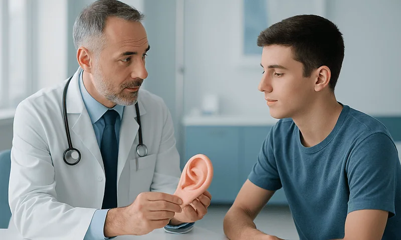
Which Ear Reconstruction Framework Is Right: Rib Cartilage or a Porous Implant?
Neither framework wins outright in ear reconstruction rib cartilage vs implant decisions. Rib cartilage is the patient's own living tissue, tolerates injury well and…
Doctors Performing This Treatment


Aesthetic Plastic & Reconstructive Surgery
Prof. Mehmet Veli Karaaltın, MD

Aesthetic Plastic & Reconstructive Surgery
Assoc. Prof. Erdem Güven, MD

Aesthetic Plastic & Reconstructive Surgery
Assoc. Prof. Ahmet Küçükçelebi, MD
Aesthetic Plastic & Reconstructive Surgery
Assoc. Prof. Mehmet Altıparmak, MD

Aesthetic Plastic & Reconstructive Surgery