

Electrophysiology (EPS )
Electrophysiology study (EPS) is a catheter-based test that maps the heart’s electrical signals to diagnose abnormal rhythms and guide treatments such as ablation or device therapy.
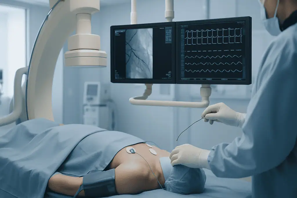
Quick answer
An electrophysiology study (EP study) is a catheter-based test that maps the heart's electrical system from inside the heart. Thin catheters record electrical signals and deliver small, controlled impulses to find where an abnormal rhythm starts. It is used to diagnose arrhythmias precisely and to guide treatment decisions, catheter ablation, medication planning, or a pacemaker or implantable defibrillator.
What Is an Electrophysiology Study?
An electrophysiology study, usually shortened to EP study or EPS, is a catheter-based test that examines the heart’s electrical system from inside the heart itself. Thin, flexible catheters are passed through a blood vessel, usually a vein in the groin, and guided into the heart, where they record electrical signals directly and deliver small, controlled impulses to test how the rhythm system responds. It is used when the heart beats too fast, too slowly or irregularly and the cause needs to be identified precisely enough to guide treatment.
The heart beats because electrical signals travel along an organised pathway. The signal begins in the sinus node, spreads through the atria, passes through the atrioventricular node and then travels into the ventricles. When there is an extra pathway, an irritable electrical focus, a scar-related circuit or a conduction delay, the rhythm can become too fast, too slow or chaotic. An EP study identifies these mechanisms with a level of detail that tests recorded from the skin cannot always provide.
If your heart suddenly races, pauses, flutters or beats out of rhythm, the experience can be frightening. Some people feel only brief palpitations. Others have dizziness, breathlessness, chest discomfort, fainting or a persistent sense that something is wrong even when standard tests look normal. Many people arrive at the decision to have an EP study after months or years of uncertainty, repeated emergency visits, medication trials, and questions about stroke risk, sudden cardiac events or simply quality of life. The value of the study is that it replaces assumption with measurement.
For many patients, an EP study answers the questions that matter most. What is causing the rhythm problem? Is it dangerous? Is medication enough? Would catheter ablation help? Is a pacemaker or an implantable defibrillator needed? The goal is not only to name the arrhythmia, but to build a treatment plan that fits your rhythm pattern, heart structure, symptoms, medical history and personal priorities. The study may be purely diagnostic, or it may be combined with treatment in the same session, most commonly catheter ablation, in which targeted energy modifies or eliminates the tissue responsible for the abnormal rhythm.
What does EP mean in cardiology?
In cardiology, EP stands for electrophysiology: the study of the heart’s electrical activity. When your cardiologist refers to the EP lab, the EP team or an EP study, they mean the specialised service that investigates and treats heart rhythm disorders. The abbreviation has other meanings elsewhere, an extended-play record in music, for instance, but in a hospital setting EP almost always refers to the heart’s electrical system.
You may also hear EPS, which simply adds the word study. EP study and EPS describe the same procedure: intracardiac recording and testing of the heart’s conduction system. Related terms include EP mapping, which means building a detailed picture of where signals travel, and EP ablation, which means treating the arrhythmia source during the same session.
What is cardiac electrophysiology?
Cardiac electrophysiology is the branch of cardiology that deals with the electrical function of the heart: how each heartbeat is generated, how the signal spreads through the chambers, and what happens when that process goes wrong. It covers the diagnosis of arrhythmias, invasive studies, catheter ablation, and the implantation and follow-up of rhythm devices such as pacemakers and defibrillators.
The field sits alongside, but is distinct from, other areas of heart care. A cardiologist treating blocked arteries deals mainly with the heart’s plumbing; cardiac electrophysiology deals with its wiring. The two often overlap: a previous heart attack can create scar tissue that becomes the source of a dangerous rhythm, which is why electrophysiology works best inside a broader cardiovascular programme rather than in isolation.
How is an EP study different from an ECG?
An EP study records the heart’s electrical activity from inside the heart, while an electrocardiogram (ECG) records it from electrodes on the skin for a short period. An ECG is quick, painless in the clinical sense and very useful, but it is a surface summary of the heart’s electrical behaviour at one moment. If your arrhythmia does not happen during the recording, the ECG may look entirely normal.
An EP study goes further in three ways. It measures signals directly from specific structures inside the heart, so conduction can be timed segment by segment. It can deliberately and safely attempt to trigger the abnormal rhythm under controlled conditions, so the mechanism can be observed rather than inferred. And it can be combined with three-dimensional mapping and, where appropriate, immediate treatment. Ambulatory monitors, Holter recorders, event monitors, implantable loop recorders, sit between the two: they extend the recording window but still watch from the outside.
Who Performs an EP Study?
An electrophysiologist is a cardiologist who has completed additional training in the diagnosis and treatment of heart rhythm disorders. This subspecialty training covers the interpretation of intracardiac signals, catheter handling, mapping systems, ablation techniques and rhythm device therapy. The distinction matters: reading electrical signals from inside a beating heart, and deciding in real time what they mean, is a skill built through dedicated training and case volume.
If you are looking for a cardiac electrophysiologist, it is worth knowing what actually determines the quality of care: the experience of the physician with your specific arrhythmia type, the equipment and staffing of the electrophysiology laboratory, and the strength of the wider cardiac programme behind it. Proximity is convenient, but for a planned, non-urgent procedure, the fit between your rhythm problem and the team’s expertise usually matters more than the distance to the hospital.
An EP study is never a one-person procedure. A cardiac electrophysiologist leads a team that typically includes specialised nurses, cardiac technicians, anaesthesiology professionals and imaging staff. The laboratory itself is built for continuous monitoring of heart rhythm, blood pressure and oxygen levels throughout the study, with resuscitation capability immediately at hand. That combination, trained people and a purpose-built environment, is what allows abnormal rhythms to be provoked, studied and stopped in a controlled way.
Who May Need an EP Study?
An EP study may be recommended when symptoms, test results or medical history point to an abnormal heart rhythm that needs a precise diagnosis. Most people who reach this point have already had an ECG, ambulatory rhythm monitoring, echocardiography, blood tests or stress testing. Sometimes those tests clearly show a cardiac arrhythmia. Sometimes they suggest a problem without fully explaining it. The EP study is designed to close that gap.
Typical reasons for referral include recurrent palpitations, sudden episodes of rapid heartbeat, unexplained fainting or near-fainting, dizziness, an irregular pulse, fatigue linked to rhythm changes, or a documented arrhythmia that needs further characterisation. The study may also be advised after a serious rhythm event, in people with certain inherited rhythm disorders, or in selected patients with heart muscle disease, a previous heart attack or abnormal findings on other cardiac tests.
One of the most common scenarios is a fast heart rhythm that starts and stops abruptly. Patients often describe a sudden switch from normal rhythm to a racing heartbeat, sometimes with chest pressure, anxiety, sweating or lightheadedness. Episodes like this may be caused by supraventricular tachycardia, an accessory pathway or atrial tachycardia. An EP study can locate the responsible pathway or circuit and establish whether ablation is an appropriate option.
Another important scenario is unexplained syncope, fainting without an identified cause. Fainting has many possible explanations, including blood pressure changes, neurological conditions and dehydration. When a rhythm cause is suspected, however, an EP study can show whether the heart’s electrical system is too slow, intermittently blocked or vulnerable to dangerous fast rhythms. The distinction matters because the treatments are entirely different: a slow, blocked system points towards pacing, while an inducible fast ventricular rhythm points towards ablation, medication or a defibrillator.
How do doctors decide an EPS is necessary?
The decision rests on a complete clinical assessment, not on symptoms alone. Your cardiologist or electrophysiologist reviews your medical history, previous emergency visits, current medications, family history, ECGs and any rhythm monitor recordings. Imaging such as echocardiography or cardiac MRI may be used to assess the heart’s structure and pumping function. Blood tests can identify thyroid disease, electrolyte imbalance or other contributors to arrhythmia that need addressing first.
Broadly, there are two routes to the EP lab. In some patients, the rhythm diagnosis is already established and invasive mapping is needed to plan or deliver treatment. In others, non-invasive monitoring has never captured the rhythm, but the pattern of symptoms strongly suggests a clinically significant arrhythmia, and the study is the most direct way to test that suspicion. In both cases, the guiding question is the same: will the result change what happens next? An EP study is most valuable when its findings will alter treatment, clarify risk or make a targeted intervention possible.
Complete records make that judgement possible. Prior ECGs, Holter reports, hospital discharge summaries and a current medication list allow the team to assess whether a study is genuinely indicated, and, if it is, whether it is likely to be diagnostic only or combined with ablation in the same session.
Conditions and Indications Addressed by EPS
An electrophysiology study can evaluate a wide range of rhythm disorders. Its exact role depends on the type of arrhythmia, your overall cardiac condition and whether treatment is planned for the same session.
- Supraventricular tachycardia: the study can identify the common mechanisms, atrioventricular nodal reentrant tachycardia, atrioventricular reentrant tachycardia and atrial tachycardia, and distinguish between them, which matters because the ablation target differs for each.
- Accessory pathways and Wolff-Parkinson-White pattern: the study assesses the location and conduction properties of an extra electrical pathway and guides ablation where appropriate.
- Atrial flutter: mapping defines the circuit responsible for the flutter and can guide catheter ablation of that circuit.
- Selected cases of atrial fibrillation: electrophysiology mapping may form part of an ablation strategy, particularly when symptoms persist despite medication or when a rhythm-control approach is preferred.
- Ventricular tachycardia: the study can evaluate abnormal rhythms arising from the lower chambers, particularly in patients with structural heart disease, a previous heart attack or cardiomyopathy.
- Unexplained fainting: when initial assessment suggests a cardiac rhythm cause, the study can test the conduction system and the heart’s vulnerability to fast rhythms.
- Conduction system disease: the study measures the pathways responsible for slow heart rhythms or intermittent block, which informs decisions about pacing.
- Risk assessment in selected patients: in certain inherited or structural heart conditions, the findings contribute to decisions about monitoring, medication, ablation or device therapy.
Not every arrhythmia needs an EP study. Some rhythm problems are appropriately managed with observation, lifestyle changes, medication or extended external monitoring. Occasional extra beats in a structurally normal heart, for example, rarely justify an invasive test. The study earns its place when the answer it provides is likely to change your management, and a careful electrophysiologist will say so plainly when it is not needed.
How an Electrophysiology Study Is Performed
The study takes place in a dedicated electrophysiology laboratory, led by a cardiac electrophysiologist with a team of nurses, technicians, anaesthesiology professionals and imaging staff. Everything in the room is arranged around one requirement: continuous, precise monitoring of your heart rhythm, blood pressure and oxygen levels while the electrical system is tested.
Before the Procedure
Preparation begins with a detailed consultation. Your physician reviews your symptoms, prior rhythm recordings, medical conditions and medications. Some rhythm medications may need to be paused before the study so that the abnormal rhythm can be provoked and studied accurately; blood thinners, diabetes medications and other treatments require individualised instructions from your treating doctor. Do not stop or change any medication on your own: the plan is set specifically for you, and it balances test accuracy against your safety.
Pre-procedure testing may include an ECG, blood work, echocardiography and further imaging if needed. If a cardiac stress test has been part of your earlier work-up, bring those results too: they help the team understand how your rhythm behaves under exertion. You will receive fasting instructions, information about the anaesthesia or sedation planned for your case, and a clear description of what to expect afterwards.
On the day of the study, an intravenous line is placed for fluids and medications. The skin over the access site, usually the groin, is cleaned and numbed with local anaesthetic. Depending on the case, you may receive moderate sedation or deeper anaesthesia. Some studies are done under light sedation deliberately, because certain rhythms are easier to provoke and assess when the patient is not deeply anaesthetised; more complex ablation procedures may involve a different anaesthesia plan. This is decided case by case, and you should know the plan before the day itself.
During the Procedure
The core of the study follows a logical sequence:
- Access: the electrophysiologist inserts thin catheters into a vein, often using ultrasound guidance to make vascular access precise.
- Positioning: the catheters are guided through the blood vessels into specific chambers of the heart under imaging guidance. You usually do not feel the catheters inside the heart, although there may be pressure at the access site.
- Recording: once positioned, the catheters record electrical signals directly from different chambers and pathways, allowing conduction to be timed segment by segment.
- Programmed stimulation: the physician delivers small, controlled electrical impulses to test conduction and attempt to reproduce your arrhythmia. If your usual rhythm problem is induced, the team can analyse its origin and mechanism directly.
- Mapping: where needed, three-dimensional mapping systems build a detailed electrical map of the heart, showing where signals begin, how they travel and where abnormal circuits sit.
- Treatment, if planned: if the study identifies an arrhythmia suitable for immediate treatment, and this was discussed and agreed beforehand, catheter ablation may follow in the same session.
During programmed stimulation you may feel palpitations similar to your usual symptoms. This is expected, reproducing the rhythm is often the point of the test, and the rhythm is monitored continuously and can usually be stopped promptly, either by pacing through the catheters or with medication.
Modern laboratories add several technologies around this sequence: three-dimensional electroanatomical mapping, intracardiac signal recording, fluoroscopic imaging with radiation-reduction techniques, and ultrasound guidance for vascular access. None of these replaces the physician’s judgement. Their purpose is to improve precision, reduce unnecessary catheter movement and support safer decisions during the procedure. Ablation, where performed, uses targeted energy, most commonly radiofrequency heat or cryothermal (freezing) energy, to interrupt the abnormal pathway or focus. If the study instead shows that a device is needed, such as a pacemaker or implantable cardioverter-defibrillator, that is usually planned as a separate, considered step based on the findings and your overall condition.
How long does an EP study take?
The duration depends on the complexity of the rhythm problem and on whether ablation is performed. A diagnostic study alone is relatively short; mapping and ablation of complex arrhythmias take considerably longer. Plan for additional time on the day for preparation beforehand and monitored recovery afterwards: the procedure itself is only part of the visit.
Complexity drives the difference. A study for a single suspected accessory pathway involves fewer catheters and less mapping than a scar-related ventricular arrhythmia, where the team may spend considerable time building a detailed electrical map before any treatment is delivered. Your team can usually give you a realistic range beforehand based on the suspected diagnosis and the planned extent of the procedure.
Does an EP study hurt?
Most people describe pressure rather than sharp pain at the access site, because the skin is numbed with local anaesthetic before the catheters are placed. The catheters themselves are usually not felt inside the heart. The most noticeable sensation for many patients is the palpitations during programmed stimulation, deliberately provoked, closely monitored and typically stopped quickly. Sedation levels are set for your case, and you can discuss the anaesthesia plan in detail beforehand.
Immediately After the Procedure
Once the catheters are removed, pressure is applied to the access site to reduce bleeding. You are then monitored in a recovery area while your heart rhythm, blood pressure and the puncture site are checked. Expect to lie flat for several hours, particularly when the groin vein was used. Mild soreness or bruising at the access site is common and usually settles on its own.
Some patients go home the same day; others stay overnight, particularly after ablation, after a complex case, or when additional monitoring is sensible. Before discharge, the team gives you specific instructions covering walking, bathing, medication, wound care, warning signs and follow-up. It is worth asking before discharge when it is safe to resume driving and longer journeys: the answer differs between a short diagnostic study and a longer ablation, and it is set for your individual case.
Why Acting Early Matters
Many rhythm disorders are treatable, but delaying evaluation allows symptoms and risks to accumulate. Recurrent rapid rhythms interfere with daily life, sleep, exercise and emotional wellbeing. Some arrhythmias can weaken the heart muscle over time if the heart beats too fast for prolonged periods: a preventable form of heart failure. Others may raise the risk of fainting, injury, stroke or serious cardiac events, depending on the rhythm type and the underlying heart condition.
Early evaluation carries particular weight when symptoms include fainting, chest pain, breathlessness, documented very fast rhythms, a family history of sudden cardiac death, or known structural heart disease. None of these automatically means a dangerous rhythm is present. They do mean the assessment deserves physicians experienced in arrhythmia management, working with complete information rather than fragments.
A timely EP study reduces uncertainty. Instead of repeated emergency evaluations with no clear plan, you get a specific diagnosis and a structured pathway. Sometimes the outcome is reassurance and continued monitoring. Sometimes it is ablation, a medication adjustment made by your treating doctor, or device therapy. The consistent value is the same in every case: decisions based on detailed electrical measurement instead of assumption.
Benefits of an Electrophysiology Study
The benefits depend on your specific rhythm problem, but the study can provide information that directly shapes diagnosis, treatment and long-term management.
| Benefit | What It Means for You |
|---|---|
| Precise rhythm diagnosis | The study can identify where an abnormal rhythm begins and how it travels through the heart, especially when external tests have not provided enough detail. |
| Guidance for ablation | If the arrhythmia is suitable for catheter ablation, mapping during the study helps target the exact area responsible for the rhythm problem. |
| Better risk assessment | For selected patients with fainting, heart disease or concerning rhythm findings, the study helps clarify whether a rhythm disorder may pose a higher risk. |
| Medication planning | Results help your physician decide whether medications are needed, should be adjusted, or can be avoided in favour of another strategy. |
| Device therapy decisions | Findings support decisions about pacemakers or implantable defibrillators when slow rhythms, conduction disease or dangerous fast rhythms are suspected. |
| Reduced uncertainty | For patients with recurrent, unexplained symptoms, the study can provide a clear explanation and a focused plan for future care. |
Recovery Timeline After EPS
Recovery is usually straightforward, though it varies depending on whether the study was diagnostic only or combined with ablation or another intervention. The table below describes a typical course; your own instructions take precedence.
| Time Period | What Patients Can Expect |
|---|---|
| Day 1 | Monitoring after the procedure, several hours of limited movement, mild groin soreness or bruising, and discharge the same day or overnight observation depending on the case. |
| First Week | Most patients resume light daily activities. Heavy lifting, intense exercise and long walks may be restricted for several days to protect the access site. |
| First Month | Follow-up may include an ECG, symptom review and medication adjustment by your treating doctor. If ablation was performed, brief palpitations can occur during the healing phase and should be discussed with the care team. |
| Longer Term | The plan may include continued monitoring, medication, lifestyle guidance, ablation follow-up or device evaluation depending on the findings. |
For patients whose arrhythmia sits alongside broader heart disease, a structured cardiac rehabilitation programme may form part of the longer-term plan, combining supervised exercise, education and risk-factor management. Whether this is appropriate depends on your underlying condition rather than on the EP study itself.
Factors That Influence Outcomes
The results of an EP study, and of any treatment performed during the same session, depend on several medical and procedural factors. The most important is the type of arrhythmia. Some rhythm disorders, many forms of supraventricular tachycardia, for example, often respond well to ablation when the abnormal pathway or circuit is clearly identified. More complex arrhythmias, including atrial fibrillation and ventricular tachycardia in patients with structural heart disease, may require more extensive mapping, staged treatment over more than one procedure, or ongoing medication alongside ablation.
The structure and function of the heart also matter. A patient with normal heart muscle and an isolated electrical pathway has different expectations from a patient with cardiomyopathy, a prior heart attack, valve disease or congenital heart disease. Imaging before the study establishes this context and shapes both the plan and the honest conversation about what the procedure can and cannot achieve.
Whether the arrhythmia can be induced during the study is another variable. The study is designed to reproduce abnormal rhythms under controlled conditions, but some intermittent rhythms simply do not appear during testing. Even then, the study still yields useful information about conduction properties and future management. Occasionally, extended monitoring afterwards or a repeat procedure is the reasonable next step, and it is better to hear that possibility beforehand than to be surprised by it.
Medication use affects the findings. Some antiarrhythmic drugs suppress the very rhythms the study is trying to provoke; others alter conduction measurements. This is why the pre-procedure medication plan is individualised by your treating doctor: the team balances the need for accurate testing against your safety and comfort, and no two plans are identical.
Procedural expertise and team coordination matter throughout. Electrophysiology demands careful interpretation of intracardiac signals, precise catheter positioning, disciplined use of mapping data and constant attention to patient-specific risk. Good outcomes are supported by experienced physicians, skilled laboratory teams, appropriate technology, careful anaesthesia planning and structured follow-up after the procedure.
Finally, your own participation contributes. Sharing complete records, describing your symptoms accurately, following the medication instructions your doctor gives you, and respecting activity restrictions after the procedure all make the study safer and more informative. Clear communication with the clinical team before the procedure and at each follow-up is particularly valuable: it is how the plan holds together over time.
Risks and Safety Considerations
An EP study is generally considered a safe procedure when performed by trained electrophysiology teams, but it is an invasive cardiac test and carries potential risks. These may include bleeding, bruising or infection at the catheter insertion site; blood vessel injury; arrhythmias that require treatment during the study; blood clots; reactions to medications or contrast agents if used; and, rarely, damage to heart structures or fluid accumulating around the heart. Radiation exposure can occur when fluoroscopy is used, although modern laboratories apply techniques to keep exposure as low as reasonably possible.
The risk profile differs between a short diagnostic study and a longer mapping and ablation procedure, more time in the heart and more energy delivery change the calculation. Patients with advanced heart disease, kidney disease, bleeding disorders or multiple medical conditions may need additional precautions. This is precisely what the pre-procedure assessment exists to establish: whether the study is appropriate for you at all, and how it should be planned if it is.
Before discharge, your care team gives you written instructions covering the access site, activity limits, medication and the specific warning signs relevant to your case. Keep those instructions and your procedure summary with you until your follow-up review is complete.
Electrophysiology Care at Acibadem
At Acibadem, electrophysiology is delivered within a broader cardiovascular programme rather than as a standalone service. This matters because arrhythmias are frequently connected to other conditions, coronary artery disease, valve disease, cardiomyopathy, congenital heart conditions, thyroid disease, medication effects, or sleep apnoea, which is itself typically investigated with an overnight sleep study. When a case is complex, it can be discussed across disciplines: cardiologists, cardiac imaging specialists, cardiac surgeons, anaesthesiologists and intensive care physicians. Patients with complicated histories or previous procedures benefit most from this kind of joint review.
Diagnostic pathways follow international, evidence-based protocols and may include ECG, ambulatory monitoring, echocardiography, stress testing, cardiac CT or MRI, laboratory testing and a full review of your previous records. The stated aim is deliberately two-sided: avoid unnecessary procedures, and at the same time make sure clinically significant rhythm disorders are not missed. An EP study is recommended when its result is expected to change your management, not as a default.
In the electrophysiology laboratory, modern mapping and monitoring technologies support the detailed evaluation of electrical activity: three-dimensional mapping to visualise arrhythmia circuits, intracardiac recordings to measure conduction intervals directly, and imaging guidance for catheter placement. These tools do not replace physician judgement; they extend what a careful physician can see and how precisely the team can act on it.
Treatment planning is individual by necessity. A young patient with sudden episodes of supraventricular tachycardia, an older patient with fainting and conduction disease, and a patient with ventricular tachycardia after a heart attack may all undergo an EP study, but their risks, goals and treatment options differ substantially. The right plan reflects the specific rhythm diagnosis, the state of the heart, your lifestyle and the follow-up arrangements available to you.
Preparing for a Specialist Consultation
If an EP study is being considered, the quality of the consultation depends heavily on the records you bring. Useful documents include recent ECGs, Holter or event monitor reports, echocardiogram results, stress test findings, cardiac imaging, blood tests, hospital discharge summaries and a complete current medication list. Recordings from a smartwatch or mobile ECG device can be genuinely helpful as supporting evidence, although they do not replace medical-grade testing.
Come with questions, and expect direct answers. Sensible things to ask an electrophysiologist include: What rhythm is suspected, and how confident are you? Is the study planned as diagnostic only, or is ablation likely in the same session? What anaesthesia approach will be used? What medication changes will be needed beforehand, and who will manage them? How long will I be in hospital, and when can I return to normal activity? What follow-up will I need, and how will it be arranged? What happens if the arrhythmia cannot be induced during the study?
A good plan covers more than the procedure date. It sets out preparation, the anaesthesia approach, the expected length of stay, recovery restrictions, warning signs, and how communication with the team will work after discharge, including who reviews your ECGs at follow-up. If any of those elements is vague, ask again until it is specific.
For many patients, an EP study provides the first clear explanation for symptoms that have been disruptive or frightening for years. For others, it refines a known diagnosis and guides treatment decisions that reduce future episodes or address more serious risks. Weigh the decision with an experienced electrophysiology team that explains the benefits, the limitations and the alternatives in terms you understand, and that is willing to tell you, plainly, when the study is not the right next step.
Preparation
- Before EPS, your cardiologist reviews your medical history, ECG results, medications, and any blood tests or imaging. You may be asked to stop certain rhythm or blood-thinning medicines and fast for several hours before the procedure. Tell your team about allergies, implanted devices, or pregnancy possibility.
Aftercare
- After the study, your heart rhythm and catheter insertion site are monitored while you rest. You may need to lie flat for several hours to reduce bleeding risk. Avoid heavy lifting and strenuous activity for a few days, and contact your doctor for chest pain, fever, swelling, or bleeding.
Frequently Asked Questions
What affects the cost of an electrophysiology study?
Cost is influenced by whether the EPS is diagnostic only or combined with ablation, the complexity of the arrhythmia, catheter lab time, mapping technology, anaesthesia, pre-procedure tests, hospital stay, specialist fees, and follow-up needs.
How can I get a personalised quote for EPS in Turkey?
You can request a free consultation and share your ECG, Holter report, echocardiogram, blood test results, medication list, previous cardiac reports, and a summary of symptoms. A cardiology team can then advise which tests or procedures may be needed and prepare a personalised estimate.
Is ablation always included with an EPS?
No. EPS may be performed only to diagnose the rhythm problem, or it may be combined with catheter ablation if the specialist finds that treatment is appropriate and consent has been obtained.
Can international patients receive help with travel and language support?
Hospitals with international patient services may help coordinate appointments, medical record review, interpreter support, accommodation guidance, transfers, and follow-up planning. The exact services included should be confirmed before travel.
Will my final cost change after evaluation?
It can change if additional tests are required, if a more complex arrhythmia is found, if ablation or device evaluation is added, or if the hospital stay is longer than expected. A specialist review is the best way to understand the likely care pathway.
Medically reviewed by the Acıbadem International Medical Board September 13, 2026
See our medical review board →
Update history
- PublishedJune 8, 2026
- Medical review approvedSeptember 13, 2026
- Last content updateSeptember 12, 2026
References1
- Cardiac Electrophysiology Test medlineplus.gov
Trusted care for international patients
From the Acibadem Blog

What an Electrophysiology (EP) Study Involves: Catheters, Rhythm Mapping and Test Pacing
During an electrophysiology (EP) study, a cardiologist threads thin, flexible catheters through a vein, usually in the groin, into the heart. Electrodes on the…

After an EP Study: Why You Lie Flat for Several Hours and When Normal Activity Resumes
After an EP study you lie flat because the catheters enter through a large vein in the groin, and that vein needs steady pressure…



































