Endoscopic Disc Surgery
Endoscopic disc surgery is a minimally invasive spine procedure that removes herniated disc material through a small incision using an endoscope. It aims to relieve nerve compression, leg pain, and numbness.

Quick answer
Endoscopic disc surgery is a minimally invasive operation that removes the fragment of a herniated spinal disc pressing on a nerve. Working through a small incision, the surgeon uses an endoscope, a thin tube with a camera, to see the nerve and remove the offending disc material while preserving surrounding muscle, bone and ligament. It is used mainly for lumbar disc herniations causing sciatica, numbness or weakness.
What Is Endoscopic Disc Surgery?
Endoscopic disc surgery is a minimally invasive spine operation that removes the fragment of a herniated disc pressing on a nerve. The surgeon works through a small incision, often small enough to close with a stitch or two, using an endoscope, a thin tube carrying a camera and a light source, to see the nerve and the disc magnified on a high-definition monitor. It is intended for people whose leg pain, numbness, tingling or weakness comes from a specific, imaging-confirmed disc herniation, not for back pain in general.
The core principle is decompression. A spinal disc sits between two vertebrae and acts as a cushion. When the soft inner material bulges or leaks through the tougher outer layer, it can narrow the space around a nerve root. The nerve responds with inflammation and, under enough mechanical pressure, with pain that travels along its path, altered sensation, weakness or changed reflexes. Endoscopic disc surgery removes the disc material responsible for that pressure while leaving as much healthy disc, bone, ligament and muscle in place as possible. The aim is never to take out the whole disc. It is to take out the part causing the problem, and to disturb everything else as little as the anatomy allows.
The procedure is most commonly performed for lumbar disc herniation, the usual cause of sciatica, pain running from the lower back into the buttock, thigh, calf or foot. In selected cases, endoscopic techniques can also be applied at other levels of the spine, depending on where the herniation sits, your individual anatomy and the surgeon’s assessment. It is worth stating plainly at the outset: endoscopic disc surgery is still spine surgery. It requires the same careful patient selection as any spinal operation, and not every herniated disc is best treated this way. Some people do better with conservative care, some with microsurgical discectomy, and some need a different form of decompression or stabilisation altogether. A complete evaluation, not the availability of a technique, determines the right plan.
When a Herniated Disc Starts to Control Your Life
Back and leg pain can become more than a physical problem. When a herniated disc presses on a spinal nerve, ordinary activities start to feel unpredictable: sitting through a flight, walking to work, sleeping through the night, lifting a child, standing in a queue. Many people describe not only pain but a loss of confidence in their own body. You may worry that movement will make the condition worse, that surgery will be too invasive, or that waiting too long will leave permanent nerve damage.
These worries are reasonable, and they deserve a precise answer rather than a generic one. The most important question is not whether endoscopic disc surgery is available: it is whether this particular operation fits your anatomy, your symptoms, your imaging findings, your general health and your personal goals. That answer comes from a structured assessment: clinical examination, modern imaging, specialist review and a treatment plan built around your case rather than around a procedure. This matters especially if you are comparing opinions from different doctors or different countries and trying to reconcile advice that does not quite agree.
How does endoscopic disc surgery differ from microdiscectomy?
Both operations remove the herniated fragment; the difference lies in how the surgeon reaches it and sees it. Microdiscectomy, the long-established standard for surgical disc removal, uses an operating microscope positioned outside the body and a somewhat larger incision, with a corridor created through or between the back muscles. Endoscopic disc surgery places the camera inside the body, at the tip of the working channel, so the access corridor can be narrower and the disturbance to muscle and bone smaller. Irrigation fluid keeps the view clear, and fine instruments pass through or alongside the endoscope to remove the fragment.
Neither technique is universally better. A narrow endoscopic corridor is an advantage when the fragment is well defined and reachable; it can be a limitation when the pathology is broad, multi-level or complicated by instability. Prior surgery, scar tissue, calcified fragments and unusual anatomy all influence the choice. An experienced spine surgeon selects the approach that gives the safest, most complete decompression for your specific herniation, and should be able to explain that reasoning to you in plain terms before you decide.
Who May Need Endoscopic Disc Surgery?
Many disc herniations improve without any operation. Physiotherapy, anti-inflammatory medication prescribed by your doctor, activity modification, targeted injections and simply time can reduce inflammation and allow symptoms to settle. Surgery enters the discussion when symptoms are severe, persistent, recurrent, or accompanied by neurological changes, and when the imaging shows a herniation that convincingly explains those symptoms.
People evaluated for endoscopic disc surgery typically have leg-dominant pain rather than back pain alone. In lumbar disc herniation, pain tends to follow a nerve pathway down the leg. Some describe it as burning, electric, stabbing or shooting; others report numbness, pins and needles, cramping, or weakness when climbing stairs or lifting the foot. Symptoms often worsen with sitting, coughing, sneezing, bending or prolonged standing. The pattern matters, because the distribution of pain and altered sensation points to the specific nerve root under pressure, and the surgeon needs that clinical picture to match what the scan shows.
Diagnosis begins with a detailed medical history and a neurological examination. The spine specialist asks where the pain travels, how long it has been present, what treatments you have already tried, and whether there are signs of nerve dysfunction. Examination includes testing strength, sensation, reflexes, walking pattern and nerve tension signs. Imaging, usually magnetic resonance imaging, then confirms whether the clinical findings correspond to the location and severity of the herniation. This correlation step is not a formality. Disc bulges appear on scans of many people who have no pain at all, so a scan finding alone is not an indication for surgery.
Endoscopic disc surgery may be considered in situations such as:
- Persistent sciatica that has not improved enough with an appropriate course of conservative treatment.
- Severe leg pain that limits walking, sitting, sleep, work or travel.
- Numbness or tingling caused by nerve root irritation from a disc herniation.
- Muscle weakness related to nerve compression, depending on its severity and how quickly it is progressing.
- Recurrent symptoms after temporary improvement with medication, therapy or injections.
- Imaging-confirmed herniation that corresponds closely with your symptoms and examination findings.
Certain findings change the timetable entirely. Progressive weakness, numbness in the saddle area, or loss of bladder or bowel control can indicate cauda equina syndrome: a severe compression of the nerve bundle at the base of the spine that spine teams treat as an emergency, because delayed decompression can leave lasting deficits. Rapidly worsening neurological symptoms are assessed with the same urgency. These situations sit outside the ordinary, measured decision-making described on this page.
How can I treat an L4 L5 disc bulge without surgery?
Most L4-L5 disc bulges are managed without an operation, and non-surgical care is the usual first step when there is no significant weakness or other urgent finding. The standard elements are structured physiotherapy to restore movement and build core support, anti-inflammatory or nerve-pain medication prescribed and adjusted by your treating doctor, modification of aggravating activities such as prolonged sitting and heavy lifting, and, where pain remains disabling, image-guided injections that deliver anti-inflammatory medication around the irritated nerve root. Weight management and stopping smoking support disc and nerve health over the longer term.
Time is itself a treatment. Herniated disc material can shrink as the body reabsorbs it, and inflammation around the nerve settles in many people over weeks to months. Surgery is reconsidered when leg-dominant symptoms persist despite a genuine trial of conservative care, when they recur repeatedly, or when neurological deficits appear or progress. A useful principle: conservative care treats the inflammation and the conditioning around the problem; surgery treats the mechanical compression itself. Which one you need depends on which of those is driving your symptoms.
Conditions and Indications Treated with Endoscopic Disc Surgery
Endoscopic disc surgery is used mainly for symptomatic disc herniation that compresses a nerve root. The precise indication depends on where the herniation lies and how it affects the spinal canal or the nerve’s exit pathway. It sits within a wider family of operations for disc disease: you can read a broader overview of the surgical options on our herniated disc surgery page, but the endoscopic technique has its own specific strengths and limits.
The most common indication is lumbar disc herniation causing sciatica or radiculopathy. A herniation at levels such as L4-L5 or L5-S1 can irritate the nerves that travel into the leg and foot. Pain follows a recognisable distribution and may be accompanied by weakness in ankle movement, toe extension or calf strength, depending on which root is compressed.
Another indication is foraminal or extraforaminal disc herniation, where disc material compresses the nerve as it exits the spine or just beyond the foramen. These herniations can be awkward to reach through some conventional posterior approaches, and the endoscopic transforaminal route can offer a direct, targeted corridor to exactly this location in selected cases.
Endoscopic methods may also address contained or migrated disc fragments. Some herniated fragments travel upward or downward from the disc space. Whether an endoscopic corridor can reach a migrated fragment safely and completely depends on its position, size and relationship to bone: the surgeon judges this on the imaging before recommending the approach.
In certain patients, endoscopic surgery forms part of the treatment for lateral recess narrowing with disc-related nerve compression. But if bone overgrowth, ligament thickening or spinal instability is the dominant problem, another form of decompression or stabilisation is usually more appropriate. Accurate diagnosis of the true pain generator comes first.
Equally important is what endoscopic disc surgery does not treat. It is generally not intended for non-specific low back pain without nerve compression. It is usually not the right operation for extensive spinal stenosis, significant deformity, marked instability, infection, tumour or severe degenerative collapse. When the scan shows several of these features together, the honest answer may be a different procedure, or no procedure at all.
How Endoscopic Disc Surgery Is Performed
Preparation Before Surgery
Preparation begins well before the day of the operation. You undergo a clinical evaluation and a review of your previous imaging, with updated scans if your symptoms have changed or the earlier studies lack detail. Magnetic resonance imaging is the main test for disc herniation because it shows discs, nerves and soft tissue clearly. Computed tomography or X-rays are added in selected cases to assess bone anatomy, alignment or instability.
The medical team also reviews your general health: current medications, allergies, previous surgery and anaesthesia history. Blood tests, cardiac evaluation or other assessments may be recommended depending on your age and medical background. If you take blood thinners, certain supplements or other medication that affects bleeding risk, the treating team gives you specific instructions, decisions about pausing or adjusting any medicine belong to them, never to a website. Smoking status, diabetes control, body weight and general conditioning are discussed openly because each influences healing and surgical risk.
The final decision for surgery is always made after in-person examination and review of complete, current imaging. If your scans are several months old, or your symptoms have changed since they were taken, updated imaging is usually requested: the size and position of a herniated fragment can change over time, with some fragments shrinking or migrating, and the surgical plan must reflect the spine as it is now, not as it was. Preparation also includes an honest conversation about expectations: which of your symptoms the operation is likely to help, which may recover more slowly, and what the plan will be if the findings at surgery differ from the picture on the scan.
The Procedure Step by Step
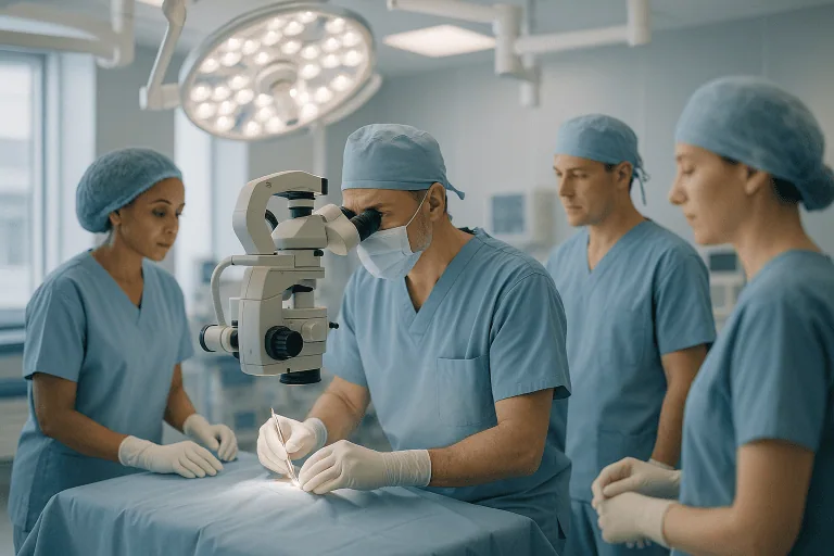
Endoscopic disc surgery takes place in an operating theatre under strict sterile technique. Anaesthesia varies with the case: many procedures use general anaesthesia, while some selected cases are done under regional or local anaesthesia with sedation. The anaesthesiology team decides the safest plan together with the spine surgeon. A typical operation follows this sequence:
- Step 1: Positioning and level check. You are positioned to give access to the affected spinal level. Fluoroscopic imaging confirms the exact level and guides the planned entry point on the skin. Operating on the correct level is verified repeatedly, not assumed.
- Step 2: Access. The surgeon makes a small incision and advances a narrow working channel toward the herniation along the approach chosen for your anatomy, splitting rather than cutting the muscle where possible.
- Step 3: Visualisation. The endoscope enters the working channel. Continuous irrigation keeps the magnified view clear on the monitor, letting the surgeon distinguish disc tissue, nerve, ligament and bone directly.
- Step 4: Decompression. Fine instruments identify and remove the fragment or protruding disc material compressing the nerve. If limited trimming of bone or ligament is needed for access or a fuller decompression, it is done sparingly, far less than in open approaches.
- Step 5: Confirmation and closure. The surgeon inspects the nerve root to confirm it is free, checks that no significant loose fragment remains, withdraws the instruments and closes the small incision.
Throughout, the team works with two sources of certainty at once: the direct endoscopic view of the structures, and the imaging guidance confirming position within the spine. The goal is a complete decompression of the affected nerve with the smallest possible footprint on everything around it.
Transforaminal and Interlaminar Approaches
Transforaminal and interlaminar approaches are the two main routes used in endoscopic disc surgery, and the choice between them is one of the most consequential planning decisions. The transforaminal approach enters from the side, passing through the foramen: the natural window where the nerve exits the spine. It reaches foraminal and far-lateral herniations directly and, at many lumbar levels, avoids entering the spinal canal from behind. Its geometry is constrained by the pelvis and the iliac crest, which can make the lowest lumbar level harder to reach in some people.
The interlaminar approach enters from the back, through the natural gap between the laminae of two vertebrae. It suits herniations at L5-S1 particularly well, where the interlaminar window is naturally wide, and it handles certain central and migrated fragments that the lateral route cannot reach. Neither route is superior in the abstract; the disc level, the fragment’s position and your bony anatomy decide. A surgeon comfortable with both can match the corridor to the pathology rather than forcing the pathology through a single preferred corridor.
Technology Used to Support Precision and Safety
Several categories of technology support the operation. The endoscope itself provides magnified visualisation through a narrow access path. High-resolution camera systems and monitors help the surgeon distinguish tissue types confidently. Fluoroscopic image guidance confirms the spinal level and the instrument trajectory. Purpose-built micro-instruments, graspers, punches, burrs and radiofrequency probes, allow controlled removal of disc material through a channel only a few millimetres wide. Modern theatres add advanced anaesthesia monitoring, careful positioning systems and structured intraoperative imaging protocols.
Be sceptical of technology presented as an outcome in itself. Equipment does not select patients, choose approaches or judge when a decompression is sufficient: the surgeon does. The value of the technology lies in how it is integrated into those clinical decisions: identifying the true source of compression, protecting the nerve while the fragment is removed, and confirming the result before closing.
How long does the operation take, and will you stay in hospital?
Operative time varies with the spinal level, the complexity of the herniation, your anatomy and whether you have had previous spine surgery; many endoscopic disc procedures are short compared with more extensive spinal operations. The full hospital experience is longer than the operation itself: preparation, anaesthesia, recovery-room monitoring, first mobilisation and a discharge assessment all take time.
Some patients go home the same day; others stay overnight for observation. The decision rests on pain control, neurological status after surgery, walking ability, your medical history and the surgeon’s judgement. Before discharge, the team confirms that you can walk safely, that pain is controlled with oral medication, and that you understand your wound care, activity limits and follow-up schedule.
Recovery After Endoscopic Disc Surgery
After surgery, you are encouraged to stand and walk with assistance as soon as it is safe. Leg pain caused by nerve compression can ease quickly in some patients, while numbness, tingling or weakness typically takes longer, because nerves heal slowly and on their own schedule. Soreness around the small incision and the deeper access path is expected and settles as the tissues recover.
You receive specific instructions on wound care, medications, walking, sitting, lifting and when to resume work or travel. Early movement is generally encouraged, but bending, twisting, heavy lifting and prolonged sitting are usually restricted for a period. Physiotherapy may be recommended depending on your symptoms, conditioning and goals. The longer-term focus is practical: restore mobility, rebuild core support, reduce the risk of recurrence and return you safely to the activities that matter to you.
| Time Period | What You Can Generally Expect |
|---|---|
| Day 1 | Monitoring after anaesthesia, pain control, neurological checks and assisted walking. Some patients go home the same day; others stay overnight. |
| First week | Incision care, short frequent walks, avoidance of heavy lifting and bending, gradual return to basic daily activities. Leg pain may improve early; numbness often lags behind. |
| First month | Increasing walking tolerance and light activity. Some patients return to desk-based work, depending on symptoms, job demands and travel requirements. |
| Six to twelve weeks | Progressive strengthening, improved flexibility and more normal daily function. Physiotherapy supports safe movement patterns where recommended. |
| Longer term | Continued nerve recovery, conditioning and prevention. Sensory symptoms can take months to improve, especially after longstanding compression. |
How long does it take to recover from L5-S1 herniated disc surgery?
Early recovery after L5-S1 disc surgery is usually measured in days to weeks, while nerve recovery is measured in weeks to months. Most patients walk on the day of surgery or the day after, manage basic daily activities within the first week or two, and return to desk-based work within a few weeks if pain control and travel allow. Physically demanding work, lifting, driving long shifts, manual labour, takes longer and returns in stages agreed with the surgical and rehabilitation team.
The L5-S1 level carries a specific practical point: the S1 nerve root supplies calf strength and contributes to the ankle reflex, so weakness or numbness in that territory recovers on nerve time, not wound time. Pain relief often comes first; sensation and strength follow more slowly, and how completely they return depends partly on how long and how severely the root was compressed before surgery. Your surgeon can give you a realistic personal timeline based on your pre-operative deficit.
How long does it take to recover from spinal decompression surgery?
Recovery from spinal decompression surgery depends heavily on which decompression you have. Endoscopic disc removal, with its small incision and muscle-sparing corridor, generally allows earlier mobilisation and a shorter early recovery than open laminectomy or decompression combined with fusion, where the disturbed tissue envelope is larger and the healing timeline correspondingly longer. As a broad pattern across decompression procedures: wound healing takes weeks, functional reconditioning takes weeks to months, and nerve recovery, where a deficit existed before surgery, can continue for many months.
Two things do not change with the size of the incision. First, the biology of the compressed nerve: a nerve irritated for a long time recovers at its own pace regardless of how elegantly it was decompressed. Second, the need for graded rehabilitation: returning to bending, lifting and sport too early risks setbacks after any decompression, minimally invasive or not.
Can you live a normal life after herniated disk surgery?
Many people return to a broadly normal life after herniated disk surgery, working, travelling, exercising and sleeping without the leg pain that drove them to the operation. The honest qualifications matter, though. The operated disc is decompressed, not made new: a recurrent herniation at the same level, or a new problem at another level, remains possible over time. Residual numbness or patches of altered sensation can persist even when pain resolves, particularly if the nerve was compressed for a long period before surgery.
What you do after surgery genuinely influences the long term. Core strengthening, sensible lifting mechanics, weight management, regular walking and stopping smoking all support the health of the remaining disc tissue and the rest of the spine. Surgery removes the mechanical problem; the habits that follow protect the result.
Risks and Limitations You Should Understand
Every spinal operation carries risk, and a smaller incision reduces some risks without eliminating any of them. Possible complications of endoscopic disc surgery include injury or irritation of the nerve root, a tear in the dura (the membrane containing spinal fluid), bleeding, infection, incomplete removal of the fragment, and recurrence of the herniation over time. Occasionally an endoscopic procedure needs to be converted to, or later followed by, a different operation if the fragment cannot be removed safely through the narrow corridor. Anaesthesia carries its own risks, assessed individually before surgery. A responsible surgical team walks you through these possibilities in relation to your specific case, not in the abstract.
Why can endoscopic disc surgery produce sensory deficit?
Sensory deficit after endoscopic disc surgery usually arises because the working corridor passes very close to nerve tissue. In the transforaminal approach, the channel travels through the foramen alongside the exiting nerve root and its dorsal root ganglion: the sensory relay station of that nerve. Contact, retraction or irritation of the ganglion during access can produce numbness, tingling or burning dysaesthesia in that nerve’s territory, even when the decompression itself goes exactly as planned. Heat from radiofrequency instruments used near the nerve, swelling of the root after it is freed, and manipulation needed to remove an awkwardly placed fragment can contribute in the same way.
There is a second, distinct cause: the deficit that was already committed before surgery. A nerve compressed severely or for a long time may have sustained damage that decompression cannot immediately reverse. In that situation the operation removes the pressure, but the sensory loss recovers slowly and sometimes incompletely, because the limiting factor is the nerve’s own healing capacity. Most procedure-related sensory disturbance improves over weeks to months as the irritated nerve settles, but it can persist, and an honest pre-operative discussion should say so. This is also part of the argument for not leaving a significantly compressed nerve unassessed for long periods.
Why Acting Early Matters
Not every herniated disc needs surgery, and when symptoms are stable, conservative treatment is usually the right first step. Persistent nerve compression, however, deserves attention rather than indefinite patience. When a nerve root stays compressed for a prolonged period, inflammation and mechanical irritation contribute to ongoing pain and declining function, and deficits such as weakness or sensory loss can become harder to reverse.
Delay also allows secondary problems to build. Pain leads to reduced activity; reduced activity leads to muscle deconditioning, stiffness, weight gain, disturbed sleep and growing reliance on pain medication. Altered walking patterns strain the hips, knees and the opposite side of the back. Work and travel become progressively harder, especially if sitting aggravates your symptoms.
Some situations do not tolerate delay at all: progressive weakness, worsening numbness, or bladder and bowel changes indicate compression that spine teams evaluate urgently. Early specialist assessment separates the symptoms that can safely be monitored from those that need prompt intervention. The aim is not to operate early on every disc herniation: it is to act at the right time for the right patient.
Potential Benefits of Endoscopic Disc Surgery
For carefully selected patients, endoscopic disc surgery offers practical advantages over both living with ongoing nerve compression and undergoing a more extensive operation. None of these benefits is automatic; each depends on correct patient selection and on the technique matching the pathology.
| Benefit | What It Means for You |
|---|---|
| Targeted nerve decompression | The disc material pressing on the nerve is removed, with the goal of reducing leg pain, numbness, tingling and nerve irritation. |
| Small incision and muscle-sparing access | Less disruption to surrounding soft tissue can support earlier mobilisation and less wound-related discomfort in suitable patients. |
| Preservation of spinal structures | The problematic fragment is removed while as much normal disc, bone, ligament and muscle as possible is left intact. |
| Potentially shorter hospital stay | Many patients need limited inpatient recovery, though the plan depends on medical condition, pain control and travel needs. |
| Earlier return to light activity | Walking usually begins soon after surgery, with daily activity increased gradually under medical guidance. |
What Influences a Good Outcome?
The result of endoscopic disc surgery depends on more than the operation itself. The single most important factor is accurate diagnosis. Patients tend to do best when symptoms, examination findings and imaging all point to the same compressed nerve root. If the main complaint is back pain without clear nerve compression, the expected benefit is far less predictable, and a candid surgeon will say so before operating.
The type and location of the herniation matter. A well-defined fragment compressing a single root is usually more straightforward than complex multi-level degeneration, severe stenosis, instability or widespread arthritic change. Previous spine surgery alters anatomy and can change both the approach and the expected recovery.
Nerve health at the time of surgery is another major factor. Pain typically improves faster than numbness or weakness. A nerve compressed for a long time recovers slowly and sometimes incompletely, which is why a significant or progressive neurological deficit should be assessed without delay rather than watched indefinitely.
General health shapes healing too. Diabetes, smoking, obesity, poor conditioning, osteoporosis, inflammatory disease and certain medications can influence recovery and complication risk. Patients who follow post-operative instructions, keep to the agreed activity limits and take part in rehabilitation when it is recommended put themselves in the best position for a good functional result.
Finally, understand what the operation does and does not do. Disc surgery relieves pressure on the nerve; it does not restore the disc to its original state. Recurrent herniation at the same level, or a new problem at another level, remains possible over the years. Prevention rests on healthy movement mechanics, core strength, weight management, stopping smoking where relevant, and avoiding sudden heavy strain during the recovery period.
How Acibadem Approaches Endoscopic Disc Surgery
Spine surgery decisions are only as good as the evaluation behind them, and Acibadem’s spine pathway is built around that principle. Assessment is individualized rather than procedure-driven: a patient with a herniated disc may be advised to continue non-surgical care, consider an injection, undergo endoscopic decompression or choose a different operation, depending on what the findings actually show. Complex cases are discussed across disciplines, drawing on the neurosurgery team together with neurologists, pain specialists, radiologists, physiatrists and rehabilitation professionals as the case requires.
Careful correlation between symptoms, examination and imaging sits at the centre of decision-making. High-quality scans define the level and location of nerve compression; the clinical examination confirms whether the scan explains what you actually feel. This discipline matters because disc bulges are common on imaging even in people without pain, and treating the scan rather than the patient is one of the most reliable routes to a disappointing spine surgery outcome.
In theatre, magnified endoscopic visualisation, fluoroscopic guidance, specialised micro-instruments and modern anaesthesia monitoring support surgeons working through small access corridors. The stated purpose of all of it is narrow and practical: find the true source of compression, remove it with minimal disturbance to healthy tissue, and support a controlled, predictable recovery.
Follow-up after endoscopic disc surgery is structured rather than left to chance. An early wound review confirms healing; later visits track pain, sensation and strength against the pre-operative picture, and the rehabilitation plan is adjusted as recovery progresses. Where physiotherapy is recommended, it is coordinated with the surgical team so that activity advances in step with tissue healing rather than ahead of it. Communication is treated as part of the treatment throughout this pathway: you should leave every conversation understanding the benefits, the risks, the alternatives, the realistic recovery timeline and the reasoning behind the recommendation you have been given, and you should feel able to ask why a particular approach was chosen for your spine rather than another.
Weighing the Decision
Endoscopic disc surgery is an effective option for selected patients with nerve compression from a herniated disc, particularly when leg pain, numbness or weakness has not improved with a genuine course of conservative care. Its minimally invasive nature supports smaller incisions, less soft-tissue disruption and earlier mobility, but none of that substitutes for the questions that actually determine the outcome: Is the diagnosis right? Does the imaging explain the symptoms? Is this the correct operation for this herniation, in this spine, in this person?
If you are living with persistent sciatica or have been told you may need disc surgery, the most useful thing you can bring to any specialist review is a clear account of your symptoms, your prior treatments and your current imaging. With those three elements in hand, a spine team can tell you honestly whether endoscopic disc surgery, a different procedure, or continued conservative care is the right next step, and just as importantly, why.
Preparation
- Preparation includes a spine specialist evaluation, neurological examination, and imaging such as MRI or CT to confirm the disc problem and surgical plan. Patients may need blood tests and anesthesia assessment, and should review medications, especially blood thinners. Fasting is usually required before the procedure.
Aftercare
- Patients are usually encouraged to walk soon after surgery and avoid heavy lifting, bending, or twisting during early recovery. Pain medication, wound care instructions, and follow-up appointments are provided. Physical therapy may be recommended to improve mobility and reduce recurrence risk.
Frequently Asked Questions
What affects the cost of endoscopic disc surgery?
The final cost depends on the disc level, complexity of nerve compression, required imaging, surgeon and hospital factors, anaesthesia, hospital stay, medications, and follow-up needs. Travel support, translation, and accommodation coordination may also affect the package.
How can I get a personalised quote?
You can request a free consultation by sharing your recent MRI, medical reports, symptoms, and previous treatments. A spine specialist reviews suitability first, then the international patient team can prepare a personalised treatment and cost estimate.
Is the lowest quote always the best choice?
Not necessarily. Patients should compare what is included, the surgeon’s experience with endoscopic spine procedures, hospital accreditation, imaging review, anaesthesia care, emergency readiness, and postoperative follow-up. This information is general and not financial advice.
What is usually included in an international patient package in Turkey?
Packages may include medical review, hospital services, surgeon and anaesthesia fees, standard preoperative tests, nursing care, and care coordination. Inclusions vary by case, so patients should confirm imaging, medications, physiotherapy, hotel, transfers, and follow-up before travel.
Will I need to travel for follow-up after surgery?
Follow-up planning depends on the procedure, symptoms, and recovery. Some checks may be coordinated remotely, while in-person review may be needed if there are concerns. Your surgeon will advise a safe follow-up plan based on your case.
Medically reviewed by the Acıbadem International Medical Board September 1, 2026
See our medical review board →
Update history
- PublishedJune 7, 2026
- Medical review approvedSeptember 1, 2026
- Last content updateSeptember 1, 2026
References2
- Herniated disk (slipped disc) nhs.uk
- Herniated disk medlineplus.gov
Trusted care for international patients
From the Acibadem Blog

Endoscopic Disc Surgery Recovery: Walking the Same Day and When Bending and Lifting Return
Endoscopic disc surgery recovery usually begins with walking within hours of the procedure, often on the same day, because the small incision spares most…