

Heart Valve Diseases Treatment
Heart valve diseases affect how blood flows through the heart and may require medication, catheter-based TAVI, or valve repair/replacement surgery. Acibadem provides multidisciplinary cardiac care in Turkey.
Quick answer
Heart valve disease treatment is the medical, catheter-based or surgical management of heart valves that no longer open or close properly. Options range from monitoring and medication to transcatheter aortic valve implantation (TAVI) and surgical valve repair or replacement. The right approach depends on which valve is affected, how severe the narrowing or leakage is, heart function, symptoms, anatomy and overall health.
Heart Valve Diseases: What They Are and How They Are Treated
Heart valve diseases are conditions in which one or more of the four valves inside the heart no longer opens or closes as it should. Treatment covers everything from regular monitoring and medication to catheter-based procedures such as transcatheter aortic valve implantation (TAVI) and open surgery to repair or replace the damaged valve. It is intended for people whose valve problem is severe enough, or progressing quickly enough, to strain the heart, cause symptoms or put long-term heart function at risk.
Heart valves are small structures with an outsized job. They open and close with every heartbeat, keeping blood moving in one direction through the heart and onward to the lungs and the rest of the body. When a valve becomes narrowed, leaky or structurally damaged, the heart has to work harder to push the same amount of blood through. Over time, that extra workload can lead to enlargement of the heart chambers, rhythm disturbances, heart failure, blood clots and other complications. This is why valve problems are followed so closely, even when they cause no symptoms at first.
Many people first hear about a valve problem after an echocardiogram performed for a murmur, breathlessness, chest discomfort, swelling in the legs, dizziness or a change in exercise tolerance. Others have lived for years with a known valve condition and are now being told it is time to consider a catheter-based procedure or heart surgery. Both situations raise the same questions: how serious is this, what are the options, and when is the right time to act?
The decision to treat is rarely based on one test alone. It requires a clear picture of the valve involved, the severity of the narrowing or leakage, the strength of the heart muscle, the symptoms you are experiencing, your age, other medical conditions, your anatomy and your own priorities. Some people need nothing more than scheduled follow-up and medication. Others benefit from a catheter-based intervention. Many need valve repair or valve replacement surgery. What matters is that the plan fits the individual patient, not the other way around.
What is heart valve disease?
Heart valve disease is any condition in which a valve of the heart does not open fully, does not close tightly, or both. Doctors describe it in two main ways. Stenosis means the valve has become narrowed or stiff, so blood struggles to pass through it. Regurgitationalso called insufficiency or leakage, means the valve does not seal properly, so some blood flows backwards with each beat. A single valve can be both narrowed and leaky at the same time, and more than one valve can be affected in the same patient. Severity ranges from mild disease that never needs intervention to severe disease that changes the structure and function of the heart itself.
What causes heart valve disease?
The most common cause in older adults is age-related calcification, in which the valve leaflets gradually stiffen and thicken over decades. But the list of causes is long. Some people are born with abnormal valves: a bicuspid aortic valve, which has two leaflets instead of three, is a well-known example and is one of several congenital heart diseases that can affect valve function later in life. Rheumatic heart disease, a late consequence of untreated streptococcal throat infection, remains an important cause of mitral and aortic valve damage in many parts of the world. Infection of the valve itself, known as endocarditis, can destroy leaflet tissue rapidly and is one of the conditions managed together with an infectious diseases team. Degenerative changes, connective tissue disorders, previous heart attacks, weakening of the heart muscle from myocardial diseases, radiation exposure to the chest and previous cardiac surgery can all damage valves as well. The underlying cause matters, because it shapes the recommended treatment, the timing of intervention and the follow-up you will need afterwards.
The Valves of the Heart and How They Work
The valves of the heart are four one-way gates: the aortic, mitral, tricuspid and pulmonary valves. The mitral and tricuspid valves sit between the upper chambers (atria) and the lower chambers (ventricles); the aortic and pulmonary valves control blood leaving the heart towards the body and the lungs. Each opens to let blood through and snaps shut to stop it flowing back. Any of the four can be affected by disease, although aortic and mitral valve disorders are the ones most commonly treated in adults.
When a valve narrows, the chamber behind it has to generate higher pressure to force blood through, and the muscle typically thickens in response. When a valve leaks, the heart has to pump the same blood twice, and the chambers tend to stretch and enlarge. Both processes can be tolerated for years, the heart is remarkably adaptable, but neither is harmless in the long run. Understanding which mechanism is at work, and how the heart is responding, is the foundation of every treatment decision that follows.
What is mitral valve prolapse heart disease?
Mitral valve prolapse is a condition in which the leaflets of the mitral valve are floppy or elongated and bulge backwards into the left atrium when the heart contracts. Is mitral valve prolapse considered heart disease? Strictly speaking, yes: it is a structural abnormality of a heart valve, and doctors classify it as a form of valve disease. In practice, however, its significance varies enormously. Many people with mild prolapse and little or no leakage live entirely normal lives and simply need periodic echocardiographic checks. The condition becomes clinically important when the prolapsing valve produces significant mitral regurgitation, and at that point it is evaluated and treated like any other cause of a leaking mitral valve, sometimes with repair of the patient’s own valve, which is often possible in this particular condition.
Symptoms and Who May Need Evaluation
Some people with valve disease have no symptoms for years. The condition may come to light when a physician hears a heart murmur during a routine examination, or when imaging is performed for another reason entirely. In other patients, symptoms develop so gradually that they are mistaken for ageing, reduced fitness, lung disease or stress. This is one reason a structured cardiac evaluation matters more than self-assessment.
Symptoms that may point towards valve disease include shortness of breath during activity or when lying flat, fatigue, reduced exercise capacity, chest pressure, palpitations, fainting or near-fainting, swelling of the feet or ankles, unexplained weight gain from fluid retention, coughing at night, and the sensation of a racing or irregular heartbeat. The changes can be subtle. A patient may notice only that they now avoid stairs, walk more slowly than their peers, or need longer to recover after routine activity. People often adjust their lives around a failing valve without realising they have done so.
Evaluation and treatment become relevant when valve disease is severe, when symptoms appear, when heart function begins to decline, when chambers are enlarging, when pressures in the lung circulation are rising, when rhythm disturbances such as atrial fibrillation develop, or when the valve condition creates a risk of serious complications. In some situations, treatment is recommended before severe symptoms occur, because waiting until the heart muscle is significantly weakened can limit how well it recovers afterwards.
How Heart Valve Disease Is Diagnosed
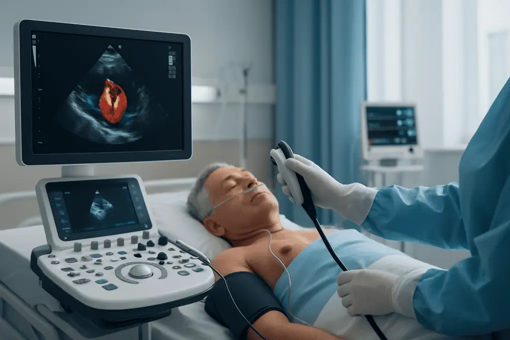
Diagnosis begins with a medical history, a physical examination and a review of any previous tests. A murmur is an important clue, but it does not define the disease, imaging does. Echocardiography is the central diagnostic test for most valve conditions. It uses ultrasound to show how the valve leaflets move, how blood flows across the valve, how large the heart chambers are, how strongly the muscle contracts, and what the pressures inside the heart are estimated to be. In selected cases, a transoesophageal echocardiogram provides more detailed images from inside the oesophagus, which sits directly behind the heart. This view is particularly useful for assessing mitral valve anatomy, suspected infection, clots or complex patterns of leakage.
Additional testing may include electrocardiography to assess heart rhythm, blood tests, chest imaging, cardiac CT to examine anatomy and calcium patterns, and cardiac MRI in selected patients. Because narrowed heart arteries frequently coexist with valve problems in older adults, coronary angiography or CT coronary imaging is often arranged to check for coronary artery disease before any intervention. Exercise testing can help when symptoms are unclear, some patients genuinely have none, while others have quietly restricted their activity to avoid them. For patients being considered for TAVI, advanced CT imaging measures the aortic valve, the aorta and the access vessels in the legs with precision, so the team can judge whether a catheter approach is anatomically suitable and plan it safely.
How is valvular heart disease graded?
Valvular heart disease is graded by severity, typically described as mild, moderate or severe, using measurements of valve opening area, flow velocities, pressure differences across the valve and the volume of any leak. Severity grading is combined with what the disease is doing to the heart: whether the ventricles are enlarging, whether pumping function is falling, whether lung pressures are rising. A severe grade does not automatically mean immediate intervention, and a moderate grade does not always mean waiting. Serial imaging over time often tells the team more than any single scan, which is why patients with known valve disease are asked to return for scheduled echocardiograms even when they feel well.
Conditions and Indications Addressed
Valve treatment may be considered for a wide range of disorders, and the most common adult indications involve the left side of the heart, where pressures are highest.
Aortic valve disease: stenosis and regurgitation
The aortic valve sits between the left ventricle and the aorta, the main artery carrying blood to the body. In aortic stenosis, the valve becomes narrowed, most often through calcification, and restricts blood flow out of the heart; the ventricle thickens under the strain, and symptoms such as breathlessness, chest pain and fainting carry particular clinical weight once they appear. In aortic regurgitation, blood leaks back into the left ventricle after each beat, gradually stretching and enlarging it. Because the aortic valve and the aorta itself are so closely connected, disease of one often needs to be assessed alongside the other; enlargement of the aorta is evaluated within the same framework as other aortic diseases.
Mitral, tricuspid and pulmonary valve disease
In mitral regurgitation, the valve between the left atrium and left ventricle leaks, increasing the workload of both chambers and, over time, raising pressure in the lung circulation. In mitral stenosisoften rheumatic in origin in some regions: the same valve narrows and limits blood flow into the left ventricle. Tricuspid valve disease can also require treatment, particularly when severe leakage causes right-sided heart strain, liver congestion, swelling and fatigue. Pulmonary valve disease is less common in adults but becomes important in congenital heart conditions and after previous cardiac procedures. Some patients have disease affecting more than one valve at once, which calls for integrated planning rather than treating each valve as an isolated problem: the order, timing and combination of interventions all matter.
How Heart Valve Disease Treatment Is Performed
Treatment begins with a complete evaluation. Before recommending medication, TAVI, valve repair or valve replacement, the cardiac team reviews your symptoms, medical history, previous operations, current medicines, allergies, kidney function, lung disease, diabetes, stroke history and other risk factors. Existing test results are assessed, and further imaging is arranged if earlier studies are incomplete or if procedural planning needs more detail. Preparation may include echocardiography, CT imaging, rhythm assessment, blood tests, a dental check in selected patients to reduce infection risk, and coronary artery assessment where appropriate. If you take blood-thinning medication, any adjustment before a procedure is decided and supervised by your treating doctor, never on your own.
Medication and monitoring
Medication has a real role in valve disease. Drugs can control blood pressure, remove excess fluid, manage rhythm disorders and treat associated heart failure, all of which reduce symptoms and the burden on the heart. What medication usually cannot do is restore heart valves that are severely narrowed or structurally damaged. When drug treatment and surveillance are the chosen path, follow-up imaging is scheduled according to the valve involved and the severity grade, and the care team explains which changes should prompt an earlier reassessment. For many patients with mild or moderate disease, this pathway continues for years without any procedure ever becoming necessary.
How is TAVI performed?
TAVI, transcatheter aortic valve implantation, also called TAVR, is a catheter-based treatment for selected patients with aortic valve disease, most commonly severe aortic stenosis. It places a new valve inside the diseased one through the blood vessels, usually without opening the chest. A typical procedure follows this sequence:
- The patient is prepared in a specialised catheterisation laboratory or hybrid operating theatre, under general anaesthesia or deep sedation depending on clinical factors and institutional protocol.
- The team gains access through an artery, most commonly in the groin.
- A catheter carrying the folded replacement valve is guided to the diseased aortic valve under imaging guidance.
- The new valve is expanded into position inside the old one, pushing the diseased leaflets aside.
- Imaging and pressure measurements confirm the valve’s position and function before the catheters are withdrawn and the access site is closed.
Many TAVI procedures are completed within a few hours, with additional time for preparation and recovery monitoring. Planning relies heavily on modern imaging: CT measurements determine valve size, map calcium distribution, assess the aorta and confirm that the leg arteries can safely accommodate the delivery system, while echocardiography assesses valve performance before and after treatment. Continuous rhythm and haemodynamic monitoring continues during and after the procedure, because some patients develop conduction changes that require extended observation or, in selected cases, a pacemaker.
Surgical valve repair and replacement
Surgical treatment is performed by a cardiovascular surgery team under general anaesthesia. Where anatomy allows, the surgeon may repair the existing valve, using techniques that restore leaflet motion and improve closure: an approach most often used in selected mitral and tricuspid conditions, and preferred in many suitable degenerative mitral cases because preserving the native valve carries important advantages. When repair is not durable or anatomically feasible, the valve is replaced with a prosthesis. Some operations are performed through a traditional chest incision; selected patients are candidates for less invasive surgical approaches. The best incision and technique depend on anatomy, the valve involved, prior operations, body structure, coexisting coronary disease and whether additional procedures are needed at the same time. Surgical aortic valve replacement remains an established treatment for many patients, especially younger patients, those whose anatomy suits surgery better, those needing additional cardiac procedures, and those for whom long-term valve durability is the central consideration.
Mechanical or biological heart valve: how is the choice made?
A replacement heart valve may be mechanical or biological, and the choice deserves an unhurried discussion. Mechanical valves are highly durable but usually require lifelong blood-thinning medication and regular monitoring of clotting levels. Biological valves, made from animal tissue, generally do not require the same long-term anticoagulation for the valve itself, but they wear out over time and may not last as long in younger patients. The decision weighs age, lifestyle, pregnancy plans, the practicality of managing anticoagulation, bleeding risk, occupation, travel habits, other medical conditions and personal preference. There is no universally correct answer, only the answer that fits your circumstances.
What happens immediately after the procedure?
After TAVI or surgery, patients are monitored in a recovery area or intensive care setting according to the procedure and their clinical status. Monitoring covers heart rhythm, blood pressure, oxygen levels, kidney function, bleeding, access sites or surgical wounds, pain control and early mobility. Echocardiography is often repeated to assess the treated valve, and the team reviews medications, including antiplatelet or anticoagulant therapy where indicated. TAVI usually involves a shorter hospital stay and faster early mobility than open surgery for appropriately selected patients. Surgical procedures require more time for the breastbone or incision to heal, with staged activity, breathing exercises, wound care and cardiac rehabilitation. Because prosthetic material can become a focus for infection, dental hygiene and routine dental care remain important throughout life for anyone with a repaired or replaced valve, and the care team explains which precautions apply before future dental or surgical procedures. In both pathways, long-term follow-up is essential, because both prosthetic and repaired valves need periodic assessment for the rest of your life.
Why Timing Matters
Disease of the heart valves can be deceptive. Symptoms may appear only after the heart has been compensating for a long time. In severe disease, the muscle thickens, stretches or weakens as it tries to maintain output. Once advanced damage has developed, treatment can still help, but the heart may not recover as completely as it would have done with earlier intervention. Delay also raises the risk of heart failure, atrial fibrillation, pulmonary hypertension, fainting, hospitalisation, blood clots, stroke and reduced exercise capacity. Severe mitral regurgitation can quietly enlarge the left ventricle and atrium before symptoms become obvious, and untreated tricuspid disease can lead to progressive fluid retention and organ congestion.
Early evaluation does not always mean early intervention. Often it confirms that careful monitoring remains the right course. What timely assessment does is identify the point at which treatment offers the best balance of benefit and risk, and that point is different for every patient, every valve and every mechanism of disease.
How do you prevent heart valve disease?
Not all valve disease is preventable, age-related calcification and congenital abnormalities cannot be avoided, but several important causes can be reduced. Prompt medical treatment of streptococcal throat infections lowers the risk of rheumatic fever and the valve damage it leaves behind, which is why this remains a public health priority in many regions. Good dental hygiene and regular dental care reduce the bacterial load that can seed valve infections, a precaution that matters even more for people who already carry a prosthetic or abnormal valve. Managing blood pressure, cholesterol and diabetes, staying physically active and not smoking protect the heart and its arteries, and healthier arteries mean less of the ischaemic damage that can secondarily affect valves. For people with known mild valve disease, the most valuable preventive step is simply attending scheduled follow-up, so that progression is caught before it harms the heart muscle.
Potential Benefits of Heart Valve Disease Treatment
The benefits of treatment depend on the valve involved, disease severity, heart function and the selected approach, but the aims are consistent: better blood flow, symptom relief and protection of the heart over time.
| Benefit | What It Means for You |
|---|---|
| Improved forward blood flow | A repaired or replaced valve can help blood move more efficiently through the heart, reducing the strain created by narrowing or leakage. |
| Relief of symptoms | Many patients notice improvement in breathlessness, fatigue, chest pressure, swelling or activity tolerance, although recovery varies by condition and overall health. |
| Protection of heart function | Treating severe valve disease at the right time may help prevent or limit chamber enlargement, weakening of the heart muscle and pressure build-up in the lungs. |
| Reduced risk of valve-related complications | Appropriate treatment may lower the likelihood of repeated heart failure episodes, rhythm-related deterioration and other complications linked to advanced valve disease. |
| A personalised treatment pathway | Monitoring, medication, TAVI, repair or replacement is chosen according to anatomy, risk profile and long-term priorities rather than a one-size-fits-all rule. |
Recovery Timeline After Valve Treatment
Recovery differs for every patient and varies considerably between medication management, TAVI and surgical repair or replacement. The timeline below reflects common expectations after an interventional or surgical valve procedure: your own course may be faster or slower, and your care team will adjust the plan accordingly.
| Time Period | What Patients Can Expect |
|---|---|
| Day 1 | Close monitoring of heart rhythm, blood pressure, breathing, bleeding and valve function. Early movement may begin when medically appropriate. |
| First week | TAVI patients often begin walking early and may leave hospital sooner if stable. Surgical patients usually need more inpatient recovery, pain control, breathing exercises and wound monitoring. |
| First month | Energy gradually improves and activity is increased step by step. Medication adjustments, wound or access-site checks and follow-up imaging may be scheduled. |
| Two to three months | Many surgical patients regain greater independence as the chest and soft tissues heal. Cardiac rehabilitation may help rebuild strength and confidence. |
| Longer term | Regular cardiology follow-up continues to monitor valve performance, heart function, rhythm, blood-thinning treatment where prescribed and overall cardiovascular health. |
Factors That Influence Outcomes
Outcomes in valve disease are shaped by more than the procedure itself. A good result begins with an accurate diagnosis and careful timing. Treating too early exposes a patient to procedural risk they did not yet need to take; treating too late allows heart changes that may not fully reverse. This is why serial imaging, symptom assessment and specialist review sit at the centre of valve care rather than at its edges.
The type and severity of disease matter. Severe calcific aortic stenosis, degenerative mitral regurgitation, rheumatic valve disease, endocarditis-related damage and multi-valve disease each behave differently and each responds differently to intervention. The condition of the heart muscle, chamber sizes, lung-circulation pressures, rhythm status and kidney or lung function all influence both procedural risk and the pace of recovery afterwards.
Anatomy plays a major role. For TAVI, the size and shape of the aortic valve area, the distribution of calcium, the height of the coronary artery openings, the dimensions of the aorta and the calibre of the access vessels must all be suitable. For mitral or tricuspid repair, leaflet quality, the supporting chordal structures, the size of the valve ring and the precise mechanism of leakage determine whether a repair is likely to be durable. Where coronary artery disease coexists, the team weighs whether it should be treated before, during or after the valve intervention.
Age and lifestyle matter, but they are not decisive on their own. A healthy older adult may do well with a catheter-based approach, while a younger patient may need a strategy built around long-term valve durability. For replacement, the mechanical-versus-biological decision should align medical facts with personal priorities: lifelong anticoagulation may be acceptable to one patient and genuinely problematic for another because of occupation, travel, bleeding risk or pregnancy plans.
Team experience and coordination influence quality of care. Valve disease sits at the intersection of imaging, interventional cardiology, cardiovascular surgery, anaesthesia and intensive care, and a multidisciplinary review reduces fragmented decision-making. Complex cases are often discussed in specialist boards where imaging and clinical data are examined together, so that the options are compared in a structured way rather than in isolation.
Finally, your own participation counts. Taking medicines as prescribed, attending follow-up appointments, reporting new symptoms, maintaining dental hygiene, managing blood pressure, stopping smoking, controlling diabetes and taking part in cardiac rehabilitation when advised all support recovery. Patients with prosthetic valves also need to understand infection precautions, anticoagulation requirements where they apply, and why lifelong surveillance is part of living well with a treated valve.
How Acibadem Organises Heart Valve Care
At Acibadem hospitals, valve conditions are managed through multidisciplinary cardiac pathways that bring together cardiologists, cardiovascular surgeons, interventional cardiologists, imaging specialists, anaesthesiologists, intensive care physicians, rehabilitation teams and nurses experienced in cardiac care. This structure matters most when the decision is not straightforward, choosing between TAVI and surgery, judging whether a mitral valve can be repaired rather than replaced, or planning treatment when more than one valve is involved.
Diagnostic pathways follow evidence-informed practice. Echocardiography, advanced cardiac imaging, catheter-based assessment where needed and laboratory evaluation define the problem before any treatment is proposed. For TAVI candidates, imaging establishes valve sizing and vascular access; for surgical candidates, it helps the surgeon understand anatomy and plan a repair or replacement strategy. Technology is used to improve precision and anticipate risk, not as a substitute for clinical judgment.
Continuity is treated as part of the treatment itself. Valve care does not end at discharge, so patients leave with a follow-up plan, medication instructions, wound or access-site guidance and activity recommendations, together with medical reports that can be shared with your own cardiologist. Second opinions are a routine part of valve medicine everywhere: when recommendations from different physicians conflict, an independent structured review of the imaging and clinical data may confirm the original plan, suggest further testing or identify a different pathway.
Making Sense of Your Options
Heart valve disease demands careful attention, but it is also an area of medicine where modern diagnosis and treatment can make a meaningful difference to how you feel and how your heart works. Whether the recommendation you have received is monitoring, medication, TAVI, valve repair or valve replacement, the most useful thing you can do is understand your own condition clearly: which valve is affected, what the mechanism is, how severe it is, what it is doing to your heart, and why a particular timing and approach have been proposed. Heart valves can fail slowly and quietly, but they can also be assessed precisely and treated well, and a plan built on accurate imaging, honest discussion of trade-offs and a team that reviews the whole picture is the strongest foundation for a good outcome.
Preparation
- Preparation starts with cardiology evaluation, echocardiography, blood tests, ECG, and imaging when needed. Your doctor reviews medications, especially blood thinners, and explains fasting instructions before the procedure. International patients may receive coordinated pre-travel assessment and scheduling support.
Aftercare
- After treatment, heart rhythm, blood pressure, wound sites, and valve function are closely monitored. Patients receive guidance on medications, activity limits, cardiac rehabilitation, and follow-up echocardiography. Seek urgent care for chest pain, severe shortness of breath, fever, fainting, or signs of infection.
Frequently Asked Questions
What affects the cost of heart valve disease treatment?
The final cost depends on the diagnosis, valve affected, treatment option, required imaging, device or valve type, hospital stay, intensive care needs and follow-up plan. Travel, accommodation and translation support may also affect the overall budget for international patients.
How can I get a personalised quote from Acibadem?
You can request a free consultation by sharing recent medical reports, echocardiography results, CT or angiography images if available, medication list and a summary of symptoms. The cardiac team can review your information and provide a personalised treatment plan and cost estimate.
Is TAVI less costly than open-heart valve surgery?
Not always. TAVI may involve a shorter hospital pathway for suitable patients, but device selection, imaging, catheter laboratory use and specialist team requirements influence the quote. Open surgery and TAVI should be compared medically first, with cost discussed after specialist assessment.
Does a treatment package include everything I need?
Package details vary by patient and procedure. A package may include consultation, tests, the procedure, hospital stay and coordination support, but additional care may be needed if new findings, complications or extended recovery requirements arise.
Can international patients receive support with language and travel planning?
Yes. International patient services can help coordinate appointments, interpreter support, hospital admission planning and practical travel information. The exact support and inclusions should be confirmed during the quotation process.
Is this information medical or financial advice?
No. This is general educational information only. A cardiologist or cardiac surgeon should decide suitability for treatment, and a personalised quote should be requested before making care or travel decisions.
Medically reviewed by the Acıbadem International Medical Board September 8, 2026
See our medical review board →
Update history
- PublishedJune 8, 2026
- Medical review approvedSeptember 8, 2026
- Last content updateSeptember 8, 2026
References2
- Heart Valve Diseases medlineplus.gov
- Heart valve disease nhs.uk
Trusted care for international patients
Compare options, costs and recovery
From the Acibadem Blog

TAVI vs Open Valve Surgery for Heart Valve Disease: How the Approach Is Chosen
TAVI and open heart valve surgery both replace a failing aortic valve, but they reach it differently: TAVI threads a new valve through a…

How Serious Is a Leaking Heart Valve, and Can It Be Fixed Without Surgery?
A leaking heart valve, or regurgitation, ranges from a trivial finding that never causes trouble to a serious condition that strains the heart over…

How Many Valves the Heart Has, and What They Do
The human heart has four valves: the tricuspid, pulmonary, mitral, and aortic valves. Each is a set of thin tissue flaps that opens to…

Heart Valve Replacement: The Average Age, How Serious It Is, and Bypass vs Valve
There is no single official average age for heart valve replacement, but most operations happen in people in their 60s, 70s, and 80s, because…



































