

Pericardial Diseases Treatment
Pericardial diseases affect the sac around the heart, causing inflammation, fluid buildup or constriction. Care focuses on diagnosis, symptom relief and preventing complications such as tamponade.

Quick answer
Pericardial diseases are conditions affecting the pericardium, the thin protective sac around the heart. They include pericarditis (inflammation), pericardial effusion (excess fluid), cardiac tamponade (dangerous pressure on the heart) and constrictive pericarditis (a stiffened sac). Treatment ranges from anti-inflammatory medication and monitoring to image-guided fluid drainage, pericardial window surgery or, in selected cases, surgical removal of the pericardium.
Pericardial Diseases: What They Are and Why They Matter
Pericardial diseases are conditions that affect the pericardium, the thin, double-layered sac that surrounds the heart and anchors it within the chest. The sac can become inflamed, fill with excess fluid, or slowly thicken and stiffen, and each of these changes disturbs the heart in a different way. Some pericardial problems settle with medication and careful follow-up. Others put dangerous pressure on the heart, interfere with its ability to fill with blood, and need urgent drainage or surgery.
Chest pain, breathlessness, a racing heartbeat or a sudden feeling of pressure in the chest are frightening symptoms, and for most people the first thought is a heart attack. In a meaningful number of cases, however, the problem does not come from the heart muscle or the coronary arteries at all. It comes from the sac around the heart. Distinguishing pericardial diseases from coronary artery diseases, lung conditions and other causes of chest pain is one of the central tasks of cardiac diagnosis, because the treatments are entirely different.
To understand pericardial diseases, it helps to know what a healthy pericardium does. The sac has two layers: an inner layer that sits directly on the heart’s surface and an outer, tougher layer that faces the chest cavity. Between them lies a small amount of lubricating fluid, which lets the heart beat smoothly with minimal friction. The pericardium also holds the heart in position, provides a physical barrier against the spread of infection from neighbouring structures, and limits sudden over-stretching of the heart chambers. When this system is disturbed, by inflammation, infection, injury, fluid or scarring, pain and impaired heart function can follow.
Accurate diagnosis matters because the right treatment depends on the cause, the amount of fluid, the severity of inflammation and whether the heart is under pressure. For many patients the uncertainty is what weighs most: whether the condition is urgent, whether medication will be enough, and whether a procedure or surgery might eventually be needed. This page sets out what pericardial diseases are, how they are diagnosed, and what each treatment realistically involves.
What is the most common pericardial disease?
Pericarditis, inflammation of the pericardium, is the most common pericardial disease. It frequently follows a viral infection, but it can also occur after heart surgery, after a heart attack, in association with autoimmune disease, kidney failure, cancer, radiation therapy or trauma, and sometimes without a clearly identifiable cause at all. Pericarditis typically causes sharp chest pain that improves when sitting forward and worsens when lying down or taking a deep breath. That pattern is a useful clue, but it is not enough on its own; confirmation still relies on examination, blood tests and imaging.
The Main Types of Pericardial Disease
Pericardial diseases are not a single condition with a single treatment. They form a spectrum, and one condition can evolve into another. An episode of acute pericarditis can produce an effusion. A large effusion can progress to tamponade. Long-standing or repeated inflammation can leave the sac scarred and rigid. Understanding where a patient sits on this spectrum is the starting point of every sound treatment plan.
Acute pericarditis
Acute pericarditis is inflammation of the pericardium that develops over days. The classic symptom is sharp, stabbing or pressure-like chest pain that may radiate to the shoulder, neck or back. Some patients also have fever, weakness, palpitations or a dry cough. Because the pain can mimic a heart attack, pulmonary embolism, pneumonia or severe reflux, expert assessment is important before the diagnosis is settled. In some patients the inflammation also involves the adjacent heart muscle, a combination known as myopericarditis, which is one reason blood tests for heart muscle injury are checked alongside inflammation markers. Most cases of uncomplicated acute pericarditis respond to anti-inflammatory treatment, although a proportion of patients experience recurrences and need a longer-term strategy.
Pericardial effusion
Pericardial effusion means excess fluid has collected in the pericardial space. A small effusion in a stable patient may simply be monitored with repeat imaging. A large effusion, or one that develops rapidly, can compress the heart and impair its filling. The clinical significance of an effusion depends not only on its size but on how quickly it accumulated: the pericardium can stretch to accommodate fluid that builds up slowly over weeks, whereas even a moderate volume arriving over hours can be dangerous because the sac has no time to adapt. This is why imaging findings must always be interpreted alongside symptoms and vital signs, never in isolation.
Is pericardial effusion heart disease?
A pericardial effusion is a disease of the sac around the heart rather than of the heart muscle itself, but it is treated within cardiology because it directly affects how the heart works. In many patients the heart muscle, valves and coronary arteries are structurally normal; the problem is external pressure from fluid. That said, an effusion is often a sign of another disease process, infection, inflammation, kidney failure or cancer, so finding fluid around the heart usually prompts a search for the underlying cause, not just treatment of the fluid itself.
Cardiac tamponade
Cardiac tamponade is the most urgent pericardial condition. It occurs when the pressure of fluid in the pericardial sac becomes high enough to prevent the heart from filling normally between beats. Less blood enters the heart, so less blood is pumped out, and blood pressure falls. Patients may experience severe breathlessness, chest fullness, lightheadedness, fainting, a rapid heartbeat or sudden weakness. Tamponade is treated as an emergency, and the definitive treatment is drainage of the fluid, most often by needle pericardiocentesis under imaging guidance. Once the pressure is relieved, circulation typically improves quickly, but the cause of the fluid still needs to be identified and managed.
Constrictive pericarditis
Constrictive pericarditis develops when the pericardium becomes thickened, scarred or calcified and loses its normal elasticity. Instead of expanding as the heart fills with blood, the rigid sac restricts the heart’s movement. The result is a distinctive pattern of symptoms: swelling of the ankles or abdomen, unexplained weight gain from fluid retention, fatigue, reduced exercise tolerance, abdominal discomfort and breathlessness when lying flat. Because the heart muscle itself may be relatively preserved, these symptoms are frequently mistaken for liver disease, kidney disease or other forms of heart failure. Distinguishing constriction from restrictive myocardial diseases, which can look very similar on first assessment, is one of the more demanding diagnostic problems in cardiology and often requires advanced imaging and sometimes catheter-based pressure measurement.
Effusive-constrictive pericarditis
Effusive-constrictive pericarditis combines two problems at once: fluid in the pericardial space and a constricting, inelastic inner layer of the sac. The condition may only be recognised after an effusion is drained, when the expected improvement in heart filling does not fully materialise because the underlying constriction remains. Recognising this pattern matters, because it changes the treatment plan from drainage alone to a strategy that also addresses the constrictive component.
Pericardial cysts
Pericardial cysts are fluid-filled sacs attached to the pericardium, most often present from birth as a benign developmental variation rather than an acquired illness. They are usually discovered by accident, when a chest X-ray or CT scan performed for another reason shows a rounded shadow next to the heart. Unlike inflammatory pericardial disease, cysts rarely cause inflammation of the sac itself, and most never need treatment beyond periodic imaging to confirm they are stable. Very large cysts, or cysts in an unfavourable position, are occasionally treated by drainage or surgical removal.
Are pericardial cysts considered heart disease?
Pericardial cysts are generally not considered heart disease in the usual sense, because they do not involve the heart muscle, valves or coronary arteries and most cause no symptoms at all. They are benign structural findings related to the pericardium. They are, however, evaluated by cardiac and thoracic specialists, because a cyst must be reliably distinguished from other masses near the heart, and because a large cyst can occasionally press on nearby structures. Since many are congenital in origin, they are sometimes discussed alongside congenital heart diseases, even though the heart itself is typically normal.
What are the common symptoms of pericardial cysts?
Most pericardial cysts cause no symptoms and are found incidentally on imaging. When symptoms do occur, usually with larger cysts, they can include chest discomfort or a sense of pressure, a persistent cough, breathlessness on exertion or, rarely, palpitations if the cyst presses on the heart or nearby airways. Because these symptoms are non-specific, imaging with CT or cardiac MRI is used to confirm that the finding is a simple cyst rather than something that needs different management.
What Causes Pericardial Disease?
Pericardial disease has many possible causes, and identifying the cause is central to choosing the right treatment. In a substantial share of pericarditis cases, particularly after a flu-like illness, the cause is presumed to be viral, and in many patients no specific trigger is ever confirmed: these cases are labelled idiopathic. Beyond viral and idiopathic disease, the main recognised causes include:
- Bacterial infection, including purulent pericarditis, which requires rapid antimicrobial treatment and often drainage, and tuberculous pericarditis, an important consideration in certain regions and patient groups. Infectious causes are typically managed together with an infectious diseases team.
- Autoimmune and inflammatory conditions, such as lupus, rheumatoid arthritis and related systemic diseases, in which the pericardium becomes a target of the body’s own immune activity.
- Kidney failure, where metabolic changes can inflame the pericardium; optimising dialysis is often part of treatment.
- Cancer, when tumours involve the pericardium directly or cause recurrent effusions that need drainage and sometimes a surgical window.
- Cardiac injury, including inflammation after a heart attack, after cardiac surgery or after catheter-based procedures: the so-called post-cardiac injury syndromes.
- Radiation therapy to the chest, which can cause pericardial inflammation early on and, years later, constrictive scarring.
- Trauma to the chest, which can cause bleeding into the pericardial space.
In many patients, more than one factor is in play, and the cause shapes the outlook. Viral and idiopathic pericarditis usually respond well to anti-inflammatory treatment. Bacterial, tuberculous, malignant and autoimmune-related pericardial disease tend to require longer, more complex care that addresses the underlying illness as well as the sac itself.
Can Lyme disease cause pericardial effusion?
Yes, although it is uncommon. Lyme disease can involve the heart, a condition called Lyme carditis, and its most frequent cardiac effect is disturbance of the heart’s electrical conduction. Pericarditis and pericardial effusion have also been reported as rarer manifestations. When a patient with a compatible tick exposure history and a pericardial effusion is evaluated, Lyme testing may form part of the work-up, and confirmed cases are treated with antibiotics directed at the infection alongside management of the effusion itself.
Does takotsubo cardiomyopathy cause pericardial disease?
Takotsubo cardiomyopathy, sometimes called stress cardiomyopathy or broken-heart syndrome, is primarily a disease of the heart muscle, not the pericardium, and belongs to the group of myocardial diseases. It does not typically cause pericardial disease, although a small pericardial effusion can occasionally accompany the acute phase as part of the surrounding inflammation. These effusions are usually minor and settle as the heart muscle recovers. If a patient with takotsubo develops a large or symptomatic effusion, it is investigated in its own right rather than assumed to be part of the cardiomyopathy.
Symptoms: Who May Need Evaluation for Pericardial Disease?
Evaluation for pericardial disease is considered when symptoms suggest inflammation or pressure around the heart, when fluid is found on imaging, or when an underlying illness raises the risk of pericardial involvement. Some people come to attention suddenly, through an emergency department. Others are referred after an echocardiogram, chest CT or cardiac MRI, often performed for another reason, shows pericardial fluid or thickening.
Symptoms vary with the type and severity of the condition. Acute pericarditis usually announces itself with chest pain that is sharp, positional and worse with deep breathing. Pericardial effusion, by contrast, may cause few or no symptoms when it develops slowly; when fluid accumulates quickly or becomes large, patients may notice breathlessness, chest fullness, lightheadedness, fainting, low blood pressure or a rapid heartbeat. Constrictive pericarditis presents differently again, with gradual fluid retention, swelling and fatigue rather than acute pain: a pattern easily misattributed to other organs.
Because the same symptoms can arise from the coronary arteries, the heart valves, the heart muscle, the lungs or the digestive system, a structured assessment is needed before the pericardium can be confirmed as the source. Conditions such as heart valve diseases and myocardial disorders sit high on the list of alternatives that must be considered and excluded.
What are the symptoms of a pericarditis flare-up?
A pericarditis flare-up typically brings back the familiar sharp chest pain, worse when lying flat or breathing deeply, eased by sitting forward, often together with fatigue, low-grade fever or palpitations. Many patients who have had pericarditis before recognise the pattern themselves. During a flare, blood tests may show inflammation markers rising again, and imaging may show fresh pericardial fluid or enhancement. Flares are assessed rather than assumed, because chest pain in a patient with previous pericarditis can still have another cause, and because the findings guide how intensively the recurrence needs to be treated.
Recurrent Pericarditis
Recurrent pericarditis means pericarditis that returns after a symptom-free interval following apparent recovery. It is one of the more frustrating patterns in pericardial disease: the first episode settles, life returns to normal, and then weeks or months later the pain comes back. Recurrences are usually less dangerous than the first episode but can significantly affect quality of life, and they call for a longer-term prevention strategy rather than repeated short courses of treatment. Management typically combines anti-inflammatory therapy tailored to the individual patient, a carefully paced return to physical activity, and follow-up testing to confirm that inflammation has genuinely resolved before treatment is tapered.
What are the common causes of recurrent pericarditis?
The most common scenario is recurrence after viral or idiopathic pericarditis, where the immune system’s response to the initial injury appears to keep reactivating: an autoinflammatory pattern rather than persistent infection. Other recognised contributors include stopping anti-inflammatory treatment too early or tapering it too quickly, underlying autoimmune disease, post-cardiac injury syndromes after heart surgery or a heart attack, and, less commonly, an undiagnosed cause such as tuberculosis or malignancy that was not identified during the first episode. This is why a recurrence often prompts a fresh look at the underlying diagnosis, not just a repeat of the previous prescription.
How Pericardial Disease Is Diagnosed
Diagnosis begins with a detailed medical history and physical examination. Doctors ask about recent infections, autoimmune disorders, tuberculosis exposure, cancer history, kidney disease, previous heart procedures, radiation therapy, medications and recent trauma. The examination may reveal characteristic heart sounds, including a pericardial friction rub in pericarditis, as well as fluid retention, low blood pressure, distended neck veins or signs of systemic illness.
Testing then proceeds in a logical sequence:
- Electrocardiogram (ECG), which can show patterns typical of pericarditis and helps exclude a heart attack.
- Blood tests for inflammation markers, heart muscle injury markers, kidney and liver function, and, where relevant, autoimmune and infection-related testing.
- Chest X-ray, which may show an enlarged cardiac silhouette when an effusion is large.
- Echocardiography, usually the central test: it shows pericardial fluid in real time, evaluates how the heart fills, and identifies the tell-tale signs of tamponade.
- CT and cardiac MRI, used to assess pericardial thickness, active inflammation, masses, calcification and complex or loculated fluid collections. MRI is particularly valuable for distinguishing reversible inflammation from fixed scarring.
- Catheter-based hemodynamic assessment, reserved for selected cases where non-invasive tests cannot fully separate constrictive pericarditis from restrictive heart muscle disease.
- Pericardial fluid analysis, when fluid is drained: cell counts, culture, cytology and biochemical studies can reveal infection, inflammation or malignancy.
Not every patient needs every test. The art of diagnosis lies in choosing the tests that answer the specific clinical question, is there fluid, is the heart under pressure, is the sac constricting, and what is driving the process, without unnecessary duplication.
Conditions and Indications Addressed by Pericardial Disease Care
Because the pericardium can be affected by so many underlying illnesses, treatment plans are individualised. Some patients need short-term anti-inflammatory therapy and monitoring; others need urgent drainage or surgery. The clinical priorities are always the same: determine whether the pericardial disease is compromising circulation, whether infection or malignancy is present, and whether a chronic constrictive process has taken hold. Pericardial disease care may address:
- Acute pericarditis, including viral, idiopathic, post-procedure, post-heart attack or autoimmune-related inflammation.
- Recurrent pericarditis, in which episodes return after apparent improvement and require a longer-term prevention strategy.
- Pericardial effusion, ranging from small incidental fluid collections to large effusions that require drainage.
- Cardiac tamponade, an emergency in which fluid pressure prevents the heart from filling properly.
- Constrictive pericarditis, caused by scarring, thickening or calcification of the pericardium.
- Effusive-constrictive pericarditis, where fluid accumulation and constrictive physiology occur together.
- Purulent or bacterial pericarditis, which requires rapid antimicrobial treatment and often drainage.
- Tuberculous pericarditis, an important consideration in certain regions and patient populations.
- Malignant pericardial disease, when cancer involves the pericardium or causes recurrent effusion.
- Pericardial cysts, usually benign findings that are monitored and, in selected cases, drained or removed.
- Post-surgical or post-radiation pericardial complications, which may appear after cardiac surgery, thoracic surgery or chest radiation.
In many patients, more than one factor must be weighed at once. A patient with cancer and breathlessness may have a pericardial effusion, pleural fluid, anaemia or lung involvement, or several of these together. A patient with kidney failure may develop pericardial inflammation related to metabolic changes. A patient with autoimmune disease may have recurrent inflammatory episodes on top of chronic medication effects. This is why a structured diagnostic pathway comes before any decision about treatment.
How Pericardial Disease Is Treated: From Diagnosis to Recovery
Preparation and initial assessment
Treatment begins with a single question: is the patient stable? Signs of cardiac tamponade, severe low blood pressure or worsening breathlessness shift the plan towards urgent intervention. In stable patients, the care team has time to complete diagnostic testing, identify the cause and choose the least invasive effective treatment.
Preparation may include blood tests, cardiac markers, inflammatory markers, kidney and liver function tests, coagulation studies and infection-related testing where indicated. Imaging is selected according to the clinical question: echocardiography for fluid size and hemodynamic effect, CT for calcification, thickening and masses, cardiac MRI for inflammation and constrictive physiology. Together, these findings determine whether medication, monitoring, drainage or surgery is appropriate.
Prior medical records carry real weight in this phase. Earlier echocardiogram reports, CT or MRI images, laboratory results, cancer treatment summaries, operative notes and medication lists help the team avoid repeating tests unnecessarily and shorten the path to a working diagnosis. At Acibadem, care for pericardial diseases is organised so that cardiologists, cardiovascular surgeons, radiologists and intensive care specialists can review a case together when the situation calls for it.
Medical treatment for inflammation
Many cases of uncomplicated acute pericarditis are treated with medication alone. Anti-inflammatory therapy is commonly used to relieve pain and settle inflammation, and colchicine may be prescribed in selected patients to reduce the risk of recurrence. The exact regimen is decided by the treating physician and depends on kidney function, gastrointestinal tolerance, drug interactions, bleeding risk and the suspected cause of the pericarditis, which is precisely why it is tailored individually rather than standardised.
If pericarditis is related to autoimmune disease, care is coordinated with rheumatology, and disease-specific anti-inflammatory or immune-modulating treatment may be used. If infection is suspected, antimicrobial therapy is chosen based on the likely organisms and confirmed by test results, in cooperation with infectious disease specialists. If kidney failure is contributing, dialysis optimisation may be part of the plan. If cancer is the underlying cause, cardiology and oncology teams work in parallel, relieving symptoms from the pericardium while the broader cancer treatment continues.
During active inflammation, doctors usually advise limiting intense physical activity, and follow-up testing is used to confirm that inflammatory markers are falling and that fluid is not increasing. The timing of a return to exercise, work and travel is set individually, based on symptoms, test results and the physician’s assessment: an early, unsupervised return to strenuous activity is one of the recognised contributors to recurrence.
Pericardiocentesis and fluid drainage
Pericardiocentesis is the procedure used to drain fluid from the pericardial space with a needle and catheter. It may be performed urgently for cardiac tamponade or electively for large, symptomatic or diagnostically unclear effusions. The typical sequence is:
- Imaging, usually echocardiography, identifies the safest access route to the fluid.
- Under local anaesthesia and continuous monitoring of heart rhythm, blood pressure and oxygen levels, a needle is advanced into the pericardial space.
- A thin catheter is placed over the needle, and fluid is drained, often gradually, to let the heart adapt.
- Samples are sent for laboratory analysis: cell count, culture, cytology and biochemical studies.
- The catheter may be left in place for a period of time to prevent rapid reaccumulation before it is removed.
Fluoroscopy or CT guidance may be used in selected cases, depending on where the fluid sits and the patient’s anatomy. Afterwards, patients are monitored in a cardiac unit or intensive care setting when appropriate. Many people notice their breathing and chest pressure improve after significant fluid is removed, but drainage relieves the pressure, it does not treat the cause, and the underlying disease still needs its own plan.
Surgical treatment: pericardial window and pericardiectomy
Some patients need surgery. A pericardial window creates an opening in the pericardium so that fluid can drain into the chest or abdominal space, where the body can absorb or manage it more effectively. A window is considered when effusions keep recurring, when fluid is loculated into pockets a needle cannot reach, when tissue diagnosis is needed, or when pericardiocentesis is not the right option for anatomical reasons. It is a smaller operation than removal of the sac and is frequently used in malignant effusions to prevent repeated drainage procedures.
For constrictive pericarditis, selected patients need pericardiectomyan operation to remove the thickened, restrictive pericardium and free the heart to fill normally. This is major cardiac surgery, and the assessment beforehand is deliberately thorough. Surgeons and cardiologists confirm that the symptoms are genuinely caused by constriction rather than by heart muscle disease, evaluate whether long-standing congestion has damaged the liver or kidneys, and judge whether the patient can safely undergo the operation. Being candid about this matters: pericardiectomy helps the right patient considerably, and the wrong patient not at all, so the selection process is as important as the surgery itself.
Surgical planning leans heavily on imaging. CT defines calcification and anatomy, including how the scarred sac relates to prior operations. Cardiac MRI helps separate active inflammation, which may still respond to medication, from fixed scarring, which will not. Echocardiography and catheter-based hemodynamic testing confirm the diagnosis when the picture remains uncertain. The surgical approach is then shaped by the extent of disease, previous chest surgery and the patient’s overall condition.
Typical duration and hospital stay
The duration of care varies widely, and it would be misleading to quote a single figure. A stable patient with mild acute pericarditis may be evaluated and treated without an extended hospital stay at all. Pericardiocentesis itself is a relatively short procedure, but monitoring afterwards is essential, particularly if the effusion was large or associated with tamponade. Pericardial window surgery adds operative and recovery time. Pericardiectomy requires the longest preparation and the most gradual recovery, with the length of stay determined by the patient’s condition before surgery, the extent of the operation and the response afterwards.
Recovery follows the same individual logic. Patients treated medically often feel their pain improve within days, though the inflammation itself can take longer to settle fully. After drainage, breathing may improve quickly, yet the cause of the effusion must be managed to reduce the risk of recurrence. After pericardiectomy, recovery is more gradual and may involve cardiac rehabilitation, wound care, medication adjustment by the treating team and close monitoring of fluid balance as the body re-equilibrates.
Why Acting Early Matters
Pericardial disease changes over time, and not always in a predictable direction. A mild inflammatory episode may resolve completely, but some patients go on to recurrent symptoms or fluid accumulation. A slowly growing effusion may remain stable for months and then begin to affect heart filling. Infections can spread. Malignant effusions tend to recur. Constrictive pericarditis progresses gradually, and the congestion it causes can affect the liver, the kidneys and overall physical capacity long before the pericardium itself is suspected.
The most urgent risk is cardiac tamponade, in which fluid pressure limits the heart’s ability to fill with blood, causing low blood pressure and, without prompt drainage, circulatory collapse. Tamponade is one of the clearest examples in cardiology of a condition where the timing of treatment determines the outcome, which is why effusions are followed attentively rather than dismissed once found.
Early evaluation also protects against the opposite error: unnecessary treatment. Not every episode of chest pain is pericarditis, and not every pericardial effusion needs drainage. Accurate diagnosis allows physicians to say with confidence when observation is safe, when medication is enough and when intervention is genuinely needed. It also uncovers underlying diseases, autoimmune conditions, infection, kidney disease, cancer, that require treatment plans of their own.
Benefits of Appropriate Pericardial Disease Treatment
The benefits of treatment depend on the specific diagnosis, but timely, correctly targeted care can relieve symptoms, protect heart function and clarify the underlying cause.
| Benefit | What It Means for You |
|---|---|
| Relief of chest pain and inflammation | Medication can reduce pericardial irritation, making breathing, resting and daily activity more comfortable. |
| Improved heart filling when fluid is drained | Removing significant fluid reduces pressure on the heart and may rapidly improve breathlessness and weakness. |
| Lower risk of urgent complications | Identifying large effusions or early tamponade allows intervention before severe circulatory compromise occurs. |
| Diagnosis of the underlying cause | Fluid analysis, imaging and blood tests can reveal infection, inflammation, malignancy or systemic disease that needs its own targeted treatment. |
| Better long-term function in constrictive disease | For carefully selected patients with constrictive pericarditis, pericardiectomy may improve congestion and exercise capacity when performed for the right indication. |
Recovery Timeline After Pericardial Disease Treatment
Recovery depends on whether treatment involved medication, drainage or surgery, but the overview below reflects what many patients can expect.
| Time Period | What Patients Can Expect |
|---|---|
| Day 1 | Assessment of symptom relief, heart rhythm, blood pressure and signs of recurrent fluid. After drainage or surgery, monitoring is more intensive. |
| First week | Chest pain and breathing often improve with effective treatment. Medication plans are adjusted by the treating team, and follow-up imaging is performed if clinically needed. |
| First month | Daily activity increases gradually. Doctors monitor inflammation, fluid recurrence, wound healing where surgery was performed, and the response to treatment of the underlying cause. |
| Longer term | Some patients need extended follow-up for recurrent pericarditis, cancer-related effusion, autoimmune disease or constrictive pericarditis. Return to exercise and travel is decided individually. |
What Influences Outcomes and a Good Result?
Outcomes in pericardial diseases depend on several factors, and being clear about them helps set realistic expectations. The most important is the underlying cause. Viral and idiopathic acute pericarditis usually respond well to appropriate anti-inflammatory treatment, although recurrences do occur. Bacterial, tuberculous, malignant and autoimmune-related pericardial disease typically require more complex, longer treatment, and the outlook is tied to the underlying illness as much as to the pericardium itself.
The speed of fluid accumulation also matters. A slowly developing effusion can become quite large before symptoms appear, because the pericardium has time to stretch. A smaller but rapidly developing effusion can cause tamponade precisely because the sac cannot adapt in time. This is why the same volume of fluid can be trivial in one patient and dangerous in another, and why imaging is always read in the context of symptoms and vital signs.
A third factor is whether constrictive physiology is present. Once the pericardium is chronically thickened or calcified, medication cannot correct the mechanical restriction. However, some cases carry an active inflammatory component that can still improve with medical therapy, sparing the patient an operation. Distinguishing reversible inflammation from fixed constriction, usually with cardiac MRI, is one of the points where specialist experience most clearly changes the treatment plan.
Beyond the disease itself, a good result depends on accurate imaging, appropriate timing of intervention and disciplined follow-up. Draining an effusion relieves pressure, but recurrence is possible if the underlying disease remains active. Surgery benefits carefully selected patients, but it must be planned with attention to surgical risk, heart function, lung health, liver and kidney function and any prior chest procedures.
Patient factors count as well: age, general fitness, kidney function, immune status, cancer history, autoimmune disease activity and current medications all shape treatment choices and recovery. Continuity after discharge is part of the outcome too: a clear discharge plan, complete copies of imaging and laboratory results, and structured communication with the physicians who will provide ongoing follow-up support safer long-term care than a discharge summary alone.
How Pericardial Disease Care Is Organised at Acibadem
Pericardial disease sits at the intersection of cardiology, imaging, surgery, infectious disease, oncology, rheumatology and intensive care. Patients whose symptoms are complex, or whose earlier evaluations did not produce a clear answer, tend to benefit most from a setting where these specialties can work on the same case without long gaps between steps.
At Acibadem, patients with pericardial diseases are evaluated by physicians who use structured diagnostic pathways to separate pericarditis, pericardial effusion, tamponade and constrictive disease from other cardiac and pulmonary conditions. When a case calls for it: a patient with cancer, autoimmune disease, active infection or a complex surgical question: it is discussed across specialties rather than handled by a single department in isolation.
Advanced cardiac imaging is central to this approach. Echocardiography assesses fluid and heart filling in real time. CT evaluates pericardial thickness, calcification and chest anatomy. Cardiac MRI characterises inflammation, scarring and constrictive features in detail. Catheter-based hemodynamic assessment is reserved for selected cases where non-invasive tests do not fully answer the clinical question. These tools are not used for their own sake; they are chosen to clarify the diagnosis and guide the least invasive effective treatment.
Treatment plans are personalised accordingly. Some patients need medication and follow-up. Others need image-guided drainage, fluid analysis, pericardial window surgery or evaluation for pericardiectomy. Both urgent and planned care can be managed, with intensive monitoring available for patients who are unstable or recovering from major intervention. When a treatment journey involves several consultations, repeat imaging or staged procedures, the aim is for it to run as a single coherent plan rather than a series of disconnected visits.
Second Opinions and Long-Term Follow-Up
Pericardial diseases are an area of cardiology where second opinions are genuinely common, and for understandable reasons. The distinction between constrictive pericarditis and restrictive heart muscle disease is technically demanding. The decision between watching an effusion and draining it involves judgement, not just measurement. And the question of whether recurrent pericarditis reflects an autoinflammatory pattern or an undiagnosed underlying disease can change the entire treatment strategy. A structured review of existing imaging, laboratory results and treatment history, before any new intervention, is a reasonable and widely practised step, not a sign of distrust in previous care.
Long-term follow-up matters just as much as the initial treatment. Patients who have had pericarditis are typically followed until inflammation has fully settled and treatment has been tapered under medical supervision. Patients with drained effusions are followed for recurrence, especially when the underlying disease remains active. Patients who have undergone pericardiectomy are followed as their circulation adapts and congestion resolves. In each case, the aim is the same: clear answers to the questions that matter, what type of pericardial disease is present, whether it poses a risk now, what the most appropriate treatment is, how long recovery will realistically take, and what follow-up is needed once daily life resumes. For most patients, that clarity is as valuable as the treatment itself.
Preparation
- Evaluation usually includes physical examination, ECG, echocardiography, blood tests and, when needed, advanced imaging. Tell your doctor about anticoagulants, chronic illnesses and recent infections. If an invasive procedure is planned, fasting and medication adjustments may be required.
Aftercare
- After treatment, follow the prescribed medication plan and attend cardiology follow-up visits with repeat imaging if advised. Avoid strenuous activity until your doctor confirms it is safe. Seek urgent care for worsening chest pain, shortness of breath, fainting or high fever.
Frequently Asked Questions
What affects the cost of pericardial disease treatment?
The main factors are the diagnosis, urgency of care, need for imaging and laboratory tests, whether drainage or surgery is required, length of hospital stay, intensive monitoring needs and treatment of any underlying disease.
How can I get a personalised quote from Acibadem?
You can request a free consultation and share your medical reports, imaging results and current symptoms. The international patient team can coordinate specialist review and provide a personalised estimate based on the recommended care plan.
Is pericardial disease always treated with surgery?
No. Many patients are managed with medication and monitoring, while others may need fluid drainage or surgery depending on the severity, recurrence and risk of complications such as tamponade. A cardiology or cardiovascular surgery specialist decides suitability.
What is usually included in an international patient package?
Depending on the case, a package may include specialist consultation, diagnostic coordination, hospital admission, procedure planning, medical interpretation and follow-up guidance. Inclusions should be confirmed in the written quote.
Can I travel to Turkey if I have fluid around the heart?
Travel safety depends on the amount of fluid, symptoms, heart function and risk of tamponade. You should have your records reviewed by a specialist before travel, and urgent symptoms require immediate local medical attention.
Medically reviewed by the Acıbadem International Medical Board August 30, 2026
See our medical review board →
Update history
- PublishedJune 8, 2026
- Medical review approvedAugust 30, 2026
- Last content updateSeptember 8, 2026
References2
- Pericardial Disorders medlineplus.gov
- Pericarditis my.clevelandclinic.org
Trusted care for international patients
From the Acibadem Blog
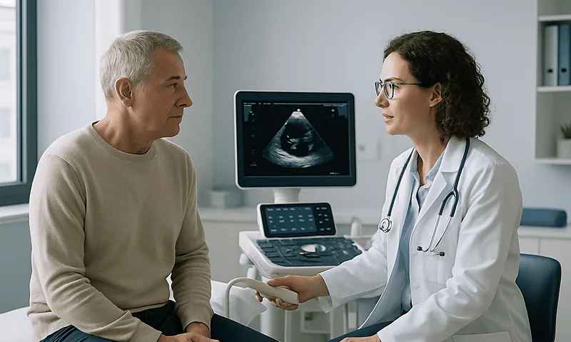
What an Echocardiogram Shows in Pericardial Effusion and How It Guides Treatment
An echocardiogram shows how much fluid surrounds the heart, where it sits, and whether it is pressing on the heart chambers. Measuring the fluid…



































