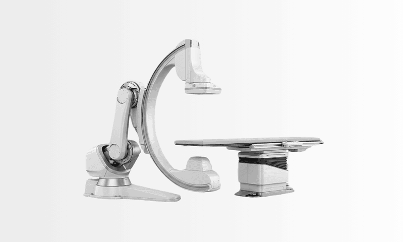
Peripheral Angio
Peripheral angio is an imaging procedure that visualizes arteries in the legs, arms, or other peripheral vessels using contrast dye to detect narrowing, blockage, or circulation problems.
Quick answer
Peripheral angio, also called peripheral angiography, is a catheter-based imaging procedure that maps blood flow through arteries outside the heart and brain, most often in the legs, arms and pelvis. Contrast dye is injected through a thin catheter while X-ray images are recorded, showing narrowings, blockages or vessel injuries. When appropriate, a blockage can be treated during the same session with balloon angioplasty or a stent.
Peripheral Angio: Understanding the Decision
Peripheral angio, also called peripheral angiography or a peripheral angiogram, is a catheter-based imaging procedure that shows how blood flows through the arteries outside the heart and brain, most often in the legs, arms and pelvis. A thin catheter delivers contrast dye into the arteries while X-ray images are recorded, producing a moving, detailed map of the circulation. Physicians use it when a narrowing, blockage, injury or abnormal vessel needs to be located precisely, usually because symptoms or earlier tests point to a problem with blood supply.
If you have been offered this procedure, the underlying question is usually a simple one: is blood reaching your tissues safely and adequately? Reduced circulation in the legs or arms can show itself in many ways. You may notice calf pain when walking, coldness in the foot, numbness, a wound that does not heal, or a change in skin colour. For other people, the concern is found during testing for diabetes, kidney disease or peripheral vascular disease, or after a previous vascular procedure. In each situation, peripheral angio is one of the most detailed ways to answer that central question.
It is normal to approach the procedure with concerns. You may be thinking about discomfort, the contrast dye, radiation exposure, or what happens if a blockage is found and needs treatment. A careful, structured vascular evaluation is designed to answer these questions before any decision is made.
At Acibadem, peripheral angio is treated not as an isolated imaging test but as one step in a broader vascular care pathway. The aim is to understand your symptoms, risk factors, anatomy and priorities, and then select the safest and most appropriate plan. In many cases, angiography provides exactly the information needed to choose between medical therapy, endovascular treatment, open vascular surgery, wound care or continued surveillance, and, just as importantly, to avoid a procedure that would not help.
What Peripheral Angio Is
Peripheral angio is an imaging procedure that visualises arteries in the legs, arms, pelvis, abdomen and other peripheral vascular regions using contrast dye and X-ray-based imaging. The contrast is injected into the bloodstream through a thin catheter, making the arteries visible in real time. This allows the physician to identify narrowed segments, complete blockages, abnormal vessel patterns, aneurysms, dissections and areas where flow is reduced or absent.
The procedure is usually performed by an interventional radiologist, a vascular surgeon, an interventional cardiologist or another physician trained in endovascular techniques. A small puncture is made in an artery, commonly at the groin or wrist, depending on which vessels need to be examined and on your individual anatomy. A catheter is then guided carefully to the target region. Contrast is injected in controlled amounts while images are taken from several angles, building a detailed picture of the vessels and of how blood moves through and beyond any diseased segment.
The study may be purely diagnostic, or it may be combined with treatment in the same session when the findings and your overall condition make that appropriate. If a significant narrowing is found and you are a suitable candidate, the physician may perform balloon angioplasty to widen the artery, place a stent to hold it open, remove clot, or use another endovascular technique. This possibility is discussed with you in advance. Not every patient needs treatment during angiography, and not every blockage can be treated safely in the same session. The decision depends on your symptoms, the imaging findings, kidney function, bleeding risk, general health and the realistic benefit of intervention.
How does peripheral angio differ from CT, MR or ultrasound imaging?
Compared with non-invasive tests such as Doppler ultrasound, CT angiography or MR angiography, catheter-based peripheral angio provides the most detailed and dynamic view of blood flow, and it is the only one of these methods that allows treatment during the same procedure. Non-invasive tests are usually performed first. Catheter angiography is reserved for situations where those tests suggest a clinically important problem, where symptoms are severe, where an intervention is likely to be needed, or where the earlier images are incomplete or conflicting. In practice, the two approaches work together: prior CT or MR images are often reviewed alongside the angiogram to understand the anatomy fully.
Is a peripheral angiogram the same as coronary or carotid angiography?
No: a peripheral angiogram uses the same basic technique but examines different arteries. Coronary angiography studies the arteries of the heart, and carotid angiography studies the arteries of the neck that supply the brain. Peripheral angio covers the arteries of the limbs, pelvis and abdomen. Because atherosclerosis often affects more than one vascular territory, a patient found to have peripheral disease may later be assessed for heart or neck artery disease as well, but each study is a distinct procedure with its own indications.
Who May Need a Peripheral Angiogram
A peripheral angiogram may be recommended when symptoms, examination findings or earlier imaging suggest that blood flow through peripheral arteries is impaired. The most common underlying condition is peripheral artery disease, in which fatty deposits, calcium and inflammation narrow the arteries, most often in the legs. The classic symptom is pain or cramping in the calf, thigh, hip or buttock during walking that eases with rest. This pattern is called claudication. In more advanced disease, pain may occur at rest, especially at night, or wounds may heal slowly because the tissues are not receiving enough oxygen-rich blood.
What are three signs of peripheral arterial disease?
Three of the most recognisable signs of peripheral arterial disease are pain in the leg muscles when walking that eases with rest, a foot or lower leg that feels persistently cold or looks pale compared with the other side, and a wound or ulcer on the foot or toes that heals slowly or not at all. Other signs can include weak or absent pulses in the leg, shiny or thinning skin, hair loss on the lower leg, and rest pain at night that improves when the leg hangs down. It is worth knowing that the disease can also be quiet: some people, especially those with diabetes or nerve damage, feel little pain even when circulation is significantly reduced. That is one reason examination findings and simple tests matter as much as symptoms.
Beyond peripheral arterial disease, patients may be referred for angiography because of signs of a limb-threatening circulation problem: persistent foot pain, ulcers on the toes or heel, blackened tissue, an infection that is difficult to control, or a foot that feels distinctly cold. In these situations, angiography helps the team judge whether restoring blood flow could support wound healing, reduce pain and lower the risk of tissue loss. Peripheral angio is also used after trauma when an arterial injury is suspected; before or after vascular surgery; to evaluate bypass grafts or stents when symptoms return or surveillance testing is abnormal; and to clarify abnormal vessel connections or aneurysms. In selected patients it assesses circulation to the arms, pelvis, intestines or kidneys, for kidney arteries specifically, see renal angiography.
How is the diagnosis made before angiography?
Diagnosis begins with a medical history and physical examination, not with a catheter. The physician asks about walking distance, pain patterns, smoking history, diabetes, blood pressure, cholesterol, kidney disease, previous vascular procedures, current medications, and any history of heart attack or stroke. The examination typically includes checking pulses in the legs or arms, inspecting the skin and nails, measuring blood pressure in different limbs, and assessing any wounds.
Non-invasive vascular tests usually come next. These may include ankle-brachial index testing, Doppler ultrasound, pulse volume recordings, CT angiography or MR angiography. Blood tests check kidney function, blood count and clotting. Catheter angiography is considered when these findings show that a precise arterial map is needed, or when there is a realistic possibility that an endovascular treatment will be performed at the same time. If the earlier tests already answer the clinical question and no intervention is planned, catheter angiography may not be necessary at all. Stating that limit clearly is part of honest vascular care.
Conditions and Indications Peripheral Angio Can Address
Peripheral angio is used to evaluate a wide range of vascular conditions. Its role is to show the location, length, severity and pattern of arterial disease so that physicians can choose a treatment strategy with confidence. That information matters most when symptoms are significant, when a limb or organ may be at risk, or when prior imaging is incomplete or conflicting.
- Peripheral artery disease: narrowing or blockage of arteries in the legs or arms, usually caused by atherosclerosis.
- Critical limb-threatening ischaemia: severe reduction in blood flow that may cause rest pain, ulcers, infection or tissue loss.
- Diabetic foot circulation problems: assessment of blood supply in patients with diabetes-related wounds or poor healing.
- Arterial occlusion: sudden or chronic blockage of an artery, related to clot, plaque or prior vascular disease.
- Aneurysms: abnormal widening of an artery that may require monitoring or repair planning.
- Vascular trauma: suspected arterial injury after an accident, fracture, penetrating injury or medical procedure.
- Bypass graft or stent evaluation: assessment of a previously treated artery or surgical graft when symptoms return or surveillance results are abnormal.
- Renal, mesenteric or upper-extremity arterial disease: evaluation of selected circulation problems affecting the kidneys, intestines, arms or hands.
- Preoperative planning: mapping of arteries before vascular reconstruction, amputation-level planning, flap surgery or complex wound treatment.
Angiography does not replace a full clinical assessment. A narrowing seen on imaging is not always the cause of symptoms, and some patients with severe-looking disease are still best managed with medication and supervised exercise. Conversely, a small-vessel problem may be very important if it affects wound healing or tissue survival. The value of angiography is greatest when the images are interpreted alongside your complete health profile, not in isolation.
How Peripheral Angio Is Performed
Preparation before the procedure
Preparation begins with a review of your medical history, current medications, allergies and previous imaging. Particular attention is paid to kidney function, because the contrast dye is processed through the kidneys. If you have chronic kidney disease, diabetes, dehydration or take certain medications, the team may add precautions: hydration protocols, medication adjustments made by your treating physician, or an imaging strategy that reduces contrast exposure.
The care team will ask about allergies to contrast dye, iodine-containing substances, latex, antibiotics or anaesthetic medications. A history of bleeding disorders, blood-thinner use, prior stroke, heart disease or implanted devices should also be discussed. Some medications, particularly anticoagulants and certain diabetes drugs, may need to be paused or adjusted before the procedure. These decisions belong to the treating physician, who weighs the risks in your individual case; they are never made by a standard checklist alone.
You will usually be asked not to eat or drink for several hours beforehand. Blood tests may be repeated shortly before the procedure to check kidney function, haemoglobin and clotting. Where recent records exist, vascular ultrasound reports, CT or MR angiography images, medication lists and wound photographs where relevant: the medical team reviews them in advance, so that the plan is settled before the day of the procedure rather than improvised on it.
Step by step: what happens during the procedure
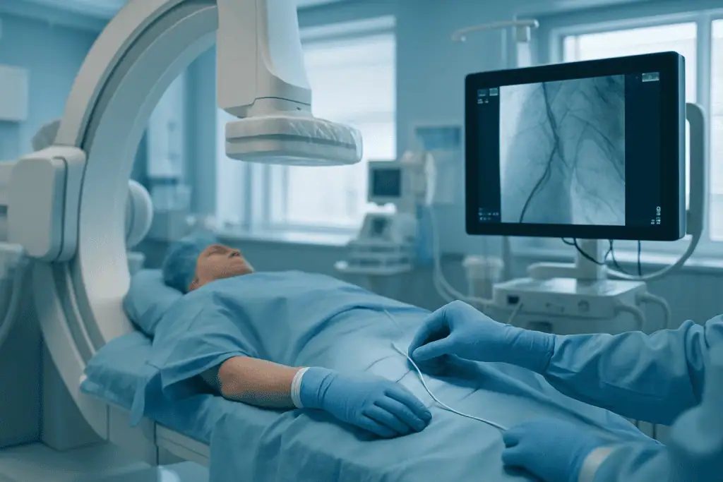
Peripheral angio is performed in an angiography suite or hybrid operating environment designed for image-guided vascular work. You lie on a procedure table while monitoring equipment tracks your heart rhythm, blood pressure, oxygen level and general condition. Most procedures use local anaesthetic at the access site plus sedation when appropriate; you are usually awake but relaxed, able to follow instructions if asked. The typical sequence is:
- Access: the physician cleans and drapes the access area, most often the groin or wrist, numbs the skin with local anaesthetic, and makes a small puncture in the artery.
- Sheath placement: a short, thin tube called a sheath is placed in the artery so that catheters and guidewires can pass safely.
- Navigation: using live X-ray imaging, the physician advances a catheter to the arteries being studied.
- Contrast injection and imaging: contrast dye is injected in controlled amounts while images are recorded from different angles. You may feel a brief warm sensation as the contrast passes; it usually settles quickly.
- Assessment: the images show where the artery narrows or stops, how collateral vessels compensate, and what the flow looks like beyond the diseased segment. Measurements of lesion length and vessel diameter may be taken. In selected cases, pressure measurements across a narrowing or intravascular imaging from within the artery add precision.
- Treatment, if planned: when a treatable blockage is found and same-session therapy has been agreed, the physician may perform balloon angioplasty, place a stent, use atherectomy to modify plaque in selected cases, deliver catheter-directed clot treatment, or perform embolisation for certain abnormal vessels or bleeding.
- Closure: the catheter and sheath are removed, and the puncture is sealed with manual pressure or a closure device.
The choice between wrist and groin access is itself an individual decision. Groin (femoral) access offers a direct route to the pelvic and leg arteries and accommodates the larger equipment sometimes needed for treatment, but it requires a period of lying flat afterwards and tends to bruise more readily. Wrist (radial) access usually allows earlier sitting and walking and is often more comfortable in the hours that follow, though the longer distance to the leg arteries limits its use for some interventions. Body build, previous surgery or scarring at either site, the pulses that can be felt on examination and the specific vessels being studied all feed into that decision.
None of the treatment steps is automatic. When intervention is performed, the benefits and risks are weighed carefully, particularly in complex disease or in patients with kidney, heart or bleeding concerns. Sometimes the safest decision is to complete the diagnostic study, discuss the findings, and plan treatment as a separate step.
How long does a peripheral angiogram take?
The diagnostic part of a peripheral angiogram often takes less than an hour in straightforward cases. Total procedure time is longer when the anatomy is complex, when several vascular regions are studied, or when treatment such as angioplasty or stenting is performed during the same session. You should also allow for preparation beforehand and a monitored recovery period afterwards, which can add several hours to the day. If groin access is used, a period of lying flat is usually required after the procedure, which extends the recovery window compared with wrist access.
How is balloon size determined in peripheral angio?
Balloon size is determined by measuring the artery itself, not by a standard formula. When angioplasty is planned, the physician measures the diameter of the healthy artery next to the narrowing, the reference vessel, using calibrated angiographic measurements on the images, sometimes supplemented by intravascular ultrasound, which views the vessel wall from inside. The balloon diameter is chosen to match the reference vessel: a balloon that is too small may not open the narrowing adequately, while one that is too large increases the risk of tearing or injuring the artery wall. Balloon length is chosen to cover the full length of the diseased segment. Vessel calibre varies widely across the body, pelvic arteries are larger than the small vessels below the knee, so sizing is always individual. Inflation pressure and duration are then adjusted to how the lesion responds, which is one reason this judgement belongs to an experienced operator rather than to a fixed protocol.
Technology used to support accuracy and safety
Modern peripheral angiography relies on high-resolution X-ray imaging, digital subtraction techniques, real-time fluoroscopy and software that enhances vessel visibility. These tools help the physician see contrast-filled arteries clearly while navigating catheters with precision. Imaging can be adjusted to focus on small vessels below the knee, larger pelvic vessels, arm arteries or other target regions, and prior CT or MR images are often reviewed alongside the live pictures.
Radiation exposure is managed through careful technique, appropriate imaging settings, shielding, and limiting imaging time to what is medically necessary. Contrast use is monitored throughout. For patients at increased risk of kidney strain, the team may use lower contrast volumes, hydration protocols, alternative imaging views, or staged procedures when that is clinically sensible; in selected patients, carbon dioxide can even be used as a contrast agent in place of some of the iodinated dye. The purpose of the technology is practical, not decorative: it exists to answer specific questions, where the blockage is, whether it explains the symptoms, and whether it can be treated safely.
Risks and Safety Considerations
What are the risks of a peripheral angiogram?
The main risks of a peripheral angiogram relate to the access site, the contrast dye and the vessels themselves. At the puncture site, bruising and tenderness are common and usually settle; less often, a larger haematoma, bleeding or a vessel injury at the access point can occur and may need further treatment. Rarely, the puncture can lead to a pseudoaneurysm, a contained leak beside the artery, or an abnormal connection between artery and vein; both are usually detected on examination or ultrasound and can most often be managed without open surgery. Contrast dye can trigger allergic-type reactions, which range from mild and self-limiting to, rarely, more serious responses that the team is prepared to treat immediately. Contrast can also strain the kidneys, particularly in people with pre-existing kidney disease, diabetes or dehydration, which is why kidney function is checked beforehand and contrast volume is kept as low as the study allows. Catheter manipulation carries a small risk of damaging a vessel, dislodging plaque or clot, or causing a blockage downstream. Infection at the puncture site is uncommon. Radiation exposure is kept to the minimum needed for the study.
When treatment such as angioplasty or stenting is added, the risk profile changes: the possibility of vessel tearing, re-narrowing over time, or clot formation on a treated segment is part of the discussion before the procedure. Your individual risk depends on your anatomy, kidney function, medications, bleeding tendency and general health, which is why the pre-procedure assessment is thorough rather than routine. The team’s role is to explain your specific risk picture plainly before you consent, not to minimise it.
What to Expect After a Peripheral Angiogram
What happens in the hours after the procedure?
After the catheter and sheath are removed and the access site is closed, you are monitored for bleeding, swelling, pain, changes in limb circulation, and any reaction to medication or contrast. Depending on the access site, you may need to lie flat for a period, especially after groin access. Nurses check your pulses, skin temperature and the puncture site at intervals. Most patients can drink fluids soon after the procedure, and hydration is encouraged unless there is a medical reason to limit it, because fluid helps the kidneys clear the contrast. Some patients go home the same day; others stay overnight, particularly after an intervention, or when complex medical conditions, limb-threatening ischaemia or an active wound require closer observation.
Before discharge, the care team explains activity restrictions, puncture-site care, medications, warning signs and follow-up appointments. You will usually be advised to avoid strenuous activity, heavy lifting and soaking the access site for a short period. Gentle walking is often encouraged gradually, guided by your symptoms and your physician’s instructions.
Recovery timeline after peripheral angio
Recovery varies depending on whether the procedure was diagnostic only or included treatment, as well as your overall health and the access site used.
| Time Period | What Patients Can Expect |
|---|---|
| Day 1 | Monitoring after the procedure, checks of the access site and pulses, gradual return to eating and drinking, and clear instructions about activity limits. Some patients go home the same day; others stay overnight. |
| First week | Mild bruising or tenderness at the puncture site may occur. Patients typically avoid heavy lifting and strenuous activity while walking lightly as advised. Medication instructions are important, especially after angioplasty or stenting. |
| First month | Follow-up may include wound assessment, pulse checks, ultrasound, medication review, or planning for additional treatment if needed. Walking capacity and pain may improve gradually when blood flow has been restored. |
| Longer term | Ongoing vascular health depends on risk-factor control, smoking cessation, exercise, diabetes and cholesterol management, and surveillance of treated vessels when recommended. |
Why Acting Early Matters
Circulation problems can progress quietly. Many people adapt without noticing, walking less, avoiding stairs, changing daily routines, so the true severity of symptoms is underestimated. In others, especially people with diabetes or nerve damage, pain signals are dulled even when tissue blood supply is poor. A wound on the foot or toe may look minor at first, yet become serious if oxygen and nutrients cannot reach the tissue well enough for healing.
Early evaluation matters because the range of options is usually broader before advanced tissue damage occurs. Lifestyle changes, medication optimisation by your physician, supervised walking programmes, diabetes control, wound care and minimally invasive vascular procedures may stabilise disease or restore adequate blood flow. When assessment waits until infection, gangrene or severe rest pain has developed, treatment becomes more urgent and more complex, and the likelihood of hospitalisation, major procedures and tissue loss rises.
Delayed assessment can also miss the wider picture. Peripheral artery disease often signals atherosclerosis in other vascular territories, including the arteries of the heart and neck. Identifying peripheral disease can therefore prompt a more complete cardiovascular prevention plan: cholesterol management, blood pressure control, antiplatelet therapy where the treating physician judges it appropriate, smoking cessation support, diabetes management and structured exercise guidance.
Two limits are worth stating plainly. Not every person with leg pain needs angiography, and not every arterial narrowing needs a procedure. Symptoms such as rest pain, non-healing wounds, rapidly worsening walking distance, sudden limb coldness, new numbness or weakness, or colour changes generally lead physicians to arrange assessment sooner rather than later, and peripheral angio becomes part of that evaluation when a detailed arterial map is needed to make a safe treatment decision. Leg numbness, it should be said, is not always vascular: nerve conditions such as peripheral neuropathy can produce similar sensations, which is another reason a proper clinical assessment comes before any catheter.
Potential Benefits of Peripheral Angio
The main benefits of the procedure relate to diagnostic clarity, treatment planning, and the opportunity to restore blood flow when intervention is appropriate.
| Benefit | What It Means for You |
|---|---|
| Detailed arterial mapping | Physicians can see the exact location and pattern of narrowing or blockage, which helps guide the most appropriate treatment plan. |
| Real-time blood flow assessment | The procedure shows how blood moves through the vessels, including collateral circulation and flow beyond a diseased segment. |
| Potential same-session treatment | If suitable, certain blockages may be treated during the same procedure, avoiding a separate appointment or an additional arterial puncture. |
| Better planning for complex disease | For patients with wounds, diabetes, prior surgery or multiple blockages, angiography helps determine whether endovascular treatment, surgery or combined care is best. |
| Support for limb preservation decisions | In severe circulation problems, the findings help wound care, vascular and surgical teams plan treatment aimed at healing and tissue protection. |
What Influences Outcomes and a Good Result
The outcome of peripheral angio depends on more than the images produced on the day. A good result begins with choosing the right patients for angiography and being clear about the question the procedure needs to answer. For one patient, the goal is to identify a blockage causing severe calf pain. For another, it is to determine whether blood flow can be improved enough to heal a diabetic foot ulcer. For a third, the purpose is to evaluate a previous stent or bypass graft. A study that answers the wrong question is not a good study, however sharp the pictures.
Several factors shape the result. The severity and location of vascular disease are central. Short, focal narrowings are generally easier to treat with endovascular methods than long, calcified, multi-level blockages. Disease below the knee, common in diabetes and kidney disease, can be technically demanding because the vessels are smaller and often more diffusely affected. Patients with active infection, advanced tissue loss or poor general health may need coordinated care across several specialties rather than a single procedure.
Kidney function matters throughout, because contrast dye places stress on the kidneys, particularly where kidney disease already exists. Careful planning, hydration, contrast minimisation and sensible timing reduce that risk. Blood-thinner use, anaemia, clotting disorders and fragile arteries affect bleeding risk and access-site management. Heart and lung conditions influence sedation choices and how closely you are monitored.
Your own participation plays a major role afterwards. If angiography leads to angioplasty, stenting or another intervention, the durability of the result often depends on medication adherence as directed by your treating physician, follow-up imaging, exercise, and risk-factor control. Smoking is one of the strongest contributors to progression and recurrence of peripheral artery disease. Diabetes control, foot protection, nutrition and wound care are especially important if you have ulcers or tissue loss.
Finally, a good outcome is not always a perfectly normal-looking artery. In vascular medicine, a meaningful result may be improving blood flow enough to relieve rest pain, supporting wound healing, preventing deterioration, making planned surgery safer, or establishing clearly that a different approach is needed. The most valuable result is the one that lines up the imaging findings with your symptoms, your risks and your goals of care.
Peripheral Angio at Acibadem
Acibadem’s approach to peripheral angio is built on multidisciplinary vascular care. Depending on your condition, evaluation may involve interventional radiology, vascular surgery, cardiology, endocrinology, nephrology, infectious diseases, orthopaedics, plastic and reconstructive surgery, wound care specialists and rehabilitation. In complex cases, multidisciplinary discussions align the imaging findings with the broader treatment plan: a structure that is particularly valuable for patients with diabetic foot wounds, critical limb ischaemia, recurrent vascular disease or several coexisting medical conditions.
For a patient undergoing angiography, established quality and safety processes shape each step: structured pre-procedure assessment, infection prevention practices, medication reconciliation, imaging safety protocols, monitoring during and after the procedure, and clear discharge instructions. These systems matter because peripheral vascular patients often carry additional risks such as diabetes, kidney disease, coronary artery disease or previous stroke, and safe care means accounting for the whole patient, not only the artery under study.
Experienced physicians are central to the process. This work requires judgement as well as technical skill: selecting the access site, minimising contrast where appropriate, interpreting complex vessel patterns, sizing balloons and stents to the individual artery, deciding whether same-session treatment is reasonable, and recognising when open surgery or conservative management is the safer path. Patients benefit when their physicians routinely evaluate the full spectrum of vascular disease, from claudication to limb-threatening ischaemia and revision cases after earlier procedures.
Personalised planning matters in this field because two patients with similar-looking angiograms may need entirely different care. A younger patient with a focal arterial injury after trauma is not the same case as an older patient with diabetes, kidney disease and a non-healing toe wound. A person whose main problem is walking pain has different priorities from someone facing infection or possible amputation. At Acibadem, angiography findings are interpreted within each patient’s medical context, functional goals and follow-up needs, not as a stand-alone image.
Continuity after discharge is treated as part of the treatment itself. Peripheral artery disease usually requires long-term management. Before discharge, the plan covers which medications to continue as prescribed, which symptoms should prompt medical review, how the puncture site should be cared for, whether follow-up ultrasound is needed, and how ongoing wound care, rehabilitation or surveillance will be organised with the teams involved in your continuing care. A clearly written plan gets followed; an unspoken one does not.
How Peripheral Angio Fits into a Vascular Care Plan
Peripheral angio provides essential clarity when circulation problems are suspected or when earlier testing has not fully explained the symptoms. It shows whether an artery is narrowed or blocked, defines the severity and pattern of disease, and gives physicians the information needed to decide among medication, endovascular treatment, surgery, wound care or watchful observation. For some patients, it also creates the opportunity to treat the problem during the same session, through balloon angioplasty or stenting sized to the individual artery.
It is, deliberately, one step in a longer pathway. The steps before it, history, examination, non-invasive testing, establish whether the procedure is needed at all. The steps after it, medication, risk-factor control, wound care, follow-up imaging, determine how durable any improvement will be. Understood that way, a peripheral angiogram is less a single event than a turning point: the moment at which a vascular problem stops being a suspicion and becomes a mapped, measurable condition with a plan attached. For the wider context of arterial and venous conditions this procedure serves, the overview of peripheral vascular diseases explains how the pieces of diagnosis and treatment fit together.
Preparation
- Your doctor may request blood tests, kidney function assessment, and a review of current medications, especially blood thinners or diabetes medicines. You may be asked to stop eating or drinking for several hours before the procedure. Inform the team about allergies to contrast dye, iodine, or previous reactions during imaging tests.
Aftercare
- After the procedure, you will be monitored while the catheter entry site is checked for bleeding or swelling. Keep the puncture area clean and avoid strenuous activity for a short period as advised. Drink fluids if permitted to help clear the contrast dye, and contact your doctor if you notice severe pain, numbness, fever, or bleeding.
Frequently Asked Questions
What affects the cost of peripheral angio?
The main factors are the imaging method, the artery area being examined, contrast dye and catheter materials, specialist fees, hospital facility fees, pre-procedure tests and whether treatment such as angioplasty or stenting is performed during the same session.
How can I get a personalised quote?
You can request a free consultation and share your medical reports, test results, medication list and any previous imaging. The clinical team reviews your case and provides a personalised plan and quote based on your needs.
Is peripheral angio always only a diagnostic test?
Not always. It may be planned as a diagnostic procedure, but in selected cases the specialist may recommend treatment during the same session if a suitable narrowing or blockage is found and consent has been obtained.
What is usually included in an international patient package?
Packages may include specialist consultation, procedure planning, basic pre-procedure tests, the angiography procedure, contrast-related supplies, standard recovery monitoring, translation support and care coordination. Inclusions vary by clinical plan, so they should be confirmed before travel.
Will insurance cover peripheral angio abroad?
Coverage depends on your insurer, policy terms, referral requirements and whether the procedure is diagnostic or therapeutic. This information is general and not financial advice; patients should confirm approval directly with their insurer.
Do I need to travel with someone for peripheral angio?
Some patients prefer to have a companion, especially if sedation is used or if a treatment is expected. The need for assistance depends on your general health, travel plans and the specialist’s recommendations.
Medically reviewed by the Acıbadem International Medical Board August 30, 2026
See our medical review board →
Update history
- PublishedJune 8, 2026
- Medical review approvedAugust 30, 2026
- Last content updateAugust 30, 2026
References1
- Peripheral Artery Disease (PAD) my.clevelandclinic.org
Trusted care for international patients
From the Acibadem Blog

After a Peripheral Angiogram: Puncture-Site Care and the Symptoms That Warrant a Call
Mild aching, bruising and a small tender lump at the puncture site are common after a peripheral angiogram and usually settle over one to…