Scoliosis Juvenile Treatment
Juvenile scoliosis is abnormal spinal curvature in children aged 4 to 10, managed with specialist monitoring, bracing, rehabilitation, and surgery for progressive or severe curves.
Quick answer
Juvenile scoliosis is a sideways spinal curve of 10 degrees or more diagnosed between the ages of 4 and 10. Because children this age still have years of growth ahead, treatment focuses on controlling progression: scheduled monitoring for mild curves, bracing and physiotherapy for progressive ones, and surgery for severe curves that threaten chest and lung development.
Scoliosis Juvenile: What the Diagnosis Means for Your Child
Juvenile scoliosis is a sideways curvature of the spine, measuring 10 degrees or more on a standing X-ray, that is diagnosed between the ages of 4 and 10. It is less common than the adolescent form, and it deserves careful attention for one simple reason: children in this age group still have years of growth ahead, and growth can drive a curve to progress. Treatment ranges from scheduled observation through bracing and scoliosis-specific physiotherapy to, in a smaller group of children, surgery.
You may see the condition indexed as scoliosis juvenile on referral letters, imaging reports or insurance paperwork. Clinicians usually say juvenile scoliosis. Both describe the same diagnosis: a structural spinal curve that appeared during the early school years, before the main pubertal growth spurt. In classification systems that sort the condition by age of onset, scoliosis juvenile in onset sits between the infantile form, which appears in the first years of life, and the adolescent form, which appears around puberty.
The diagnosis often arrives unexpectedly. A parent notices a shoulder blade that sticks out, a school nurse flags an uneven waistline, or a paediatrician spots asymmetry during a routine check. From that moment, most families carry the same questions. Will the curve get worse? Will my child need a brace? Can surgery be avoided? Will this affect school, sport, sleep or confidence? Children carry their own version of these worries: how they will look, whether treatment will hurt, whether a brace will make them feel different from their friends.
The goal of treatment is not only to change a number on an X-ray. It is to protect your child’s spinal growth, posture, chest and lung development, physical comfort and long-term function. Some children only need observation with scheduled imaging and clinical examinations. Others benefit from bracing and structured rehabilitation. A smaller group, usually children with larger or rapidly progressive curves, may need surgery to control the deformity while preserving as much growth and mobility as possible. Understanding which group your child belongs to is the real work of the first specialist visits.
At what age does scoliosis start?
Scoliosis can start at any age, and specialists group it by when the curve first appears. Infantile scoliosis is diagnosed in the first years of life. Juvenile scoliosis is diagnosed between 4 and 10. Adolescent scoliosis, the most common form, appears from around age 10 onwards, typically during the pubertal growth spurt. The age labels matter because they signal how much growth remains, and remaining growth is the main engine of curve progression. A curve found at age 6 has far more growing years in front of it than the same curve found at 14, which is why juvenile curves are followed more closely and for longer.
Is it normal for a 7-year-old to have scoliosis?
Scoliosis at age 7 is not “normal” in the sense of being an expected part of development, but it is a recognised condition that spine specialists see and manage regularly. A curve at this age warrants a proper specialist evaluation rather than alarm. Many curves found in 7-year-olds are mild and need nothing more than monitoring. What makes this age group different is the amount of growth still to come: a mild curve at 7 has years in which it could either stay stable or progress, so the evaluation is thorough and the follow-up is structured. Specialists also look more carefully for an underlying cause in this age group than they typically do in teenagers, because curves that begin earlier are somewhat more likely to have an identifiable explanation.
What Juvenile Scoliosis Treatment Involves
Juvenile scoliosis treatment is a structured approach to diagnosing, monitoring and managing spinal curvature in children aged 4 to 10. It may include observation, bracing, physiotherapy and rehabilitation, and in selected cases surgery. The right plan depends on the size and pattern of the curve, whether the curve is progressing, how much growth remains, and whether there are associated conditions affecting the spine, nerves, muscles, chest wall or lungs.
Scoliosis is measured using the Cobb angle, calculated from standing spinal X-rays. A curve of 10 degrees or more is generally considered scoliosis. In juvenile scoliosis, the curve may involve the thoracic spine, the lumbar spine, or both. Some curves are flexible and mild; others are structural and more likely to progress. The same Cobb angle can carry a different meaning in different children: a moderate curve in a 5-year-old is a different clinical problem from the same curve in a 9-year-old, because the younger child has more growing time in which the curve can change.
Treatment is usually staged. Mild curves may be followed closely with scheduled imaging. Moderate curves in a growing child may require a brace to reduce the risk of worsening. Physiotherapy may support posture, strength, breathing mechanics and brace adaptation, although exercise alone is not usually considered a substitute for bracing when a progressive curve requires it. Surgery is considered when non-surgical methods are unlikely to control progression, when the curve becomes severe, or when the deformity may interfere with chest growth, lung development or overall function.
What is juvenile idiopathic scoliosis?
Juvenile idiopathic scoliosis is a spinal curve of 10 degrees or more, diagnosed between ages 4 and 10, in which no underlying cause is found after appropriate evaluation. “Idiopathic” is the medical word for “without an identified cause”: the child is otherwise healthy, the vertebrae formed normally before birth, and the nerves and muscles work normally, yet the spine has developed a curve. It is the most common form of scoliosis in this age group. The label is only applied after other explanations have been considered and reasonably excluded, which is why the diagnostic work-up in young children is deliberately careful.
What causes juvenile idiopathic scoliosis?
By definition, no single cause is identified in juvenile idiopathic scoliosis. Research points towards a combination of genetic predisposition and growth-related factors, the condition sometimes runs in families, but there is no test that identifies one trigger in an individual child. What is well established is what does not cause it: idiopathic scoliosis is not caused by poor posture, heavy school bags, sitting habits, sports or anything a parent did or failed to do. That matters, because parents often blame themselves, and children sometimes blame their own habits. Before applying the idiopathic label, physicians also consider and rule out other explanations, including congenital vertebral differences, neuromuscular conditions, syndromic disorders, connective tissue conditions, spinal cord abnormalities and prior injury.
What is the difference between juvenile and adolescent idiopathic scoliosis?
The difference between juvenile and adolescent idiopathic scoliosis is the age at which the curve is diagnosed: between 4 and 10 for the juvenile form, and from around 10 onwards for the adolescent form. That single difference has practical consequences. A child with juvenile-onset scoliosis has more growth remaining, which means more time for the curve to progress before the skeleton matures, so juvenile curves are monitored more closely, treated more readily, and followed for longer. Specialists are also more likely to recommend an MRI in juvenile-onset cases, because curves that appear earlier are somewhat more likely to have an underlying spinal cord finding than curves that first appear in adolescence. The treatments themselves overlap heavily; it is the surveillance intensity, the diagnostic depth and the length of the treatment horizon that differ.
How fast does scoliosis progress in teens?
Scoliosis progresses fastest during periods of rapid growth, and in teenagers that means the pubertal growth spurt. Progression speed varies enormously from child to child and cannot be predicted precisely from a single X-ray; that is exactly why follow-up imaging is scheduled at intervals rather than performed once. This matters for juvenile scoliosis families in a specific way: a child diagnosed at 6 or 8 will eventually pass through that same adolescent growth spurt, and the years around it are the highest-risk window for a juvenile curve to accelerate. Because scoliosis juvenile in onset appears before that spurt rather than during it, follow-up typically continues for years, through the spurt and until skeletal maturity, rather than for months.
Who May Need Juvenile Scoliosis Care
Many children with juvenile scoliosis feel entirely well and have no pain. The first sign is usually visual, noticed by a parent, paediatrician, teacher or sports coach, or picked up during a routine health examination. Look for uneven shoulders, a shoulder blade that appears more prominent on one side, an uneven waistline, one hip that seems higher, or a visible rib prominence when the child bends forward. Clothing may hang unevenly, and parents sometimes notice that one trouser leg appears longer even though the legs are the same length.
Some children report back fatigue, muscle tightness or discomfort after activity. Significant pain, however, is not typical of idiopathic scoliosis and is evaluated carefully when present, because it can point towards a different underlying problem. Shortness of breath is uncommon with mild or moderate curves, but severe thoracic curves can affect chest shape and respiratory mechanics over time. Neurological findings, weakness, numbness, changes in walking pattern, changes in bladder or bowel control, or persistent night pain, prompt a more detailed work-up, typically including MRI, because they suggest the spine and spinal cord need closer examination than a routine scoliosis check provides.
How is juvenile scoliosis diagnosed?
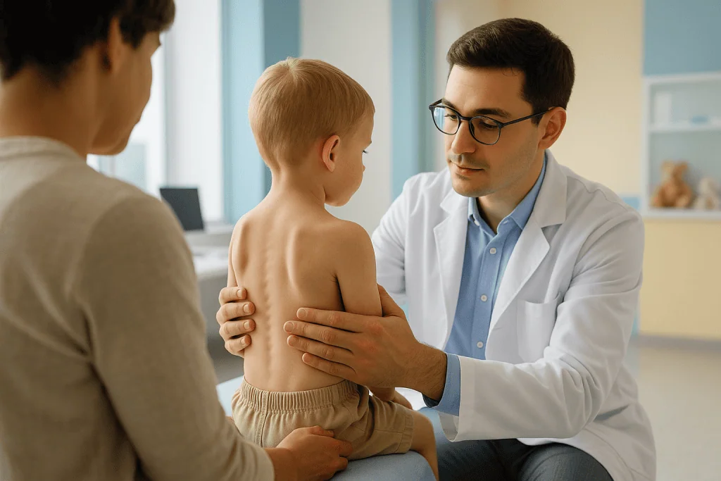
Juvenile scoliosis is diagnosed through a clinical examination confirmed by standing full-spine X-rays. The physician takes a detailed medical history, then evaluates posture, shoulder and pelvic balance, spinal rotation, flexibility, neurological function, gait and signs of growth and maturation. The forward bend test, the child bends forward at the waist while the examiner looks along the spine, may reveal a rib hump or lumbar prominence caused by vertebral rotation. Imaging then confirms the diagnosis, measures the Cobb angle and assesses alignment from the neck to the pelvis. Because children in this age group may need repeated imaging over years of follow-up, modern low-dose imaging protocols are preferred to keep cumulative radiation exposure as low as reasonably possible.
Additional tests are recommended in specific situations. MRI may be used when the child is very young, the curve pattern is atypical, neurological findings are present, the curve progresses unexpectedly, or pain is out of proportion to the spinal findings. Pulmonary function testing may be considered for larger thoracic curves or when surgery is being planned. In some children, genetic, neurological, cardiopulmonary or broader paediatric evaluations are needed to understand the full medical picture before the spine plan is set.
Families usually seek specialist care when a curve is newly diagnosed, appears to be progressing, requires a brace, has not responded to earlier treatment, or may require surgery. A second opinion is common in juvenile scoliosis, and reasonable, particularly when two clinicians have given different recommendations, or when a family is weighing observation against bracing, or bracing against surgery.
Conditions and Indications Juvenile Scoliosis Treatment Addresses
Juvenile scoliosis treatment addresses abnormal spinal curvature that develops during the early school-age years. The main indication is a measurable scoliosis curve in a child between 4 and 10, especially when there is evidence that the curve may progress during growth. Within that broad indication, the underlying diagnosis shapes the plan considerably.
Juvenile idiopathic scoliosis is the most frequent condition. The child is otherwise healthy and the curve has no identified cause. Treatment focuses on monitoring growth, controlling progression and preserving function. Because these children have substantial growth remaining, even moderate curves require careful, scheduled follow-up. A note on naming: “idiopathic” appears in several unrelated paediatric diagnosesjuvenile idiopathic arthritis, for example, is a joint condition that shares only the naming convention, not the disease. If your child’s records mention both terms, they describe separate conditions.
Congenital scoliosis occurs when one or more spinal bones formed differently before birth. These curves may behave quite differently from idiopathic curves. Depending on the anatomy and the progression pattern, they may need closer monitoring or earlier surgery, and they are less likely to respond to bracing, because the curve is built into the shape of the vertebrae themselves.
Neuromuscular scoliosis may occur in children with conditions that affect muscle tone, strength or motor control. These curves can progress more steadily and may be linked to sitting balance, respiratory function, hip position and daily care needs. Management often involves orthopaedic surgeons, paediatric neurologists, rehabilitation physicians, physiotherapists and other specialists working from a shared plan.
Syndromic scoliosis can occur as part of broader genetic or connective tissue conditions. Here, treatment planning must consider the whole child, heart, lung, eye, joint, bone and anaesthesia-related factors where relevant. The spine is treated within the context of the child’s overall health, not in isolation.
Juvenile scoliosis care may also be needed for children with progressive thoracic curves, double curves, curves causing trunk imbalance, curves associated with chest wall changes, or curves that have continued to worsen despite bracing. The plan is individualised in every case, because a diagnosis of scoliosis juvenile in type tells you when the curve appeared, not, on its own, how it will behave or what it will need.
How Juvenile Scoliosis Treatment Is Performed
Treatment begins with careful assessment rather than immediate intervention. The specialist’s first task is to understand the curve: where it sits, how large it is, how flexible it appears, how fast the child is growing, and whether anything suggests an underlying condition. In practice, the assessment pathway usually runs through a consistent sequence:
- Record review. Previous X-rays, MRI scans, brace prescriptions and therapy notes are gathered and compared, because the trajectory of a curve over time is more informative than any single image. Reviewing existing records first also helps avoid repeating imaging unnecessarily.
- Clinical examination. The spine specialist examines posture, balance, shoulder height, pelvic level, trunk shift and spinal rotation, and checks neurological function, reflexes, strength, sensation and walking pattern. Growth assessment is central, because remaining growth defines progression risk.
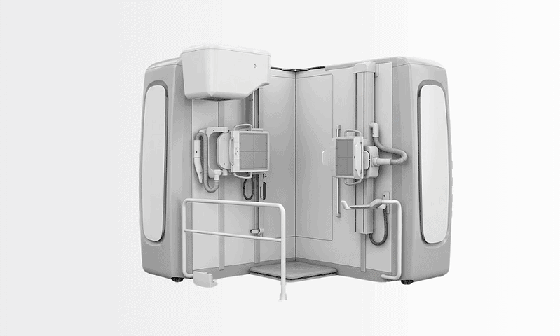
- Imaging. Standing full-spine X-rays show the curve in the position that matters for posture and daily function. The Cobb angle is measured and alignment is evaluated from neck to pelvis, using child-focused low-dose protocols where repeated imaging is expected. MRI is added when neurological signs, unusual curve patterns, significant pain, rapid progression or the child’s young age warrant a look at the spinal cord.
- Plan and schedule. Based on curve size, pattern, flexibility and growth stage, the team recommends observation, bracing, rehabilitation, surgery, or a combination, with a defined follow-up schedule rather than an open-ended “come back if worried”.
Observation: monitoring without intervening
If the curve is mild and the child is growing, observation is often the most appropriate first step. Observation is not passive neglect; it is scheduled surveillance. Follow-up visits are timed according to the curve size, growth stage and progression risk, and the physician compares measurements over time, adjusting the plan if the curve changes. For many families this is the entire treatment: regular checks, normal life in between, and a clear threshold agreed in advance for when the plan would change.
Does mild scoliosis always progress?
No, mild scoliosis does not always progress, and many mild curves remain stable through growth. But in a child aged 4 to 10, the honest answer has a second half: it is not possible to know in advance which mild curves will stay mild, and remaining growth is the main risk factor for progression. That combination, most curves behave, some do not, and prediction is imperfect, is precisely why observation in this age group is structured rather than casual. The purpose of scheduled imaging is to catch the minority of curves that start to move while there is still time to respond with a brace rather than an operation.
Bracing: how it works and what it asks of your child
Bracing is commonly recommended for moderate curves, or for curves that show progression in a child with growth remaining. A brace applies external support to the trunk to reduce the likelihood that the curve worsens during growth. It does not usually make a structural curve disappear; its job is containment through the growing years. The type of brace depends on the curve location, flexibility, the child’s size and the treatment goal. Some children wear a thoracolumbosacral orthosis under their clothing. Others need a design that controls higher thoracic curves, or a nighttime brace suited to selected curve patterns. Daily wear hours are prescribed individually, and adherence is one of the most important factors in whether bracing works.
A brace programme includes measurement, fitting, adjustment and follow-up imaging to confirm the brace is applying corrective forces where intended. Children need time to adapt, physically and emotionally. Pressure areas, skin irritation, heat, sleep disruption and clothing concerns should be raised openly with the brace team rather than endured silently, most can be improved with adjustment. A well-run programme also includes practical coaching for school, travel, hygiene, sport and social life, because a brace that a child refuses to wear helps no one.
Physiotherapy and rehabilitation
Scoliosis-specific exercise programmes support the treatment plan by improving postural awareness, core endurance, spinal mobility, breathing mechanics and muscular balance. Therapy can also help children tolerate a brace and stay active. Two honest caveats apply. First, in juvenile scoliosis, exercise works best when integrated with orthopaedic monitoring, not as an isolated substitute for bracing when a progressive curve requires it. Second, the aim is never to make the child responsible for controlling the curve alone, exercises build strength, awareness and confidence while the medical team manages the scoliosis.
What is severe juvenile scoliosis?
Severe juvenile scoliosis describes a large structural curve, typically one that causes visible trunk imbalance, continues to progress despite appropriate bracing, or threatens chest and lung development. There is no single degree threshold that defines “severe” in isolation, because severity in a growing child is a combination of curve size, curve behaviour, location and remaining growth: a thoracic curve that is progressing quickly in a 6-year-old may be treated as severe at a Cobb angle that would be watched calmly in an older, near-mature child. Severe curves are the group in which surgical planning is most likely to enter the conversation, because a large deformity established early can reshape the chest during the years when the lungs are still developing.
When surgery is considered, and how it is done
Surgery is considered when the curve is severe, continues to progress despite appropriate non-surgical treatment, threatens chest or lung development, causes significant imbalance, or is likely to create long-term functional problems. Surgical planning in young children needs particular care because the spine and chest are still growing. Depending on the child’s age, curve size and underlying condition, options include growth-friendly procedures that guide the spine while allowing continued growth, or spinal fusion in selected children who are older or have specific curve characteristics. You can read more about surgical techniques, instrumentation and hospital care on our scoliosis surgery page.
Before an operation, the team performs a detailed evaluation: updated imaging, MRI where indicated, pulmonary testing, anaesthesia assessment, blood tests and consultations with other paediatric specialists as needed. Families meet the surgical team to discuss the planned approach, the expected hospital stay, pain control, mobility after surgery, scar location, activity restrictions and the follow-up schedule. Risks and benefits are reviewed plainly, including infection, bleeding, neurological injury, implant-related issues, anaesthesia risks, the possible need for additional procedures, and the reality that in a growing child, treatment may need to evolve over time.
During surgery, the child is under general anaesthesia. Modern spinal surgery uses detailed imaging, specialised instruments, spinal implants and continuous monitoring of nerve function to support precision and safety. The surgeon corrects the curve as appropriate for the child’s anatomy and treatment goal while protecting the spinal cord and nerve roots. In growth-friendly procedures, implants may require later lengthening or adjustment, depending on the technique. In fusion procedures, selected vertebrae are stabilised so they heal together, reducing progression in that segment of the spine. Procedure length varies widely with the type of surgery and the complexity of the curve, and so does hospital stay, but children are helped to sit, stand and walk with assistance as soon as it is medically appropriate. Pain is managed through a planned strategy set by the treating team, which also monitors neurological function, wound healing, breathing, nutrition and mobility. Before discharge, families receive instructions covering bathing, incision care, activity, school return and follow-up imaging.
Recovery differs by pathway. With observation, “recovery” is really a schedule of monitoring while the child lives normally. With bracing, the first weeks focus on adaptation and building a routine. With surgery, recovery moves through stages, hospital care, home healing, gradual return to school, and later return to selected physical activities under medical guidance.
Why Acting Early Matters
Early evaluation matters because juvenile scoliosis can progress silently. A child may feel completely normal even as the curve increases. Since children between 4 and 10 still have substantial growth ahead, a curve that seems moderate today may become more significant during growth spurts if it is not followed appropriately.
Timely diagnosis gives your family more options. A mild curve can be monitored before it becomes larger. A brace can be introduced at the stage when it is most likely to help control progression. Therapy can begin before posture habits and muscle imbalance become entrenched. And if surgery is eventually necessary, early specialist involvement lets the team choose timing and technique deliberately, rather than responding to an advanced deformity under pressure.
Delay carries specific costs. A progressive curve left unmonitored can become larger, stiffer and harder to manage. Severe thoracic curves may affect chest development and respiratory function over time. Significant trunk imbalance can influence posture, comfort, appearance and activity. Delay can also reduce the window in which bracing is effective and increase the likelihood that a more complex surgical approach will be needed.
None of this means every curve requires immediate treatment. Unnecessary intervention should be avoided just as firmly as unnecessary delay. The purpose of early specialist care is to separate the children who can be safely observed from the children who need active management: a distinction best made through measured follow-up, careful imaging and experienced paediatric spine judgement, not through worry or guesswork.
Benefits of Juvenile Scoliosis Treatment
The benefits depend on your child’s diagnosis and plan, but the central aim is constant: protect growth, function and long-term spinal health.
| Benefit | What It Means for You |
|---|---|
| Earlier understanding of the curve | Your family receives a clear explanation of the curve size, pattern, growth-related risk and the most appropriate next steps. |
| Reduced risk of progression | Monitoring and bracing, when indicated, may help control worsening during growth and may reduce the need for more invasive treatment later. |
| Protection of posture and balance | Treatment aims to preserve shoulder, trunk and pelvic alignment so your child can move, sit and stand more comfortably. |
| Support for chest and lung development | For larger thoracic curves, appropriate treatment helps reduce the risk that spinal deformity will affect chest growth and breathing mechanics. |
| Personalised decision-making | The plan is adjusted to your child’s age, growth potential, curve behaviour, medical history and family priorities. |
| Confidence and a workable daily routine | Practical support with bracing, rehabilitation and school activities helps children stay active and socially engaged during treatment. |
Recovery and Follow-Up Timeline
What recovery looks like depends on whether your child is being observed, wearing a brace, working through rehabilitation or recovering from surgery. The table below sets out what families can typically expect at each stage across those pathways.
| Time Period | What Patients Can Expect |
|---|---|
| Day 1 | After diagnosis, families usually receive an explanation of the curve, the imaging findings and the treatment options. After surgery, the first day focuses on monitoring, pain control, breathing support and early movement when appropriate. |
| First Week | Children starting a brace work on fit, comfort and daily routine. Therapy may begin or continue. After surgery, walking with assistance, wound care and discharge planning become the priorities. |
| First Month | Brace wear becomes more structured, with adjustments as needed. Children recovering from surgery usually increase walking and daily activities while avoiding bending, lifting and high-impact activity as instructed. |
| First 3 to 6 Months | Follow-up imaging checks curve behaviour or surgical healing. School participation usually improves. Activity recommendations are updated according to progress and treatment type. |
| Longer Term | Monitoring continues through growth. Braces are modified as the child grows. Some growth-friendly surgeries require planned adjustments. Long-term follow-up protects alignment and function through to skeletal maturity. |
Factors That Influence Outcomes
A good result in juvenile scoliosis care depends on several factors, most of which are assessed at the first specialist visit. The child’s age and remaining growth are central. Younger children have more time for a curve to progress, which is why close monitoring is often necessary even when the initial curve is not severe.
Curve size and curve pattern matter. Larger curves are more likely to progress than smaller ones. Thoracic curves, double curves and curves with significant rotation may require different strategies from lumbar curves. Flexibility matters too: a flexible curve may respond differently to bracing or surgical correction than a stiff one.
The rate of progression is one of the most telling signs of all. A curve that stays stable across several visits is managed differently from one that increases over a short period. This is why follow-up timing is never arbitrary: it is planned around growth stage and risk, and shortened when the curve starts to move.
Brace adherence strongly influences non-surgical outcomes. A brace can only help when it is worn as prescribed and fits correctly. Tell the care team about discomfort, skin problems, social concerns or sleep disruption rather than quietly reducing wear time; most brace problems can be improved through adjustment and coaching, and an unreported problem usually becomes an unworn brace.
Rehabilitation outcomes depend on consistency, correct technique and integration with the orthopaedic plan. Exercises should be age-appropriate and understandable for the child. A programme a 6-year-old cannot follow is not a programme; it is a source of frustration for the whole household.
When surgery is needed, outcomes are influenced by the underlying diagnosis, curve severity, lung function, bone quality, nutritional status, neurological findings and the type of procedure. Surgical planning in juvenile scoliosis is particularly nuanced because the spine, chest and lungs are still developing. The objective may differ from adolescent scoliosis surgery: in some children, the priority is to guide growth safely over time rather than to complete a final correction in a single operation.
Finally, family understanding is a genuine clinical factor. Juvenile scoliosis treatment often runs for months or years. Parents who understand the purpose of monitoring, the role of the brace and the signs that warrant reassessment support their child better, and without transmitting unnecessary fear. Good communication between family, child, physician, brace team and rehabilitation team improves both the experience and the follow-through on which results depend.
How Juvenile Scoliosis Care Is Organised at Acibadem
At Acibadem, juvenile scoliosis care is planned around the child’s age, growth potential, curve type, underlying diagnosis, emotional readiness and family priorities. Paediatric orthopaedic, spine surgery, radiology, anaesthesia, rehabilitation and paediatric specialty teams work within the same hospital structure, so that a spine surgeon leading the treatment plan can draw on imaging specialists, rehabilitation physicians, physiotherapists, paediatricians, neurologists, pulmonologists or cardiology specialists as the child’s findings require. Complex cases may be discussed in specialist boards so that recommendations reflect more than one perspective, particularly valuable when scoliosis is associated with neuromuscular disease, congenital vertebral differences or syndromic conditions.
Diagnostic pathways follow contemporary practice for paediatric spine conditions: detailed standing spinal imaging, MRI when medically indicated, growth assessment and functional evaluation. In surgery, this extends to advanced imaging guidance, spinal instrumentation systems and intraoperative nerve monitoring. In bracing and rehabilitation, precise measurement, fit assessment and therapy planning translate the physician’s recommendation into a daily routine the child can realistically follow.
Treatment recommendations rest on evidence-informed principles: observe stable mild curves, brace appropriate progressive curves in growing children, integrate rehabilitation where it helps, and reserve surgery for curves that are severe, progressive or likely to compromise long-term health and function. The aim is not to rush treatment, but to match the level of care to the level of risk, at the right time. Children are not small adults, and the clinical environment, anaesthesia planning, pain management, rehabilitation guidance and communication style are all adapted to the child’s age and emotional needs.
Living With Juvenile Scoliosis: The Long View
Juvenile scoliosis is a diagnosis that deserves attention, but it does not automatically mean surgery, and it does not mean your child’s life must shrink around the condition. Most children continue school, sport and normal activities while being monitored or treated. The essential step is understanding the curve accurately and following a plan matched to your child’s growth and risk, then living normally between check-ups.
It helps to know what the years ahead usually look like. Follow-up continues through growth, with visit frequency set by curve behaviour and growth stage. Braces are refitted as the child grows. Activity is generally encouraged rather than restricted, with specific limits only where the treating team advises them. The adolescent growth spurt is the period the whole plan is built around: it is when a juvenile curve faces its greatest test, and when the years of structured monitoring pay off, because the team already knows how this particular curve behaves.
Second opinions have a legitimate place in this condition. They are most useful when recommendations from different clinicians conflict, when a curve is progressing and the next step is unclear, or when a family is weighing whether to continue bracing or move towards surgical planning. Whoever provides your child’s care, useful questions to bring to any specialist visit include: how large is the curve now and how has it changed; what growth remains; what would trigger a change in the plan; what the brace is expected to achieve and for how long; and which parts of follow-up can be handled locally. Clear answers to those questions, more than any single treatment decision, are what turn a worrying diagnosis into a manageable plan.
Our Specialists Explain
 What is (anterior) Vertebral Body Tethering? (VBT)
Preparation
- Before treatment, pediatric spine specialists review growth stage, curve pattern and prior imaging. Standing spine X-rays, neurologic assessment and sometimes MRI or pulmonary tests may be requested. Families receive guidance about bracing schedules, exercise goals and, if surgery is considered, medication and fasting instructions.
Aftercare
- Children are monitored with scheduled visits and repeat imaging to track growth and curve progression. Braces should be worn as prescribed, with skin checks and physiotherapy to support posture, strength and breathing. After surgery, activity limits, wound care and rehabilitation plans are individualized.
Frequently Asked Questions
What affects the cost of juvenile scoliosis treatment?
The main factors are curve severity, progression risk, treatment type, imaging needs, brace design, rehabilitation, surgeon and hospital factors, and whether surgery or intensive follow-up is required.
How can my family get a personalised quote?
A personalised quote is usually prepared after a specialist reviews the child’s medical history, recent imaging and any previous treatment details. Acibadem International can arrange a free consultation pathway for an individual care plan and cost estimate.
Is bracing less costly than surgery?
Bracing is usually a non-surgical pathway, but the total cost depends on brace type, fitting, adjustment visits, monitoring and rehabilitation. Surgery involves additional hospital, anaesthesia, implant and recovery-related costs.
Are travel and accommodation included in scoliosis treatment packages?
Package contents vary. Some international patient programmes may help coordinate appointments, interpreter support and travel logistics, while accommodation and flights may be separate.
Why do quotes differ between countries and hospitals?
Quotes differ because hospital pricing systems, surgeon fees, implant policies, diagnostic pathways, accreditation standards, rehabilitation services and billing models are not the same in every country.
Can the final cost change after evaluation?
Yes. The plan may change if updated imaging shows progression, if a different brace or surgical method is recommended, or if additional tests, hospital care or follow-up are needed.
Medically reviewed by the Acıbadem International Medical Board September 13, 2026
See our medical review board →
Update history
- PublishedJune 8, 2026
- Medical review approvedSeptember 13, 2026
- Last content updateSeptember 13, 2026
Trusted care for international patients
From the Acibadem Blog

How Juvenile Scoliosis Is Monitored: Standing X-Rays, Growth Checks and When MRI Is Added
Juvenile scoliosis monitoring tracks a child's spinal curve with standing X-rays, measured by the Cobb angle, repeated at intervals set by how much growth…