Selective Dorsal Rhizotomy
Selective dorsal rhizotomy (SDR) is a neurosurgical procedure designed to permanently reduce spasticity, the muscle stiffness that limits movement in many people with cerebral palsy. Using electrical nerve monitoring, the surgeon cuts…

Quick answer
Selective dorsal rhizotomy (SDR) is a spinal operation that permanently reduces leg spasticity, most often in children with spastic cerebral palsy. Under general anesthesia, a surgeon uses nerve monitoring to cut a portion of the sensory nerve rootlets driving stiffness. Benefit depends on careful candidate selection and months of intensive physical therapy.
What is selective dorsal rhizotomy?
Selective dorsal rhizotomy (often shortened to SDR) is a neurosurgical operation used to reduce spasticity. Spasticity is a form of muscle stiffness caused by damage to the brain or spinal cord; it makes muscles tight, resistant to movement, and prone to sudden contractions. The operation is called selective because the surgeon cuts only a portion of the nerve fibers, dorsal because those fibers are the sensory nerve roots at the back of the spinal cord, and rhizotomy because rhizotomy simply means cutting a nerve root.
In a healthy nervous system, sensory nerves send signals from the muscles to the spinal cord, and the brain constantly adjusts how strongly the spinal cord responds. When the brain is injured, this control is weakened and the spinal cord overreacts, producing spasticity. By cutting a carefully chosen fraction of the sensory nerve rootlets, SDR surgery reduces the abnormal input that drives the stiffness, while leaving enough sensory function for movement, balance, and touch.
Selective dorsal rhizotomy for cerebral palsy is by far the most common use. Cerebral palsy is a group of lifelong movement disorders caused by injury to the developing brain before, during, or shortly after birth. The spastic type, particularly spastic diplegia (stiffness mainly affecting both legs), is the form most often considered for SDR. Less commonly, the operation is discussed for spasticity from other causes, such as certain spinal cord injuries or hereditary spastic paraplegia, but these uses are more individualized and less well studied.
SDR is a permanent procedure. Unlike medicines or injections that wear off, the nerve rootlets that are cut do not regrow, so the reduction in spasticity is lasting. This is one reason careful candidate selection is so important. In many hospitals, including Acibadem, the operation is managed by the Neurosurgery department in close cooperation with pediatric neurology, rehabilitation medicine, and physical therapy teams.
Who is a candidate
Deciding whether SDR surgery is appropriate involves a detailed, multidisciplinary assessment. Doctors typically look at the type of movement problem, the pattern of muscle involvement, the child or adult’s strength and balance, and the goals of the family. Common features of a suitable candidate include:
- A diagnosis of spastic cerebral palsy, most often spastic diplegia, where spasticity is the main barrier to movement.
- Spasticity that is limiting function, comfort, or care, or that is expected to cause worsening joint and bone problems over time.
- Reasonable underlying muscle strength and some ability to control voluntary movement, since SDR removes stiffness but does not create strength.
- Adequate trunk control and balance, or the potential to develop them with therapy.
- A history of prematurity is common among suitable candidates, because the pattern of brain injury linked with prematurity often produces spasticity without severe involuntary movements.
- Age most often between roughly two and ten years, although some centers treat older children, adolescents, and selected adults.
- Ability and willingness of the family to commit to an intensive rehabilitation program lasting many months.
SDR is generally not considered suitable when:
- The dominant movement problem is dystonia (sustained involuntary twisting movements), athetosis (slow writhing movements), or ataxia (poor coordination), rather than spasticity. Cutting sensory roots does not treat these problems and may worsen function.
- Muscle weakness is severe, because the stiffness may be partly helping the person stand or transfer.
- There are fixed joint contractures (permanent shortening of muscles or tendons) or major bone deformities; these may need orthopedic surgery first or instead.
- There has been extensive previous spine surgery, or there are spinal abnormalities that make the approach unsafe.
- Spasticity is caused by a progressive condition that is expected to change significantly over time.
- Intensive follow-up therapy will not be possible.
A candidacy decision is never made from a single appointment. It usually requires examination by several specialists, gait analysis, and sometimes imaging of the brain and spine.
How the procedure works
Before surgery. The patient has a full medical review and anesthesia assessment. The team documents baseline movement, muscle tone, strength, and walking ability, often with video and formal gait analysis, so that progress after surgery can be measured objectively.
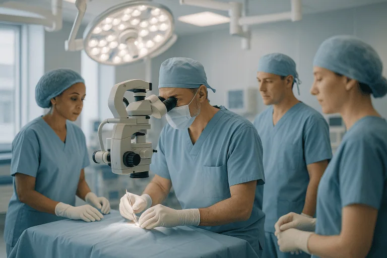
During surgery. SDR is performed under general anesthesia, meaning the patient is fully asleep and feels nothing. The patient lies face down. The surgeon makes an incision in the lower back and removes or temporarily lifts a small piece of bone from one or more vertebrae (a laminectomy or laminotomy) to reach the spinal canal. Many surgeons now use a single-level approach that limits the amount of bone removed. The protective covering of the spinal cord, called the dura, is opened, and the surgeon identifies the sensory (dorsal) nerve roots that serve the legs, keeping them separate from the motor roots that control muscle movement.
Each sensory root is then divided into several smaller bundles called rootlets. Using a technique called intraoperative electromyography (EMG), which records electrical activity in the leg muscles, the surgeon stimulates each rootlet with a tiny electrical current and watches the muscle response. Rootlets that produce an abnormal, spreading, or sustained response are considered to be contributing most to spasticity and are cut. Rootlets with normal responses are preserved. Typically only a portion of the rootlets is cut, and the exact proportion is tailored to the individual. The dura is then closed with fine sutures, the bone may be replaced, and the skin is closed.
After surgery. The patient is moved to a recovery area and then to a ward or pediatric intensive care unit for monitoring. Pain control, often including medication delivered through a catheter or intravenous line during the first days, is an important part of early care. Most protocols require the patient to lie flat for a period of time to protect the dural closure and reduce the risk of spinal fluid leakage, after which sitting and gentle movement begin under the guidance of physical therapists.
Preparation for SDR surgery
Preparation begins weeks or months before the operation and is as much about the family and therapy team as about the patient.
- Multidisciplinary evaluation. Expect appointments with neurosurgery, pediatric neurology or rehabilitation medicine, orthopedics, and physical therapy. Brain and spine imaging may be ordered.
- Baseline measurements. Standardized tests of spasticity, strength, range of motion, and walking are usually recorded before surgery.
- Pre-surgical strengthening. Therapists often recommend a program in the weeks beforehand to build strength and endurance, which may make early recovery smoother.
- Medication review. Tell the team about all medicines and supplements. Some anti-spasticity medicines may be adjusted around the time of surgery; blood thinners and certain other drugs may need to be paused as instructed.
- Anesthesia instructions. Fasting rules before general anesthesia will be given and must be followed exactly.
- Planning for rehabilitation. Arrange local physical therapy for after discharge, discuss school or work absence, and think about home accessibility, equipment such as walkers or braces, and travel logistics if care is far from home.
- Emotional preparation. Children benefit from age-appropriate explanations. Many hospitals offer child-life specialists or pre-admission tours.
Recovery and aftercare after SDR surgery
SDR surgery recovery is a gradual process measured in months rather than days. The hospital stay itself often lasts about a week, though this varies by center and by how quickly pain is controlled and mobility resumes.
First days. Patients typically lie flat for a couple of days. Pain, muscle spasms, and temporary changes in skin sensation such as tingling or heightened sensitivity in the legs are common during this period and are usually managed with medication and positioning. A urinary catheter is often used at first. Physical therapy usually starts with gentle range-of-motion exercises while still in bed.
First weeks. Once upright, many patients feel weak and unsteady. This is expected: the stiffness that previously provided some support is gone, and muscles that were never able to work normally must now learn to. Sitting balance, transfers, and then supported standing are practiced progressively. Intensive physical therapy, often several sessions per week, generally begins soon after discharge. Braces (orthoses) and walking aids are commonly adjusted or newly prescribed.
First months. Improvements in walking pattern, posture, and endurance tend to appear gradually. Most programs recommend a high-intensity therapy schedule for at least six months, with continued therapy at a lower intensity thereafter. Return to school is often possible after several weeks, depending on comfort and stamina, but heavy play and sports are usually restricted for longer to protect the spine while it heals.
Longer term. Functional gains are often described as continuing for one to two years after surgery as strength and motor control develop. Regular follow-up with the surgical and rehabilitation teams is used to monitor spine health, hip development, and any need for orthopedic procedures.
Practical aftercare points include keeping the incision clean and dry until cleared, watching for signs of infection or fluid leakage, following lifting and bending restrictions, and maintaining the home exercise program between therapy sessions.
Risks and side effects
Every operation carries risk, and SDR is a procedure on the spinal cord’s nerve roots, so a frank discussion with the surgical team is essential. Possible risks and side effects include:
- Temporary sensory changes in the legs and feet, such as numbness, tingling, or oversensitivity to touch. These are common in the early weeks and usually settle, but occasionally persist.
- Weakness. Some degree of apparent weakness is expected because spasticity was masking it; rarely, true weakness may be greater than anticipated and functionally limiting.
- Bladder or bowel changes. Temporary difficulty with urination is fairly common; lasting bladder or bowel dysfunction is uncommon but is a recognized serious risk, since the nerve roots controlling these functions lie near those treated.
- Cerebrospinal fluid leak from the dural closure, which may cause headache and sometimes requires further treatment.
- Infection of the wound or, rarely, meningitis (infection of the membranes around the spinal cord).
- Pain, including back pain and muscle spasms in the first weeks; long-term back pain is reported in some patients.
- Spinal problems later in life. Because bone is removed from the spine, there is a possible increased risk of spinal deformity such as scoliosis or excessive curvature, particularly with multilevel approaches. Ongoing monitoring is recommended.
- Hip or foot changes. Altered muscle balance may unmask or influence joint problems, and orthopedic procedures may still be needed later.
- General surgical and anesthetic risks, including bleeding, blood clots, and reactions to anesthesia.
- Unmet expectations. SDR reduces spasticity but does not cure cerebral palsy or restore normal movement. Outcomes depend heavily on rehabilitation and on the individual’s underlying abilities.
Results and outlook
The evidence base for selective dorsal rhizotomy in cerebral palsy includes randomized trials and long-term follow-up studies spanning several decades. Taken together, these studies generally show that SDR combined with intensive physical therapy produces a lasting reduction in lower-limb spasticity and that, for well-selected children, improvements in walking pattern, range of motion, and gross motor function tend to be greater than with therapy alone. Many families also report easier daily care, more comfortable positioning, and improved tolerance of braces.
Long-term studies following patients into adolescence and adulthood suggest that the reduction in spasticity is durable and that functional gains are often maintained, although some decline in walking ability with age can occur in cerebral palsy regardless of treatment. The need for later orthopedic surgery appears to be reduced in some reports but is not eliminated.
Outcomes vary widely. Children who walk independently before surgery tend to see refinements in gait quality and endurance; children who walk with support may gain efficiency or independence in some settings; and for those who are not walking, the goals are usually comfort, positioning, ease of care, and prevention of contractures rather than walking. Your doctor may use standardized classification systems to help set realistic, individualized expectations.
It is important to understand that results depend on committed rehabilitation. The operation removes a barrier; the therapy builds the new abilities.
Cost considerations
Selective dorsal rhizotomy cost varies considerably between countries, hospitals, and individual cases, and this page does not provide figures. The main factors that influence the overall expense include:
- Pre-operative assessment, including specialist consultations, imaging such as MRI, and formal gait analysis.
- The operation itself, covering surgeon and anesthesia fees, operating room time, and the intraoperative nerve monitoring equipment and staff that are central to the procedure.
- Hospital stay, which is usually several days and may include time in an intensive care unit.
- Rehabilitation, which is often the largest ongoing element: many months of intensive physical therapy, plus braces, walking aids, or other equipment that may need replacement as a child grows.
- Follow-up care, including repeat imaging, spine and hip monitoring, and any additional orthopedic procedures.
- Travel and accommodation if treatment takes place far from home, especially given the length of the rehabilitation period.
Insurance coverage and public health funding rules differ widely. Families are usually advised to obtain an itemized estimate and written information about what is and is not included before proceeding.
Frequently asked questions
What is the best age for selective dorsal rhizotomy?
Many centers consider children between roughly two and ten years old to be in the most favorable window, because spasticity has become clearly established but fixed contractures and bone deformities are usually not yet severe, and the nervous system is still highly adaptable. That said, older children, adolescents, and some adults have been treated with reported benefit. Age is only one factor; movement pattern, strength, and rehabilitation capacity matter more.
Is SDR surgery a cure for cerebral palsy?
No. Cerebral palsy results from a permanent brain injury, and SDR does not repair the brain. The operation permanently reduces spasticity in the legs, which can make movement easier and therapy more effective, but coordination, balance, and strength still depend on the underlying condition and on ongoing rehabilitation.
How long does sdr surgery recovery take?
The hospital stay is often about a week. Many patients return to school or light daily routines after several weeks, but the meaningful recovery period is much longer: intensive physical therapy typically continues for at least six months, and functional improvements are often described as continuing for one to two years. Timelines vary with age, pre-surgical function, and how consistently therapy is followed.
How is selective dorsal rhizotomy for cerebral palsy different from botulinum toxin injections or a baclofen pump?
Botulinum toxin injections temporarily weaken specific spastic muscles and wear off after a few months. A baclofen pump delivers anti-spasticity medicine continuously into the spinal fluid and can be adjusted or removed, but requires a device and refills. SDR is a one-time operation with permanent effect and no implanted hardware. Each option suits different patterns of spasticity and different goals, and some patients receive more than one over their lifetime.
Does SDR surgery affect the arms or upper body?
The nerve roots treated in a standard SDR serve the legs, so the direct effect is on lower-limb spasticity. Some families report modest secondary improvements in trunk control or arm use, likely related to better posture and easier movement, but these are not the primary aim and cannot be guaranteed.
What drives selective dorsal rhizotomy cost?
The total cost is shaped mainly by the pre-operative assessment, the operation with its specialized nerve monitoring, the length of hospital stay, and especially the many months of intensive rehabilitation, braces, and follow-up that follow. Insurance and funding arrangements differ between regions, so an itemized estimate from the treating hospital is the only reliable guide.
Can adults have SDR surgery?
Some adults with spastic cerebral palsy or other causes of lower-limb spasticity have undergone SDR, and reports suggest it can reduce spasticity and pain in selected cases. The evidence in adults is more limited than in children, and considerations such as fixed contractures, existing spine problems, and rehabilitation capacity are weighed carefully.
When to see a doctor
Anyone living with spasticity that limits movement, causes pain, interferes with sleep or daily care, or seems to be leading to tightening joints should be assessed by a specialist team experienced in movement disorders. In children with cerebral palsy, signs that warrant a specialist review include worsening stiffness, increasing difficulty with walking or transfers, toe-walking or scissoring legs that is progressing, growing difficulty with hygiene or dressing because of tight muscles, and reduced tolerance of braces. A multidisciplinary clinic can help determine whether SDR, another treatment, or a combination is most appropriate.
After SDR surgery, some symptoms need urgent medical attention:
- Fever, chills, or increasing redness, swelling, warmth, or discharge at the incision.
- Clear fluid leaking from the wound, or a severe headache that is worse when sitting up and eases when lying flat, which may indicate a spinal fluid leak.
- New or worsening weakness or numbness in the legs, or loss of movement that was present before.
- Inability to pass urine, new incontinence, or loss of bowel control.
- Severe or rapidly worsening back or leg pain not controlled by prescribed medication.
- Stiff neck, sensitivity to light, drowsiness, or confusion, which can be signs of meningitis.
- Calf pain or swelling, chest pain, or shortness of breath, which may indicate a blood clot.
Non-urgent but important reasons to contact the treating team include persistent sensory changes beyond the expected early period, difficulty keeping up with the therapy program, new back curvature, or changes in hip position noted by therapists. Regular scheduled follow-up remains essential for years after the procedure.
Preparation
- Expect a multidisciplinary evaluation with neurosurgery, neurology or rehabilitation medicine, orthopedics, and physical therapy, often including imaging and gait analysis. Follow fasting instructions for general anesthesia and tell the team about all medicines, since some may need adjusting. Arrange post-discharge physical therapy, school or work absence, and any home equipment in advance.
Aftercare
- Follow lying-flat and activity restrictions in the first days, and keep the incision clean and dry until cleared. Begin the prescribed physical therapy program promptly and maintain daily home exercises; braces and walking aids are commonly adjusted. Attend all follow-up visits for spine and hip monitoring, and report fever, wound leakage, new weakness, or bladder changes without delay.
Medically reviewed by the Acıbadem International Medical Board September 8, 2026
See our medical review board →
Update history
- PublishedSeptember 8, 2026
- Medical review approvedSeptember 8, 2026
- Last content updateSeptember 8, 2026
References3
From the Acibadem Blog

Who Is a Candidate for Selective Dorsal Rhizotomy? What the Multidisciplinary Team Assesses
Selective dorsal rhizotomy is usually considered for children with spastic cerebral palsy, most often the diplegic pattern affecting both legs, who walk independently or…

Selective Dorsal Rhizotomy or a Baclofen Pump for Spasticity: How Do the Approaches Differ?
Selective dorsal rhizotomy (SDR) and an intrathecal baclofen pump both reduce spasticity but in opposite ways. SDR is a one-time spinal surgery that permanently…

How Selective Dorsal Rhizotomy Works: Nerve Monitoring and Choosing Which Rootlets to Cut
Selective dorsal rhizotomy (SDR) treats spasticity by cutting a portion of the sensory nerve rootlets in the lower spine that drive overactive stretch reflexes.…