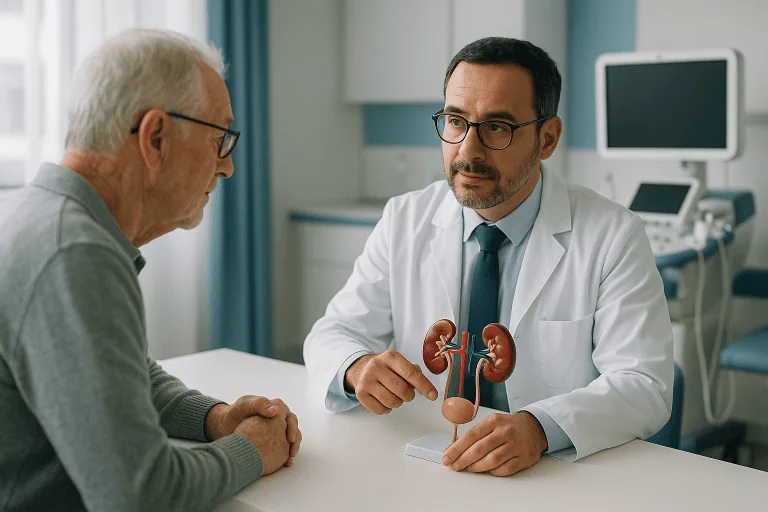
Urology Diagnostic Methods
Urology diagnostic methods assess urinary and male reproductive system symptoms using urine tests, blood tests, ultrasound, imaging, uroflowmetry, cystoscopy, or biopsy when needed.

Quick answer
Urology diagnostic methods are the tests used to find the cause of urinary and male reproductive symptoms. They range from urine and blood analysis and ultrasound to CT or MRI scans, uroflowmetry, urodynamic studies, cystoscopy and biopsy. A urologist selects tests step by step, starting with the least invasive, to reach a reliable diagnosis and guide treatment.
Urology Diagnostic Methods: What They Are and Why They Matter
Urology diagnostic methods are the tests and examinations used to find the cause of symptoms in the urinary system and the male reproductive system. They range from simple urine and blood analysis to ultrasound, CT and MRI scans, functional tests such as uroflowmetry and urodynamics, endoscopic examination of the bladder, and tissue biopsy. They exist for anyone with urinary or reproductive symptoms, an abnormal test result, or a finding on imaging that needs to be explained.
Urinary and male reproductive symptoms can be difficult to talk about, even with a physician. Frequent urination, blood in the urine, pelvic discomfort, erectile or ejaculatory problems, kidney stone pain, urinary leakage, weak urine flow, recurrent infections, infertility concerns or an abnormal prostate test all raise questions that feel personal and often urgent. On top of the symptoms themselves, there is usually another layer of uncertainty: how extensive the evaluation will be, whether invasive testing is really necessary, and how quickly a clear plan can be made.
The purpose of urology diagnostic methods is not to “run tests” for their own sake. It is to identify the underlying cause of your symptoms, estimate how serious the condition is, and guide treatment choices that fit you as an individual. In some cases a urine test and an ultrasound are enough. In others, advanced imaging, flow measurement, cystoscopy, prostate assessment, urodynamic testing or biopsy is needed before anyone can honestly say what is wrong.
Accuracy matters because many urological conditions look alike at first. Urinary frequency can be caused by infection, prostate enlargement, an overactive bladder, diabetes, stones, neurological disease or, less commonly, a tumour. Blood in the urine may come from a stone or an infection, but it can also be an early sign of bladder, kidney or prostate disease. A weak urinary stream may reflect benign enlargement of the prostate, narrowing of the urethra, weakness of the bladder muscle, or a combination of problems. The right diagnostic pathway protects you from both directions of error: it helps avoid undertreatment of something serious and unnecessary procedures for something benign.
At Acibadem, diagnosis in urology is treated as a structured clinical process rather than a menu of tests. Physicians combine a detailed medical history, physical examination, laboratory analysis, imaging, functional testing and, when indicated, endoscopic or tissue-based evaluation. Complex cases may be reviewed with specialists in radiology, pathology, oncology, nephrology, gynaecology, endocrinology or reproductive medicine, depending on the condition. This multidisciplinary model matters most for patients who arrive with incomplete or conflicting results and want one coherent assessment rather than a stack of unrelated reports.
What Are Urology Diagnostic Methods?
Urology diagnostic methods are medical tests and examinations used to evaluate the urinary system and the male reproductive system. The urinary system includes the kidneys, ureters, bladder, urethra and, in men, the prostate. The male reproductive system includes the testes, epididymis, vas deferens, seminal vesicles, prostate and penis. Because these organs are closely connected, a symptom in one area may reflect a problem elsewhere in the system, which is exactly why the evaluation is planned as a whole rather than test by test.
What happens at a first urology consultation?
A first urology consultation is a structured conversation and examination, not a battery of tests. The urologist asks about your symptoms, their timing, pain, urinary habits, sexual function, previous infections, stone history, medications, surgeries, family history and relevant lifestyle factors. A physical examination may include abdominal, genital, pelvic or prostate assessment depending on the concern. Only then does the physician select tests, based on the most likely causes and on how urgent the situation appears to be. A consultation that starts with listening tends to end with fewer, better-chosen tests.
Which tests are used most often?
The most frequently used urology diagnostic methods are urine tests, blood tests and ultrasound; more specialised tests are added only when they answer a specific question. Urine tests detect infection, blood, protein, crystals or abnormal cells. Blood tests evaluate kidney function, inflammation, hormones and prostate-related markers. Ultrasound assesses the kidneys, bladder, prostate, testes and the urine left behind after voiding. Cross-sectional imaging, computed tomography or magnetic resonance imaging, is used when more detailed anatomy is needed. Functional tests such as uroflowmetry measure how well urine passes, while urodynamic studies evaluate how the bladder stores and empties. Cystoscopy allows direct inspection of the urethra and bladder with a thin camera. Biopsy is performed when a tissue diagnosis is needed, most commonly for suspected prostate, bladder, kidney or testicular disease.
Not every patient needs every test. A well-run evaluation is selective, evidence-based and focused on a defined clinical question. A young patient with uncomplicated urinary symptoms may need urine testing and a targeted ultrasound and nothing more. An older patient with visible blood in the urine usually requires urine cytology, imaging of the whole urinary tract and cystoscopy, because intermittent bleeding can be the first sign of something that imaging alone would miss. A man with an elevated prostate-specific antigen may need repeat blood testing, prostate imaging, formal risk assessment and, only if appropriate, biopsy. The pathway is adapted to the patient rather than applied as a fixed checklist.
Who May Need a Urology Diagnostic Evaluation?
You may need urology diagnostic methods when symptoms suggest a disorder of the kidneys, bladder, prostate, urethra, testes or male reproductive tract. Some people seek evaluation because symptoms are disruptive but not severe, night-time urination that ruins sleep, leakage that limits daily life, discomfort that comes and goes. Others need prompt assessment because their symptoms may indicate infection, obstruction, bleeding or cancer. In both situations, early clarity reduces anxiety and makes decisions easier.
What symptoms should prompt a urological assessment?
The symptoms that most often lead to urological testing are changes in urination, pain in the urinary tract or genitals, and blood in the urine. Typical urinary symptoms include frequent urination, waking at night to urinate, urgency, urinary leakage, burning or pain during urination, difficulty starting the stream, weak or interrupted flow, a feeling that the bladder does not empty fully, or inability to urinate at all. Pain may occur in the side, lower back, groin, pelvis, testicle, penis or lower abdomen. Blood in the urine, whether visible or found only on laboratory testing, is always a reason for medical evaluation, even if it appears once and then disappears.
Men may also need assessment for prostate-related concerns, erectile dysfunction, premature ejaculation, penile curvature, symptoms of low testosterone, infertility, testicular pain or swelling, an abnormal semen analysis, or a mass in the scrotum. Each of these has its own diagnostic pathway, and several of them overlap: a hormonal problem, for example, can affect both sexual function and fertility, which is why the initial history is taken so carefully.
Do women need urology diagnostic tests?
Yes, urology is not a male-only specialty, and many diagnostic pathways apply equally to women. Women are commonly referred to urology for recurrent urinary tract infections, urinary incontinence, bladder pain, kidney stones, blood in the urine, or suspected structural problems of the urinary tract. The same core methods apply, urine analysis, culture, ultrasound, imaging, functional testing and, when needed, cystoscopy, with the examination and interpretation adapted to female anatomy. Conditions that sit at the border between urology and gynaecology are covered in more depth on the female urology page.
Diagnosis begins by matching symptoms with objective findings. A urine analysis may reveal infection, microscopic bleeding, crystals or inflammation. A urine culture identifies the bacteria involved and shows which antibiotics they respond to. Blood tests may show impaired kidney function, raised inflammatory markers, hormonal imbalance or a change in prostate markers. Ultrasound can reveal stones, swelling of the kidney, changes in the bladder wall, prostate size, testicular abnormalities or urine remaining after voiding. When these first-line results suggest something more complex, the urologist recommends further imaging, endoscopy or tissue sampling, in that order of invasiveness wherever possible.
When is a second opinion worthwhile?
A second opinion is most useful when a significant decision hangs on a single test result or a single interpretation. Common examples include an elevated PSA, a proposed prostate biopsy, recurrent kidney stones, repeated infections that keep coming back despite treatment, persistent pelvic pain, unexplained infertility, or a previous diagnosis of a urological cancer. A second opinion does not automatically mean repeating everything. It may involve expert review of existing reports and images, repeating only selected tests, or adding more detailed diagnostic methods to refine the picture before treatment is chosen. Sometimes the outcome is a different plan; sometimes it is confirmation of the original one, and both are valuable.
Conditions and Indications Urology Diagnostic Methods Can Address
Urological diagnostic testing helps identify a broad range of benign, infectious, functional, reproductive and oncological conditions. Which tests are used depends on the clinical concern, the patient’s age and sex, medical history, and whether symptoms are acute or long-standing.
Which conditions can these tests identify?
The conditions most commonly evaluated with urological testing include urinary tract infections, recurrent cystitis, pyelonephritis, kidney stones, ureteral stones, bladder stones, an enlarged prostate, urinary retention, overactive bladder, urinary incontinence, urethral stricture, neurogenic bladder, chronic pelvic pain, prostatitis, erectile dysfunction, male infertility, varicocele, testicular disorders, and congenital or acquired abnormalities of the urinary tract. In most of these, the diagnosis rests on combining two or three complementary tests rather than any single result.
Diagnostic methods are also central to evaluating possible urological cancers. Blood in the urine leads to investigation for bladder, kidney, ureteral or prostate disease. Abnormal prostate tests may require further assessment for prostate cancer. Testicular swelling or a mass calls for timely ultrasound and laboratory evaluation. Kidney masses found incidentally on scans performed for other reasons need careful characterisation to determine whether surveillance, biopsy, ablation or surgery is the appropriate next step, and imaging quality makes a real difference to that judgement.
Urological testing in children and adolescents
In children and adolescents, urological diagnostics are used to evaluate urinary tract infections, bedwetting, an undescended testis, congenital kidney or ureter problems, reflux of urine back towards the kidneys, urinary obstruction, or scrotal swelling. Paediatric evaluations are adapted to the child’s age, comfort and family circumstances. Wherever possible, the least invasive test that answers the question is chosen first, and parents are involved in each step of the plan.
Testing when you live with a chronic condition
If you have diabetes, a neurological disorder, a spinal cord injury, kidney disease or a history of pelvic surgery, urological testing can clarify how your bladder and urinary tract are actually functioning, which is not always obvious from symptoms alone. Some patients lose bladder sensation or develop incomplete emptying without noticing anything wrong. In these situations, functional studies and imaging can detect problems before they cause infections, stones or kidney strain. Because bladder function is closely linked to the nervous system, findings are sometimes reviewed together with colleagues in neurology when a neurological cause is suspected.
How Urology Diagnostic Testing Is Performed
The diagnostic process is planned around your symptoms and the urgency of the situation. Many patients arrive with previous records: laboratory results, ultrasound reports, CT or MRI images, pathology reports or earlier treatment recommendations. Reviewing these documents before or during the first visit lets the team avoid unnecessary repetition and concentrate on the information that is still missing. This is one of the simplest ways the evaluation is kept efficient: not by testing faster, but by not testing twice.
How should you prepare for urology tests?
Preparation depends on the specific test, but the common thread is information: bring your medication list, allergy details, previous operation notes, imaging files, laboratory results and any pathology reports. It also helps to note the timing of your symptoms, your fluid intake pattern, night-time urination, pain episodes, fever, visible bleeding, sexual symptoms and previous antibiotic courses.
- Urine testing: you may be asked to provide a clean-catch urine sample. If infection is suspected, a culture may be collected before antibiotics are started when this is clinically appropriate.
- Ultrasound: the bladder may need to be partially or fully filled, particularly when the team is measuring bladder volume and post-void residual urine.
- CT or MRI: fasting, kidney function blood tests or contrast safety screening may be required. Tell the team in advance if you have kidney impairment, a contrast allergy, a possible pregnancy, an implanted device, or if you take blood thinners.
- Cystoscopy or biopsy: preparation is more specific. Urine testing may be needed beforehand to reduce infection risk. Any adjustment of blood thinners is coordinated by the physician who prescribes them, never make changes on your own. Biopsy planning may involve imaging review, an antibiotic strategy, an anaesthesia discussion and a clear explanation of expected after-effects such as mild bleeding or urinary discomfort.
What does a urine test show?
A urine analysis shows whether there is blood, infection, inflammation, protein, glucose or crystal formation in the urine: a remarkable amount of information from a single sample. It is one of the most frequently used tests in urology because so many conditions leave a trace in the urine: infection, stones, kidney disease and inflammation among them. A urine culture goes a step further, identifying the organism causing an infection and showing which treatment will work against it. Urine cytology examines the sample for abnormal cells and is used mainly in patients being evaluated for blood in the urine or bladder cancer risk.
What is a PSA test and what does the result mean?
Prostate-specific antigen (PSA) is a blood marker used to assess prostate cancer risk: it is not, by itself, a cancer diagnosis. A raised PSA must be interpreted in context: age, prostate size, recent infection, recent procedures, certain medications, family history and the trend of previous PSA values all change what the number means. PSA can rise temporarily after infection, urinary retention, instrumentation or ejaculation, which is why timing matters and why a single elevated reading often leads to a repeat test rather than an immediate biopsy. Alongside PSA, blood testing in urology may include kidney function markers, electrolytes, blood count, inflammatory markers, testosterone and fertility-related hormones, depending on the question being asked.
What tests are used for male fertility evaluation?
Male fertility evaluation usually begins with a semen analysis, which measures sperm count, movement and shape along with semen volume and other physical characteristics. Because results vary naturally from sample to sample, an abnormal analysis is typically repeated after a short interval before conclusions are drawn. Depending on the findings, the urologist may add hormone blood tests, including testosterone and the pituitary hormones that regulate sperm production, a scrotal ultrasound to look for varicocele or structural abnormalities, and genetic testing in selected cases. The underlying goal is to distinguish problems of sperm production from problems of sperm transport, because the two lead to very different treatment options.
Ultrasound: the first-line imaging test
Ultrasound is usually the first imaging step in urology because it is non-invasive, widely available and uses no ionising radiation. It can evaluate the kidneys for swelling, cysts, masses or stones; the bladder for wall changes, stones or residual urine; the prostate for size and related obstruction; and the testes for masses, inflammation, torsion, varicocele or fluid collections. Many diagnostic journeys begin and end here: an ultrasound that confirms a straightforward finding can spare you every test that follows.
When is a CT or MRI scan needed in urology?
A CT or MRI scan is needed when ultrasound and laboratory tests cannot fully answer the clinical question, typically for stones, complex infections, suspicious masses or detailed treatment planning. Computed tomography is particularly useful for kidney and ureteral stones, trauma and detailed mapping of the urinary tract. Magnetic resonance imaging is used for prostate assessment, characterisation of kidney and pelvic findings, and soft-tissue detail. In selected cases, contrast-enhanced studies show blood supply, obstruction, tumours or anatomical relationships before a procedure is planned. How these scans are performed and interpreted in urological practice is explained in detail on the CT and MRI for urology page.
Good imaging makes the diagnosis specific. It can show whether a stone is likely to pass on its own, whether a kidney is obstructed, whether a prostate lesion needs further evaluation, or whether a mass looks benign or suspicious. But images are never read in isolation: they are interpreted together with your symptoms, laboratory results and physical examination, because the same picture can mean different things in different patients.
Other imaging and specialised tests
Beyond ultrasound, CT and MRI, urologists sometimes use additional studies to answer specific questions. A plain abdominal x-ray can track certain kidney stones over time. Nuclear medicine renography assesses how each kidney drains and how much each contributes to overall function, which is particularly useful when obstruction is suspected. Retrograde studies, performed during endoscopy, outline the ureters and the collecting system of the kidney with contrast when other imaging leaves doubt. And when a stone is retrieved or passed, laboratory analysis of its composition helps guide prevention, because different stone types call for different dietary and medical strategies. None of these tests is routine; each is chosen because it answers a question the earlier tests could not.
What is uroflowmetry?
Uroflowmetry is a simple, non-invasive test that measures the speed and pattern of your urination. You urinate into a specialised device, and the resulting flow curve helps distinguish obstruction from weak bladder contraction or an irregular voiding pattern. It is commonly used for men with enlarged prostate symptoms, patients with suspected urethral stricture, and anyone with a sense of incomplete emptying. The test itself takes only as long as urinating does; the main requirement is arriving with a comfortably full bladder.
Post-void residual measurement usually accompanies uroflowmetry. Using ultrasound, the team measures how much urine remains in the bladder after you have finished. A high residual may indicate obstruction, bladder muscle weakness, neurological dysfunction or a medication effect. This matters because persistent incomplete emptying quietly contributes to infections, stones and kidney stress over time.
What is urodynamic testing?
Urodynamic testing is a detailed functional study of how the bladder stores and releases urine, measuring pressure, capacity, sensation, contractions, leakage and coordination with the urinary sphincter. It is recommended for complex incontinence, neurological bladder problems, symptoms that persist after previous treatment, or urinary complaints that remain unexplained after simpler tests. The test can feel unfamiliar, because thin catheters are used to record pressures while the bladder fills and empties, but it provides functional information that no scan can supply. For a small group of patients, it is the single test that changes the treatment decision.
What is a cystoscopy and why is it done?
Cystoscopy is a direct examination of the urethra and bladder using a thin instrument with a camera. It can be performed with local anaesthetic gel in an outpatient setting, or with sedation or anaesthesia depending on your needs and whether a procedure is planned at the same time. Cystoscopy is used to investigate blood in the urine, recurrent infections, suspected bladder tumours, urethral narrowing, bladder stones, foreign bodies, or urinary symptoms that remain unexplained after the first round of testing.
During the examination, the physician inspects the lining of the bladder, identifies inflammation or suspicious areas, assesses the urethra, and can sometimes perform minor interventions or take samples in the same session. Afterwards you may notice temporary burning with urination, mild frequency or small amounts of blood; these usually settle within a short period, and the team explains beforehand what is expected and what is not.
When is a biopsy needed?
A biopsy is needed when only a tissue sample can confirm or exclude a disease, most often when cancer is a realistic possibility. In urology, biopsy may involve the prostate, bladder, kidney or testis. The most common scenario is prostate biopsy after risk assessment, PSA trend, examination findings and MRI, suggests that a tissue diagnosis is justified. Prostate MRI can identify specific areas that need targeted sampling, and modern biopsy planning often combines those targeted samples with systematic ones to improve diagnostic accuracy.
Bladder biopsy is usually taken during cystoscopy if an abnormal area is seen. Kidney biopsy is less common in cancer evaluation but is considered for selected renal masses or medical kidney disease, often with nephrology input. Testicular biopsy is used in certain infertility evaluations or when tissue information is clinically necessary. In every case, the pathology report, not the biopsy itself, is the product: it determines what the tissue shows, and treatment recommendations rest on it.
How long does urological testing take?
Basic urine and blood tests are completed quickly, often on the day of the consultation, while imaging, endoscopy and biopsy take longer to schedule, perform and interpret. Uroflowmetry takes only minutes once the bladder is full. Cystoscopy itself is brief, although preparation and observation add time. Biopsy duration depends on the organ involved, the anaesthesia plan and recovery monitoring, and pathology results take additional days because tissue must be processed and examined properly. The practical aim is to sequence the evaluation efficiently, grouping compatible tests together where possible, while still allowing enough time for careful interpretation and an unhurried discussion of the results.
Why Acting Early Matters
Many urological conditions are easier to manage when they are identified early. A urinary tract infection evaluated promptly can usually be treated before it spreads to the kidneys. A stone causing obstruction may need timely treatment to prevent infection or kidney damage. Persistent urinary retention stretches the bladder and can impair its function over time. Blood in the urine deserves evaluation even if it appears only once, because serious conditions are often intermittent in their early stages: the bleeding stops, but the cause does not go away.
Delay also complicates treatment decisions. Recurrent infections can become resistant to commonly used antibiotics when the underlying cause is never found. Long-standing prostate obstruction contributes to bladder changes, stones, infections and kidney strain. Untreated testicular torsion, an emergency, can threaten the viability of the testicle within hours. A suspicious mass may become harder to treat if diagnosis is postponed.
It is worth stating the other side plainly: early evaluation does not mean aggressive treatment. In many cases the outcome of a thorough diagnostic work-up is reassurance, monitoring, lifestyle guidance, medication or a minimally invasive option. The value lies in knowing what is happening and whether action is needed at all. Early planning has a practical benefit too: it avoids fragmented testing across different providers and lets diagnostics connect directly to treatment if treatment turns out to be necessary.
Benefits of a Structured Urology Diagnostic Evaluation
A well-planned diagnostic pathway moves you from uncertainty to a clear, medically grounded plan. The table below summarises what a structured approach actually delivers.
| Benefit | What It Means for You |
|---|---|
| More accurate diagnosis | Tests are selected to clarify the cause of symptoms rather than relying on assumptions or incomplete information. |
| Appropriate treatment planning | Medication, observation, procedures, surgery or cancer care can be recommended based on objective findings. |
| Reduced unnecessary procedures | When the diagnosis is clear, you may avoid treatments that are unlikely to help your specific condition. |
| Earlier detection of serious disease | Warning signs such as blood in the urine, abnormal imaging or elevated risk markers are investigated before symptoms progress. |
| Better follow-up decisions | Baseline results let physicians monitor change over time and decide when further testing or treatment is needed. |
Recovery and What to Expect After Testing
Most diagnostic tests in urology require little or no recovery time. More invasive tests, cystoscopy and biopsy in particular, involve temporary symptoms and short-term precautions, which the team explains before the procedure rather than after it.
| Time Period | What Patients Can Expect |
|---|---|
| Day 1 | After urine tests, blood tests, ultrasound or uroflowmetry, most patients return to normal activities immediately. After cystoscopy or biopsy, mild urinary discomfort, light bleeding or temporary pelvic soreness may occur. |
| First week | Results are reviewed as they become available. Patients who had a biopsy receive specific instructions about activity, hydration, medications and follow-up. |
| First month | The urologist discusses the diagnosis, treatment options and follow-up plan. Additional tests may be recommended if findings are unclear or a complex condition is identified. |
| Longer term | Some conditions require surveillance: repeated urine tests, imaging, PSA monitoring, stone prevention evaluation, bladder function follow-up or coordinated care with other specialists. |
One point deserves emphasis: “after testing” is when the real work of diagnosis happens. Results are compared against each other and against your history, and a report that looks alarming in isolation may be entirely explained by context, or vice versa. Allow time for that interpretation. It is the difference between having test results and having a diagnosis.
Factors That Influence Diagnostic Accuracy and Outcomes
A good result in urology diagnostics means more than receiving a test report. It means the information is reliable, interpreted correctly, and translated into a plan that fits your medical condition and your priorities. Several factors influence how well that works.
Completeness of medical history. Symptoms such as bleeding, pain, urinary frequency, fever, weight loss, sexual dysfunction or fertility concerns mean different things depending on their timing and what accompanies them. Share previous infections, stones, cancers, surgeries, radiation, catheter use, neurological disease, family history, smoking history and medication use, even details that seem unrelated. Diagnostic errors more often come from missing history than from missing technology.
Quality of imaging and laboratory interpretation. A small kidney stone, early obstruction, a bladder lesion, a prostate abnormality or a testicular finding may require careful imaging technique and experienced eyes to be recognised. Pathology review is particularly important when a biopsy has been performed, because treatment recommendations depend directly on the tissue diagnosis and its grading.
Choosing the right test at the right time. PSA can be temporarily elevated after infection, urinary retention, instrumentation or ejaculation, so timing and context matter. Contrast-enhanced imaging is highly informative in one patient and inappropriate in another with reduced kidney function. Cystoscopy is necessary for the evaluation of blood in the urine, but not every mild urinary symptom requires endoscopy. Sequencing tests sensibly is a clinical skill in its own right.
Coordination among specialists. A patient with blood in the urine and impaired kidney function needs both urology and nephrology input. A man with infertility may benefit from collaboration between urology, reproductive medicine and endocrinology. A patient with suspected cancer may need radiology, pathology, medical oncology, radiation oncology and surgery reviewing the findings together in a tumour board. Complex decisions are safer when they are shared.
Patient factors. Age, overall health, kidney function, pregnancy status, blood thinner use, infection risk, pain tolerance, anxiety and scheduling all influence which tests are appropriate and how they are performed. The team also weighs how quickly results are needed, whether treatment might follow during the same period of care, and how follow-up will be organised afterwards.
From Diagnosis to Treatment: How the Findings Are Used
The end point of the diagnostic process is not a report: it is a decision. Once the cause of your symptoms is established, the same findings that made the diagnosis shape the treatment: how large a stone is and where it sits, how much a prostate obstructs, how a bladder stores and empties, what a biopsy shows and how it is graded. This is why diagnostic quality and treatment quality are inseparable.
For many conditions, an accurate diagnosis opens the door to less invasive treatment rather than more. Precise imaging and functional testing allow procedures to be planned through minimally invasive urology techniques, including laparoscopic approaches, where these suit the condition. In selected cases, particularly in prostate and kidney surgery, detailed pre-operative imaging supports planning for robotic urology procedures. None of this replaces clinical judgement; it depends on it. The technology is only as useful as the diagnosis behind it.
At Acibadem, urology diagnostics may draw on high-resolution ultrasound, advanced cross-sectional imaging, functional urinary testing, endoscopic assessment, targeted biopsy planning and detailed pathology evaluation when indicated. Findings in complex cases can be reviewed across departments, laboratory medicine, radiology, anaesthesia, pathology, outpatient clinics, endoscopy units and, when needed, surgical or oncology services, so that each part of the evaluation contributes to one final clinical decision rather than sitting in a separate file. Patients with suspected or confirmed cancer may be discussed in tumour boards where urologists, radiologists, pathologists, medical oncologists, radiation oncologists, nuclear medicine specialists and other experts review the findings together. Patients with stone disease, urinary dysfunction, male infertility or complex infection receive focused specialist input in the same way.
The practical side of the diagnostic journey is organised alongside the medical side: appointment coordination, preparation guidance and review of existing medical records. Every patient’s timeline depends on which tests are actually required, so the honest promise is not speed for its own sake: it is an evaluation that is organised, understandable and thorough.
Personalised treatment starts with personalised diagnosis. A patient with urinary frequency does not need the same pathway as a patient with kidney stones or a man with an elevated PSA. Someone seeking a second opinion may need expert review of existing images rather than a full repeat of every test. A patient who is anxious about cystoscopy or biopsy may need more explanation, anaesthesia options or a staged, step-by-step approach to deciding. Matching the diagnostic plan to the medical question, and to the person asking it, is the standard the whole process is built around.
Moving From Uncertainty to a Clear Plan
Urological symptoms affect daily life, relationships, sleep, work and emotional wellbeing. They also create a particular kind of uncertainty, especially when symptoms come and go, or when previous testing has produced results without answers. A careful diagnostic evaluation resolves that uncertainty: it identifies what is causing the problem, how serious it is, and which options exist.
The same logic applies whether the starting point is a new symptom, an abnormal test result, recurrent infections, blood in the urine, stone pain, a prostate concern or a possible cancer diagnosis. In each case, the appropriate combination of diagnostic methods, chosen deliberately, sequenced sensibly and interpreted in context, is what turns a worrying question into a workable plan. That plan may be reassurance and routine follow-up. It may be medication, a procedure or multidisciplinary treatment. Whatever it turns out to be, it starts with a diagnosis you can trust.
Preparation
- Preparation depends on the planned test and your symptoms. You may be asked to provide urine or blood samples, arrive with a full bladder for ultrasound, or stop certain medications before invasive tests. Bring previous scans, lab results, and a list of current medications.
Aftercare
- Most urology diagnostic tests allow same-day return to normal activities. After cystoscopy or biopsy, mild burning, spotting, or temporary discomfort may occur, and your doctor may recommend fluids, medication, or short activity restrictions. Follow-up is arranged to review results and plan treatment if needed.
Frequently Asked Questions
What affects the cost of urology diagnostic methods?
The final cost depends on the symptoms being investigated, the type and number of tests required, whether imaging or cystoscopy is needed, pathology requirements, anesthesia use, and whether follow-up consultation is included. A personalised plan can be prepared after reviewing your medical history and current complaints.
How can I get a personalised quote from Acibadem?
You can request a free consultation and share your symptoms, previous test results, imaging reports, medication list, and any referral notes. The international patient team can then coordinate with the relevant urology specialist to outline the recommended diagnostic pathway and provide a personalised quote.
Are all urology tests included in a diagnostic package?
Not always. A package may include the initial consultation and commonly required tests, while advanced imaging, cystoscopy, biopsy, pathology, anesthesia, or additional specialist reviews may be added if the urologist considers them necessary.
Can diagnostics be completed during a short visit to Turkey?
Many routine urology evaluations can be coordinated efficiently, but the exact timeline depends on appointment availability, preparation requirements, the need for imaging or endoscopy, and whether pathology results are required. The care team can advise on a realistic schedule before travel.
Do I need to repeat tests already done in my home country?
Recent and clear results may help avoid unnecessary repetition, but the specialist may request repeat or additional tests if results are outdated, incomplete, inconsistent with symptoms, or not sufficient for safe decision-making.
Is this information medical or financial advice?
No. This is general educational information. A urology specialist should decide which diagnostic methods are suitable, and a personalised quote should be requested through a free consultation before making travel or financial decisions.
Medically reviewed by the Acıbadem International Medical Board September 13, 2026
See our medical review board →
Update history
- PublishedJune 8, 2026
- Medical review approvedSeptember 13, 2026
- Last content updateSeptember 12, 2026
References1
- Urodynamic Testing my.clevelandclinic.org
Trusted care for international patients
From the Acibadem Blog

When Urinary Symptoms Need a Urologist: Which Diagnostic Tests Usually Come First
Most people can start with their primary care clinician, who usually orders a urinalysis and, if needed, a urine culture and basic kidney blood…