Craniosynostosis Surgery
Craniosynostosis surgery treats the premature closure of skull sutures in infants and young children, a condition that can restrict brain growth and cause an abnormal head shape. Depending on age and severity,…
Quick answer
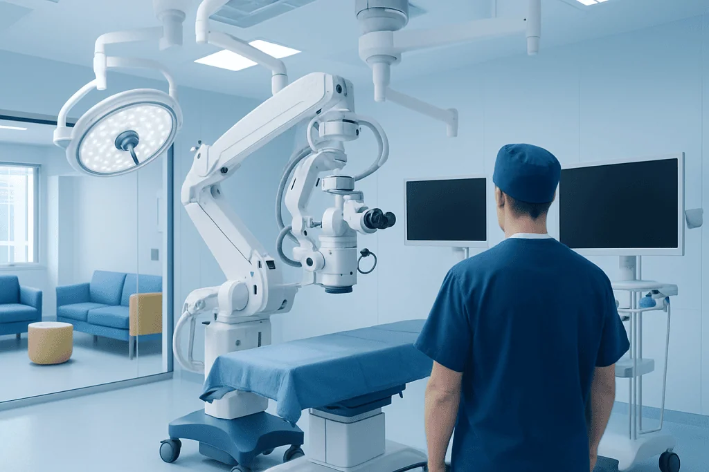
Craniosynostosis surgery corrects the early fusion of one or more skull sutures in infants, giving the brain room to grow and improving head shape. It is done under general anesthesia using either an endoscopic strip craniectomy followed by helmet therapy in young infants, or open cranial vault remodeling for older children and complex cases.
What is craniosynostosis surgery?
Craniosynostosis surgery is an operation on the skull of an infant or young child to correct a condition called craniosynostosis. Craniosynostosis happens when one or more of the sutures (the flexible, fibrous joints between the bony plates of a baby’s skull) close too early. Normally these sutures stay open during the first years of life so the skull can expand as the brain grows quickly. When a suture fuses early, the skull cannot grow in that direction, so it grows more in other directions instead. This produces an unusual head shape and, in some cases, may limit the space available for the growing brain.
The goals of craniosynostosis surgery are to release the fused suture, allow the brain enough room to grow, and restore a more typical head and face shape. The operation is usually planned and carried out by a team that includes a pediatric neurosurgeon (a surgeon who operates on the brain, skull, and nervous system in children) and a craniofacial plastic surgeon (a surgeon who specializes in the bones and soft tissues of the head and face). In hospital groups such as Acibadem, this care is typically coordinated through the neurosurgery department together with plastic surgery and pediatrics.
Two broad approaches are used:
- Endoscopic craniosynostosis surgery: a minimally invasive technique that uses small incisions and a thin camera (an endoscope) to remove a strip of bone along the fused suture. It is usually followed by several months of wearing a custom-molded helmet that guides skull growth.
- Cranial vault remodeling: an open operation, through a longer incision across the top of the scalp, in which the surgeon removes, reshapes, and repositions sections of skull bone to correct the head shape directly.
Craniosynostosis surgery is used for non-syndromic craniosynostosis (fusion of a single suture with no other associated condition) and for syndromic craniosynostosis, in which the early suture closure is part of a genetic syndrome such as Apert, Crouzon, or Pfeiffer syndrome and may involve several sutures and the facial bones.
Who is a candidate
Not every baby with an unusual head shape needs surgery. The most common cause of a flattened or asymmetric head is positional plagiocephaly (flattening caused by pressure from lying in one position), which does not involve a fused suture and is not treated with surgery. A specialist distinguishes between the two through physical examination and, when needed, imaging such as a CT scan (a detailed X-ray-based picture of the skull bones).
Craniosynostosis surgery is generally considered when:
- Imaging or clinical examination confirms that one or more sutures have fused early.
- The head shape is noticeably abnormal and expected to become more pronounced as the child grows.
- There are signs or concerns about raised pressure inside the skull (intracranial pressure), which can occur when the skull restricts brain growth, particularly when several sutures are involved.
- The child has a craniosynostosis syndrome, in which surgery is often part of a longer, staged treatment plan.
The timing and type of surgery depend heavily on the child’s age. Endoscopic craniosynostosis surgery is usually offered only to young infants, most often under about six months of age, because it relies on the rapid brain growth of early infancy and helmet therapy to reshape the skull. Older infants and children are generally treated with open cranial vault remodeling.
Surgery may not be suitable, or may be delayed, when:
- The head shape difference is due to positional flattening rather than a fused suture.
- The fusion is very mild with minimal cosmetic or functional impact, in which case observation may be recommended.
- The child has other serious medical problems that make general anesthesia or a longer operation unsafe at that time.
- The family cannot commit to the frequent follow-up and helmet adjustments required after the endoscopic approach, in which case an open procedure may be preferred instead.
The decision is individualized. Your child’s surgical team will explain which option they recommend and why.
How the procedure works
Before the operation
Once craniosynostosis is confirmed, the team reviews imaging to identify which suture or sutures are fused and plans the approach. A pediatric anesthesiologist (a doctor who specializes in anesthesia for children) assesses the child’s general health. Blood tests are usually taken, including a blood type and crossmatch, because blood transfusion may be needed, especially during open surgery. For an endoscopic procedure, arrangements are often made in advance for the child to be measured for a molding helmet.
During the operation
Both approaches are performed under general anesthesia (the child is fully asleep and feels nothing). Steps differ by technique:
- Endoscopic craniosynostosis surgery: The surgeon makes one or two small incisions in the scalp near the fused suture. Using the endoscope for visualization, a narrow strip of bone along the fused suture is removed, separating the bony plates so they can grow apart. The procedure is relatively short, and blood loss is typically lower than with open surgery.
- Cranial vault remodeling: The surgeon makes a longer, wavy incision across the top of the scalp from ear to ear, usually placed so it is hidden by hair. Sections of the skull are carefully removed, reshaped, and repositioned to create a more typical contour and to give the brain room. The bone pieces are secured with small plates and screws made of resorbable (dissolving) material or with sutures. In some cases, the forehead and the upper rims of the eye sockets are also advanced or reshaped, particularly for fusion of the sutures at the front of the skull.
After the operation
The child is taken to a recovery area and then usually to a pediatric intensive care unit or a monitored ward for the first night, depending on the type of surgery and the hospital’s practice. After open surgery, swelling of the face and eyelids is expected and often peaks in the first two to three days. Pain is managed with age-appropriate medication. After endoscopic surgery, the molding helmet is typically fitted within the first one to two weeks and worn for most of the day for several months, with regular adjustments as the head grows.
Preparation for craniosynostosis surgery
Preparation is guided by the surgical and anesthesia team and generally includes the following:
- Medical assessment: A general health check, review of any other conditions, and blood tests. Some children also have an eye examination to check for signs of raised pressure inside the skull.
- Imaging: A CT scan, sometimes with three-dimensional reconstruction, is commonly used to map the sutures and plan the operation. Some centers use ultrasound or low-dose imaging protocols where appropriate.
- Fasting instructions: You will be told when to stop breast milk, formula, and other feeds before anesthesia. Following these instructions precisely is important for safety.
- Illness check: Surgery is often postponed if the child has a cold, fever, or chest infection, because these raise anesthesia risks.
- Medications and supplements: Tell the team about anything your child takes, including vitamins or herbal products.
- Helmet planning: For the endoscopic approach, an appointment with an orthotist (a specialist who makes and fits medical devices such as helmets) is usually arranged before or shortly after surgery.
- Practical planning: Arrange time off work, care for siblings, and transportation, since a hospital stay of several days is common after open surgery.
Many parents find it helpful to ask the team in advance what the child will look like immediately after surgery, since facial swelling and a large head dressing can be alarming if unexpected.
Recovery and aftercare
Craniosynostosis surgery recovery varies with the technique used, the child’s age, and how many sutures were treated. The following is a general guide; your child’s team will give specific instructions.
In the hospital
- After endoscopic surgery, many children stay one or two nights and go home once they are feeding well and pain is controlled.
- After cranial vault remodeling, a stay of roughly three to five nights is common. The first night is often spent in intensive care for close monitoring.
- Swelling around the eyes can be significant after open surgery and may temporarily prevent the child from opening the eyes; this typically improves within several days.
- Blood transfusion may be given during or after open surgery if blood loss is substantial.
At home in the first weeks
- Pain is usually manageable with medications recommended by the team; many children are noticeably more comfortable within a week.
- Incisions are typically closed with dissolving stitches. Keep the area clean and follow instructions on when hair washing can resume.
- Most infants return to their normal feeding, sleeping, and play patterns within two to four weeks, though this varies.
- Avoid activities that risk a blow to the head, and follow guidance on tummy time and positioning.
Longer term
- After endoscopic craniosynostosis surgery, helmet therapy typically continues for several months, often until around the first birthday, with regular fitting visits.
- Follow-up with the craniofacial team usually continues for years to monitor head growth, development, and any signs of raised pressure or need for further surgery.
- Resorbable plates dissolve gradually over months; small lumps under the scalp may be felt during this time and usually settle.
Risks and side effects
Craniosynostosis surgery is a major operation, and although serious complications are uncommon in experienced centers, all surgery carries risk. Your child’s team will discuss these in detail before you give consent. Possible risks include:
- Bleeding and transfusion: The scalp and skull bleed readily in infants. Significant blood loss is more likely with open surgery, and transfusion is sometimes needed.
- Infection: Wound infection or, rarely, infection involving the bone or the lining of the brain. Antibiotics are usually given around the time of surgery.
- Anesthesia risks: General anesthesia in infants carries small risks, which are assessed individually.
- Cerebrospinal fluid leak: A tear in the dura (the tough membrane covering the brain) can allow fluid to leak; this may need repair.
- Injury to the brain or blood vessels: Rare but serious, given the closeness of the skull to the brain and large veins.
- Bone defects: Small gaps in the skull may persist if new bone does not fully fill in, occasionally requiring a later procedure.
- Incomplete correction or re-fusion: The head shape may not correct as fully as hoped, or the suture may fuse again, which can require additional surgery. Children with syndromic craniosynostosis are more likely to need staged or repeat operations.
- Scarring: A visible scar is expected after open surgery, though it is usually hidden within the hair.
- Helmet-related issues: Skin irritation or pressure sores from the molding helmet after endoscopic surgery, which are usually managed with adjustments.
Results and outlook
The evidence generally shows that both endoscopic craniosynostosis surgery and open cranial vault remodeling are effective at relieving the restriction caused by a fused suture and improving head shape when performed at an appropriate age by an experienced team. For most children with single-suture, non-syndromic craniosynostosis, one operation is sufficient, and long-term outcomes for head shape and brain growth are generally good.
Comparisons between the two techniques suggest that the endoscopic approach is associated with shorter operating time, less blood loss, and shorter hospital stays, but it depends on early diagnosis and consistent helmet use. Open cranial vault remodeling offers more direct, immediate reshaping and remains the standard for older children and complex cases. Studies comparing final head shape and developmental outcomes between the approaches have generally found broadly similar results when each is used in appropriately selected children, though research continues.
Children with syndromic craniosynostosis usually need a longer-term, multidisciplinary plan that may include further skull or facial surgery, monitoring of breathing and vision, and developmental support. Outcomes in this group depend on the underlying syndrome and the number of sutures involved.
Regular follow-up matters because the skull continues to grow for years, and rare late problems such as raised pressure can develop even after a successful operation.
Cost considerations
The cost of craniosynostosis surgery varies widely and depends on several factors rather than a single fixed fee. Elements that typically influence the overall cost include:
- Type of procedure: Open cranial vault remodeling generally involves longer operating time, more surgical staff, and more intensive monitoring than the endoscopic approach.
- Hospital stay: Time in intensive care and the total number of nights in hospital.
- Implants and devices: Resorbable plates and screws used in open surgery, and the custom molding helmet, with its repeated adjustments or replacements, after endoscopic surgery.
- Imaging and tests: Pre-operative CT scans, blood tests, and any additional evaluations such as eye or genetic testing.
- Blood products: Transfusion, when needed.
- Follow-up care: Multiple visits over years with the surgical team and, for syndromic cases, other specialists.
- Anesthesia and surgical team fees, which vary by institution and country.
Coverage by public health systems or private insurance differs by country and policy. Families are usually advised to obtain an itemized estimate from the treating hospital’s administrative team.
Frequently asked questions
At what age is craniosynostosis surgery usually done?
Timing depends on the technique and the child’s situation. Endoscopic craniosynostosis surgery is usually performed in early infancy, often before about six months of age, because it relies on rapid brain growth and helmet molding to reshape the skull. Open cranial vault remodeling is more often performed later in the first year or in early childhood, when the bone is thicker and easier to reshape and hold in place. Your child’s team will recommend a timing based on the suture involved and overall health.
Is endoscopic craniosynostosis surgery better than open surgery?
Neither approach is universally better; each has advantages in different situations. The endoscopic method involves smaller incisions, less blood loss, and a shorter hospital stay, but it is generally limited to young infants and requires months of helmet therapy. Open cranial vault remodeling provides direct reshaping and is used for older children, multiple fused sutures, and many syndromic cases. In appropriately selected children, published outcomes for the two approaches appear broadly comparable.
How long does craniosynostosis surgery recovery take?
Many children recover more quickly than parents expect. After endoscopic surgery, hospital stays are often one or two nights, and infants frequently return to normal routines within a week or two, although helmet wear continues for several months. After open surgery, a hospital stay of several nights is typical, facial swelling settles over one to two weeks, and most children are back to usual activities within a few weeks. Full bone healing and follow-up continue for much longer.
What does cranial vault remodeling involve?
Cranial vault remodeling is an open operation in which the surgeon makes an incision across the top of the scalp, removes sections of skull bone, reshapes them, and reattaches them in a new position using dissolving plates or sutures. The aim is to correct the head shape and provide room for the brain. It is performed under general anesthesia and typically requires a stay of a few nights in hospital, with the first night often in intensive care.
Will my child need more than one surgery?
Most children with a single fused suture and no syndrome need only one operation. Children with syndromic craniosynostosis or several fused sutures more often require staged procedures over childhood, including possible surgery on the midface or forehead. Occasionally, a suture re-fuses or a bone gap persists after a first operation, and a second procedure is recommended. Regular follow-up allows the team to identify these situations early.
Does craniosynostosis surgery affect brain development?
The purpose of surgery is to remove the restriction on skull growth so the brain can develop with adequate space. Untreated craniosynostosis, particularly when several sutures are involved, can be associated with raised pressure inside the skull and, in some children, developmental concerns. Surgery itself is not expected to harm the brain when performed by an experienced team. Research on development after single-suture craniosynostosis is ongoing, and many children develop typically; your team can discuss what is known.
Will there be a visible scar after craniosynostosis surgery?
After endoscopic surgery, scars are small and usually well hidden in the hair. After open cranial vault remodeling, there is a longer scar across the top of the scalp, typically placed in a zigzag or wavy line so that hair conceals it as the child grows. Scars tend to fade over time, though their final appearance varies from child to child.
When to see a doctor
Arrange for your baby to be assessed by a pediatrician or a craniofacial specialist if you notice:
- An unusual head shape, such as a long and narrow skull, a triangular forehead, or flattening on one side that does not improve with changes in sleeping position.
- A hard, raised ridge along a suture line.
- A soft spot (fontanelle) that seems absent, very small, or unusually bulging, or one that closes much earlier than expected.
- Slow head growth compared with the rest of the body, as measured at routine checkups.
- Persistent irritability, poor feeding, vomiting, or unusual sleepiness, which can sometimes indicate raised pressure inside the skull.
- A family history of craniosynostosis or a known genetic syndrome affecting the skull or face.
After craniosynostosis surgery, seek urgent medical attention if your child has:
- A fever, especially if combined with redness, warmth, swelling, or discharge from the incision.
- Clear or watery fluid leaking from the wound or from the nose, which could indicate a cerebrospinal fluid leak.
- Repeated vomiting, extreme sleepiness, difficulty waking, or a seizure.
- A bulging, tense soft spot or rapidly increasing head swelling.
- Sudden pale skin, breathing difficulty, or unusual limpness.
- Increasing rather than decreasing pain or swelling after the first few days.
Routine concerns about the incision, helmet fit, or feeding can be raised at scheduled follow-up visits, but any of the red flags above should be assessed without delay.
Preparation
- Your child will have a health assessment, blood tests including blood typing in case transfusion is needed, and usually a CT scan to map the fused sutures. Follow fasting instructions for anesthesia exactly and tell the team about any medications, supplements, or recent illness, since a cold or fever may postpone surgery. For the endoscopic approach, a helmet fitting is typically arranged before or shortly after the operation.
Aftercare
- Expect facial swelling for several days after open surgery and keep the incision clean as instructed. Give pain medication as directed and avoid activities that risk a blow to the head. After endoscopic surgery, the molding helmet is usually worn for most of the day for several months with regular adjustments. Attend all follow-up visits so the team can monitor head growth and development.
Medically reviewed by the Acıbadem International Medical Board — September 8, 2026
See our medical review board →
Update history
- PublishedSeptember 8, 2026
- Medical review approvedSeptember 8, 2026
- Last content updateSeptember 8, 2026
References2
Doctors Performing This Treatment

Prof. Dr. Altay Bedük
Neurosurgery
Prof. Dr. Müfit Kalelioğlu
Neurosurgery
Prof. Dr. Memet Özek
Neurosurgery
Prof. Dr. Mehmet Zafer Berkman
Neurosurgery
Prof. Dr. Elif Ilgaz Aydınlar
Neurology
Prof. Dr. Sertaç İşlekel
Neurosurgery
Prof. Dr. Ayşe Sağduyu Kocaman
Neurology
Prof. Dr. Sarenur Gökben
Pediatric Neurology
Prof. Dr. Murat Aksu
Neurology
Prof. Dr. Kenan Koç
Neurosurgery
Prof. Dr. Koray Özduman
Neurosurgery
Prof. Dr. Dilaver Kaya
Neurology
Prof. Dr. Deniz Konya
Neurosurgery
Prof. Dr. Kayıhan Uluç
Neurology
Prof. Dr. Hüseyin Hayrı Kertmen
Neurosurgery
Prof. Dr. Melih Bozkurt
Neurosurgery
Prof. Dr. Çağın Şentürk
Interventional Neuroradiology
Prof. Dr. Akın Sabancı
Neurosurgery
Prof. Dr. Erkin Sönmez
Neurosurgery
Prof. Dr. Muammer Doygun
Neurosurgery
Prof. Dr. Hakan Murat Göksel
Neurosurgery
Prof. Dr. Ali Kurtsoy
Neurosurgery
Prof. Dr. Kağan Tun
Neurosurgery
Prof. Dr. Kamil Kadir Topalkara
NeurologyMedical Units
Available at These Hospitals