How Neurological Disorders Are Diagnosed: Tests, Imaging, and Examinations
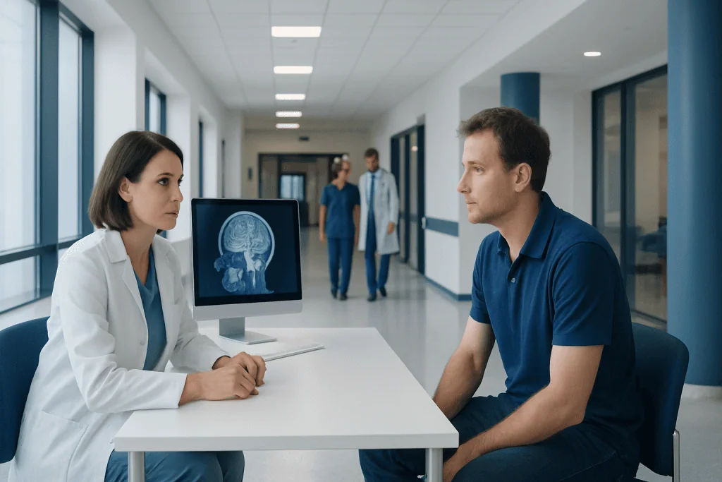
Diagnosis usually begins with symptom review, medical history, and a detailed neurological exam. Imaging tests such as CT and MRI help doctors look at the brain, spine, and blood vessels.
Key Takeaways
- Diagnosis usually begins with symptom review, medical history, and a detailed neurological exam.
- Imaging tests such as CT and MRI help doctors look at the brain, spine, and blood vessels.
- Specialized studies like EEG, EMG, and nerve conduction tests assess brain and nerve function.
- Blood tests, lumbar puncture, and cognitive testing may be used when infection, inflammation, or memory problems are suspected.
- Emergency symptoms such as sudden weakness, seizure, or severe sudden headache need urgent medical attention.
Neurological disorders are diagnosed through a careful combination of medical history, physical and neurological examination, imaging, and specialized tests. The goal is to identify where the problem is in the nervous system, what may be causing it, and which treatment plan is most appropriate.
Overview: How neurological disorders are diagnosed
Neurological disorder diagnosis is often a step-by-step process rather than a single test. The nervous system includes the brain, spinal cord, and peripheral nerves, and many different conditions can cause similar symptoms. Because of this, doctors usually combine several sources of information to understand what is happening.
The first aim is to localize the problem. In simple terms, this means deciding whether symptoms are likely coming from the brain, spinal cord, nerves, muscles, or the blood vessels that supply them. The second aim is to identify the likely cause, such as a structural change, reduced blood flow, inflammation, infection, autoimmune disease, degeneration, metabolic disturbance, or injury.
A diagnosis may sometimes be made quickly, especially in clear situations such as stroke or Bell’s palsy. In other cases, symptoms develop gradually and require follow-up visits, repeat examinations, or more than one type of test. This can feel frustrating for patients, but it is often the safest and most accurate way to reach the right conclusion.
Medical history and the neurological examination
The medical history is one of the most important parts of a neurological assessment. A doctor will ask when symptoms began, whether they started suddenly or gradually, how they have changed over time, and what makes them better or worse. Details such as headaches, dizziness, weakness, numbness, tremor, memory changes, sleep disturbance, balance problems, speech difficulty, visual symptoms, or episodes of loss of awareness can all provide valuable clues.
Past medical conditions, medications, family history, recent infections, alcohol or substance use, and exposure to toxins may also matter. For example, a family history may be relevant in some movement disorders or inherited neurological conditions such as Huntington disease. In children, pregnancy, birth history, and developmental milestones may also be important.
The neurological exam checks how the nervous system is working. It commonly includes assessment of mental status, speech, cranial nerves, muscle strength, muscle tone, reflexes, sensation, coordination, and walking pattern. The findings may suggest whether the problem involves one area of the brain, the spinal cord, a peripheral nerve, the inner ear, or the muscles themselves.
Even when advanced scans are planned, the physical examination remains essential. It helps the doctor choose the most useful tests and avoid unnecessary ones. In many cases, this careful clinical assessment provides the framework that makes later imaging or laboratory results meaningful.
Imaging tests: CT, MRI, and vascular studies
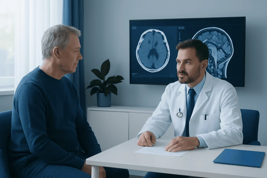
Imaging tests allow doctors to look at the structure of the brain, spine, and nearby blood vessels. A CT scan is often used in urgent settings because it is fast and widely available. It can quickly detect bleeding, major injury, some tumors, hydrocephalus, and many causes of acute neurological symptoms.
MRI provides more detailed images of the brain and spinal cord than CT in many situations. It is especially useful for conditions involving multiple sclerosis, small strokes, tumors, inflammation, spinal cord disorders, and some causes of chronic headache, seizures, or memory problems. Depending on the question being asked, MRI may be done with contrast dye to improve visibility of certain tissues.
When symptoms suggest a problem with blood vessels, doctors may use CT angiography, MR angiography, carotid ultrasound, or conventional angiography. These tests can help identify narrowing, aneurysms, malformations, or blocked vessels. This is particularly important in urgent cerebrovascular conditions such as transient ischemic attack or suspected subarachnoid bleeding.
Imaging is interpreted together with symptoms and examination findings. An abnormality seen on a scan does not always explain a person’s symptoms, and a normal scan does not rule out every neurological condition. In specialized centers, advanced neuroradiology assessment and interventional neuroradiology may support both diagnosis and, in selected cases, treatment planning.
Functional tests: EEG, EMG, nerve conduction, and evoked potentials
Some neurological problems affect how the nervous system works rather than how it looks on a scan. Functional tests help measure electrical activity in the brain, spinal cord, nerves, or muscles. These studies are often used when symptoms include seizures, blackout episodes, muscle weakness, numbness, tingling, muscle twitching, or suspected nerve injury.
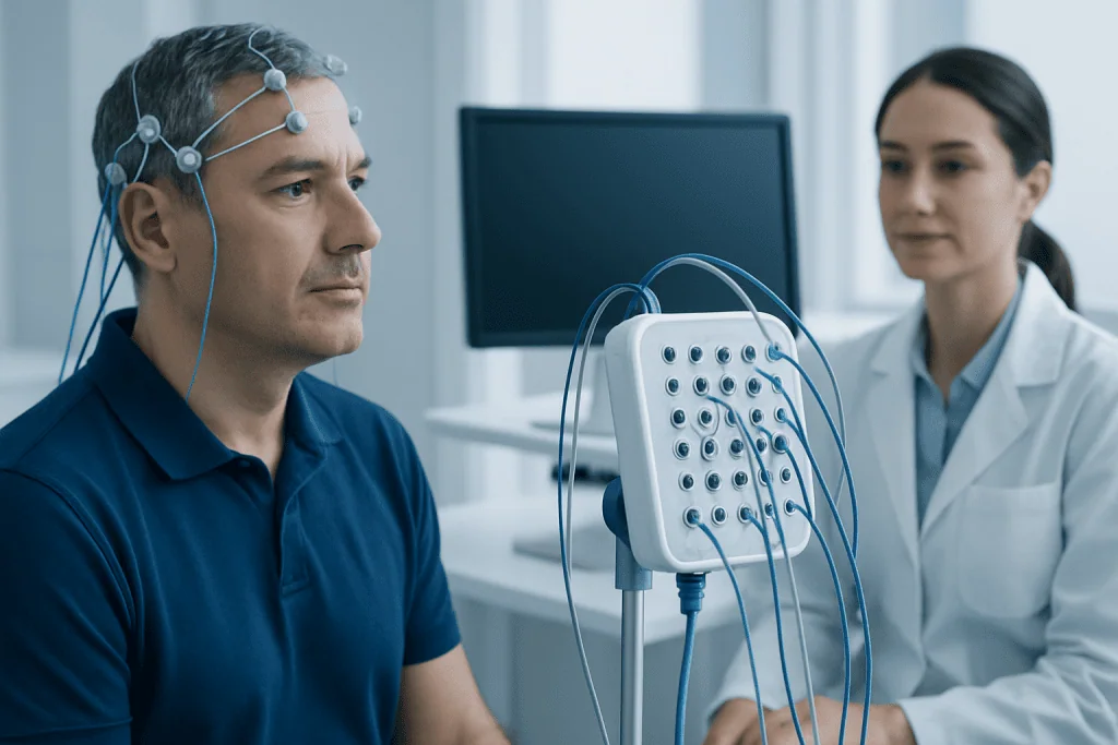
An electroencephalogram, or EEG, records electrical activity from the scalp. It is commonly used when epilepsy or other seizure disorders are suspected, but it may also be helpful in episodes of confusion, unexplained loss of consciousness, or certain sleep-related conditions. Sometimes a routine EEG is enough, while in other cases longer monitoring is needed to capture events.
Electromyography, or EMG, and nerve conduction studies evaluate how well nerves send signals and how muscles respond. They are useful in peripheral neuropathy, nerve compression, radiculopathy, and disorders of the muscle or neuromuscular junction. These tests can help distinguish whether weakness is related to nerve damage, muscle disease, or a problem affecting communication between nerve and muscle.
Evoked potential tests measure how quickly and effectively the brain responds to visual, sound, or sensory signals. They may be used in selected cases when disorders affecting sensory pathways are suspected. In many neurology services, these studies are part of a broader neurophysiology evaluation and may be combined with other tests for more accurate diagnosis.
Laboratory tests, lumbar puncture, and cognitive assessment
Blood tests are commonly used to look for underlying causes that can affect the nervous system. Depending on symptoms, doctors may check for infection, inflammation, autoimmune markers, vitamin deficiencies, thyroid problems, electrolyte imbalance, diabetes, liver or kidney disease, and medication effects. These tests are especially helpful because some neurological symptoms can be caused by treatable medical conditions outside the nervous system.
A lumbar puncture, sometimes called a spinal tap, is used to examine cerebrospinal fluid. This fluid surrounds the brain and spinal cord, and testing it can help diagnose meningitis, encephalitis, inflammatory disorders, bleeding not clearly seen on imaging, and some autoimmune or neurodegenerative conditions. The decision to perform a lumbar puncture depends on the clinical picture and imaging results.
When memory, attention, language, planning, or behavior changes are present, cognitive screening or formal neuropsychological assessment may be recommended. These evaluations can help distinguish normal aging from neurological disease, support diagnosis in dementia, and identify the pattern and severity of cognitive difficulties. In some cases, mood symptoms such as anxiety or depression are also assessed because they can overlap with or worsen neurological complaints.
Sleep studies, eye examinations, or hearing and balance testing may also be relevant in selected patients. For example, dizziness may require assessment for conditions such as vestibular neuritis, while visual symptoms may lead to ophthalmic or neuro-ophthalmic testing. The exact combination of tests is tailored to each person’s symptoms and examination findings.
How doctors combine results to reach a diagnosis
Neurologists rarely rely on a single test result. Instead, they build a diagnosis by comparing the history, examination, imaging, and laboratory findings. This process is sometimes called clinical correlation. A symptom such as numbness, for example, might come from a peripheral nerve, the spinal cord, or the brain, so doctors look for a pattern that best fits the whole picture.
In some cases, the diagnosis is confirmed quickly. In others, the initial goal may be to narrow down a list of possibilities and monitor how symptoms evolve. Follow-up can be especially important in conditions that change gradually, such as movement disorders, neuropathies, or memory disorders. Repeat imaging or repeated neurological examinations may show features that were not obvious at the beginning.
Doctors also consider whether symptoms might reflect more than one condition. Someone with headache may have a common primary headache disorder such as tension-type headache, but they may also need evaluation for less common secondary causes if red flags are present. This careful approach helps avoid both overtesting and missed diagnoses.
At specialized centers, diagnosis may involve a multidisciplinary team. Depending on the problem, this can include experts in headache medicine, movement disorders, neuroradiology, rehabilitation, neuropsychology, or neurosurgery. Near the end of the diagnostic journey, patients and families should also receive a clear explanation of what the results mean and what the next steps are.
What patients can do before and after testing
Good preparation can make neurological evaluation more efficient. Patients are often encouraged to bring a list of symptoms, when each one started, how often it happens, and whether anything triggers it. A medication list, previous scan reports, family history, and videos of intermittent symptoms such as tremor, abnormal movements, or seizure-like episodes can also be helpful.
Before some tests, special instructions may be given. For example, fasting may be needed for certain blood tests, while caffeine, sleep patterns, or specific medications may affect EEG or other studies. Patients should follow the instructions provided by their care team and ask questions if anything is unclear. It is usually helpful to arrange support for travel if a procedure or sedating medication might affect alertness.
After testing, the most important step is follow-up. Not every result can be interpreted in isolation, and some findings require discussion in context. Patients should ask what the results show, whether further tests are needed, and what symptoms should prompt urgent medical attention.
For international patients seeking coordinated neurological care, Acibadem International’s multidisciplinary specialists in JCI-accredited hospitals evaluate and treat a wide range of neurological conditions. A structured approach can be especially useful when symptoms are complex or when several specialties need to work together.
When to seek urgent medical attention
Some neurological symptoms should never be ignored. Sudden weakness or numbness on one side of the body, facial drooping, trouble speaking, sudden vision loss, severe imbalance, or a sudden severe headache may be signs of a medical emergency. A first seizure, prolonged seizure, severe confusion, sudden loss of consciousness, or head injury with neurological symptoms also needs prompt assessment.
Urgent evaluation is also important for fever with neck stiffness, rapidly worsening weakness, new bowel or bladder problems with back pain, or sudden inability to walk. These patterns can suggest infection, spinal cord compression, inflammation, or other conditions that benefit from early treatment.
Not every headache, dizzy spell, or tingling sensation is an emergency, but it is wise to contact a doctor if symptoms are new, persistent, worsening, or interfering with daily life. Early assessment can help clarify the cause, relieve uncertainty, and guide appropriate treatment or monitoring.
Frequently asked questions
What is the first step in neurological disorder diagnosis?
The first step is usually a detailed medical history and neurological examination. This helps the doctor understand the pattern of symptoms and decide which tests, if any, are most useful next.
Is an MRI always needed for neurological symptoms?
No, an MRI is not always necessary. The need for MRI depends on the symptoms, examination findings, and whether another test such as CT, blood work, or EEG may answer the question more appropriately.
What is the difference between EEG and EMG?
EEG records electrical activity in the brain and is often used for seizures or episodes of altered awareness. EMG evaluates muscles and, together with nerve conduction studies, helps assess nerve and muscle disorders.
Why might a neurologist order blood tests?
Blood tests can identify treatable causes of neurological symptoms, such as infection, inflammation, vitamin deficiency, thyroid disease, or metabolic imbalance. They are also useful for ruling out medical conditions that can mimic neurological disease.
Does a normal brain scan mean there is no neurological disorder?
Not always. Some neurological conditions affect function more than structure, so scans may appear normal even when symptoms are real. In those cases, other tests such as EEG, EMG, lab studies, or follow-up examination may provide the answer.
When should someone seek emergency care for neurological symptoms?
Emergency care is important for sudden weakness, trouble speaking, facial drooping, severe sudden headache, seizure, or sudden vision loss. Rapidly worsening weakness, severe confusion, or new loss of bladder or bowel control also require urgent evaluation.
References
- World Health Organization
- National Institute of Neurological Disorders and Stroke
- American Academy of Neurology
- National Institute for Health and Care Excellence
- Mayo Clinic
This article is for general information only and is not a substitute for professional medical advice. Please consult a qualified doctor about your individual situation.
Joint and spine care in Turkey — expert assessment & treatment
JCI-accredited · board-certified surgeons · reply within 24h
Add us as a Preferred Source to see more of our trusted health content across Google Search, AI Overviews and Discover.

Neurology
Diagnosis and treatment of disorders of the brain, spinal cord, nerves and muscles.
51 specialists in this unit