Calcinosis: Symptoms, Causes, and Treatment Options

Calcinosis is a buildup of calcium deposits in skin, muscles, or other soft tissues, often linked to local tissue damage, connective tissue disease, or kidney and metabolic disorders rather than dietary calcium. See a doctor promptly if a lump becomes painful, drains chalky material, limits movement, or shows signs of infection such as redness or fever.
Key Takeaways
- Calcinosis refers to abnormal calcium deposits in soft tissues, most often in or beneath the skin.
- It may be associated with autoimmune connective tissue diseases, tissue injury, abnormal mineral levels, certain medical treatments, or no identifiable cause.
- Symptoms range from small, firm painless nodules to painful deposits that ulcerate or limit joint movement.
- Diagnosis usually combines medical history, examination, blood tests, and imaging; a biopsy is occasionally needed.
- Treatment focuses on the underlying cause, protecting affected skin, relieving symptoms, and considering medicines or removal for selected deposits.
You notice a hard little lump under the skin on a fingertip or an elbow. It doesn’t hurt, so you leave it alone — and then one day it does hurt, or the skin over it opens.
That lump may be calcinosis: calcium deposits that form in the skin, muscles, joints, or other soft tissues. Some are painless. Others cause discomfort, restrict movement, break down the skin, or become infected, depending on how big they are and where they sit.
Calcinosis overview
Calcinosis means calcium salts have built up in tissue where they don’t belong. Most often that’s the skin and the layer just beneath it — a form called calcinosis cutis — but deposits can also form near muscles, tendons, and joints. Under your fingers they may feel like small hard grains, flat plaques, or larger lumps.
This is not about eating too much calcium. In many people, blood calcium is perfectly normal and the deposits form because of local tissue damage or inflammation. In others, an imbalance in calcium or phosphate metabolism plays a part.
No two cases behave the same way. Some deposits sit quietly for years and cause almost no symptoms; others turn painful, limit movement, press on nearby structures, or break through the skin. That’s why you need an individual assessment — the right care depends on the type, location, severity, and underlying cause.
What calcinosis can look and feel like
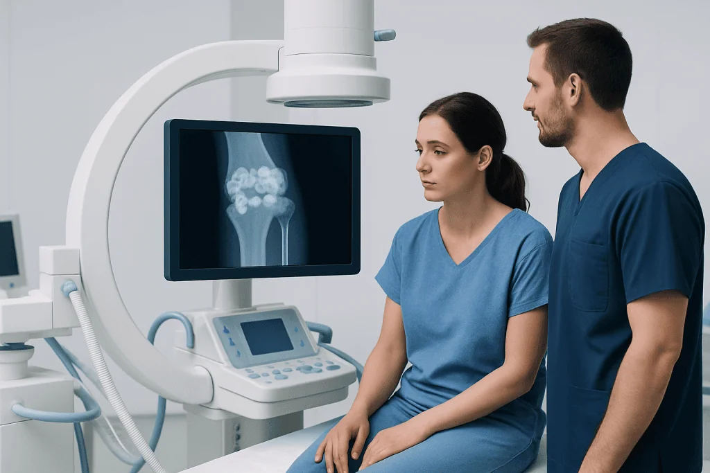
Calcinosis commonly appears as firm white, yellowish, or skin-colored bumps beneath the skin. Deposits may occur at pressure points or near joints, including the fingertips, elbows, knees, hips, shoulders, or buttocks. In people with connective tissue disease, deposits can also affect the hands and other areas exposed to repeated minor injury.
Small deposits may not cause symptoms. Larger or deeper deposits can produce tenderness, aching, stiffness, swelling, or reduced movement in a nearby joint. Calcium deposits can sometimes be seen on an X-ray before they are obvious on the skin surface.
When a deposit irritates or breaks through the skin, a chalky white material may drain from the area. The opening can be slow to heal and may become infected. Signs that warrant prompt medical assessment include increasing redness, warmth, swelling, worsening pain, pus-like drainage, fever, or a rapidly enlarging lesion.
- Hard lumps or plaques under the skin
- Pain or pressure around a deposit
- Joint stiffness or limited movement
- Skin ulceration or chalky drainage
- Recurrent inflammation or infection around affected skin
Types, causes and risk factors

Doctors sort calcinosis by why the deposits form in the first place. The most common form, dystrophic calcinosis, appears in damaged or inflamed tissue even though blood calcium and phosphate are normal. It may develop after injury, surgery, burns, repeated pressure, infections, or longstanding inflammation.
Dystrophic calcinosis is also associated with autoimmune connective tissue diseases, particularly systemic sclerosis, dermatomyositis, and some overlap syndromes. Persistent inflammation, tissue injury, reduced blood flow, and repeated trauma may all play a role. Not everyone with these conditions develops calcinosis, and deposits may appear years after the underlying illness begins.
Metastatic calcinosis occurs when calcium, phosphate, or both are elevated in the bloodstream. Chronic kidney disease and disorders of the parathyroid glands are among the possible contributors. Iatrogenic calcinosis can result from calcium-containing medical treatments entering surrounding tissues, while idiopathic calcinosis describes deposits that arise without a clear cause.
A separate and serious condition called calciphylaxis involves calcium deposition in small blood vessels, usually in people with advanced kidney disease. It can cause very painful skin changes and ulcers and requires urgent specialist care. Although its name is similar, calciphylaxis is distinct from the more localized deposits commonly described as calcinosis cutis.
How calcinosis is diagnosed
Your doctor starts by going through your symptoms, other medical conditions, medicines, past injuries, and when the deposits first appeared. A physical examination helps identify whether lesions are in the skin, under the skin, or near a joint, and whether there is ulceration, inflammation, or evidence of infection.
Imaging can show the size, depth, and distribution of calcium deposits. Plain X-rays are often useful, while ultrasound, CT, or MRI may be considered when deposits are deeper or when the care team needs to understand their relationship to muscles, tendons, nerves, or joints.
Blood tests may check calcium, phosphate, kidney function, parathyroid hormone, vitamin D status, and markers related to inflammation or an underlying autoimmune disease. Normal blood calcium does not rule out calcinosis. If the diagnosis remains uncertain, a small tissue sample may be examined to distinguish calcium deposits from other skin lumps or growths.
For people with known autoimmune disease or kidney disease, diagnosis should also include an assessment of whether the underlying condition is adequately controlled. This helps guide a care plan that addresses both the deposits and factors that may encourage new deposits to form.
Treatment options and day-to-day care
There is no single treatment that reliably clears every calcium deposit. Your care is shaped around the cause, your symptoms, where the deposits are, and how much they affect your daily life. When an underlying condition is present, managing it carefully is an important part of treatment, although improved disease control does not always make existing deposits disappear.
For small, painless deposits, observation and regular review may be appropriate. Helpful self-care measures include avoiding repeated pressure or trauma to affected areas, using protective padding when practical, keeping skin clean and moisturized, and covering any open area with a suitable dressing as advised by a healthcare professional. Deposits should not be squeezed, cut, or picked at because this can cause bleeding, infection, and delayed healing.
Specialists may consider medicines intended to reduce inflammation, alter mineral handling, or help soften deposits. Depending on the clinical setting, these may include drugs such as calcium-channel blockers, bisphosphonates, colchicine, minocycline, or sodium thiosulfate. Evidence for these approaches is still limited, and potential benefits, side effects, and suitability need to be considered individually.
Surgical removal may be an option when a deposit causes substantial pain, repeated infection, nerve compression, severe movement limitation, or persistent ulceration. However, surgery can be difficult if deposits are extensive or close to important structures, and recurrence is possible. Dermatologists, rheumatologists, nephrologists, orthopedic specialists, plastic surgeons, and wound-care teams may work together when needed.
Can calcinosis be prevented?
Calcinosis cannot always be prevented, particularly when it is linked to an autoimmune condition or past tissue injury. However, early management of conditions that affect calcium and phosphate balance, including kidney and parathyroid disorders, may reduce the risk of mineral-related deposits. Regular monitoring should be directed by the treating clinician.
People with connective tissue diseases may benefit from protecting the hands and other vulnerable areas from repeated injury and extreme cold, especially if they also have circulation problems. Comfortable footwear, cushioned supports, gloves for manual activities, and avoiding unnecessary pressure on established deposits can reduce irritation.
A balanced diet remains important for general health, but people should not independently restrict calcium or stop prescribed vitamin supplements solely because of calcinosis. Calcium and vitamin D have essential functions, and changes may be unsafe for some individuals. A doctor or dietitian can advise whether supplements or dietary choices need adjustment based on blood tests and the underlying cause.
When to seek medical care
Medical advice is appropriate for any new hard lump under the skin, especially if it grows, becomes painful, affects movement, or is associated with a chronic inflammatory, autoimmune, kidney, or hormone-related condition. A clinician can confirm whether calcinosis is likely and exclude other causes of a skin or soft-tissue lump.
Prompt assessment is important if a deposit opens through the skin, drains material, or shows possible infection. Increasing redness, warmth, swelling, severe tenderness, foul-smelling discharge, fever, or feeling unwell may require timely treatment. Sudden severe pain with dark, purple, or black skin changes requires urgent medical evaluation, particularly in a person with kidney disease.
People who have persistent pain, recurrent wounds, or difficulty using a hand, walking, or moving a joint may benefit from specialist review. Acibadem International’s multidisciplinary specialists at JCI-accredited hospitals can assess and treat calcinosis for international patients, coordinating care according to the underlying condition and each person’s needs.
Frequently asked questions
Is calcinosis the same as high calcium in the blood?
No. Many cases of calcinosis, especially dystrophic calcinosis, occur when blood calcium and phosphate levels are normal. The deposits may form in areas of longstanding inflammation, tissue damage, or repeated pressure.
Can calcinosis go away on its own?
Small deposits may remain unchanged for long periods, but complete spontaneous disappearance is not common. Treating an underlying condition and avoiding local trauma may help limit symptoms or progression, but existing deposits can be difficult to remove.
Is calcinosis painful?
It can be painless, particularly when deposits are small and away from joints or pressure points. Pain may occur when deposits become large, inflamed, ulcerated, infected, or positioned near nerves, tendons, or joints.
What doctor treats calcinosis?
The most suitable specialist depends on the cause and location of the deposits. Dermatologists and rheumatologists commonly assess calcinosis cutis, while nephrologists may be involved when kidney disease affects mineral balance; surgeons may be consulted for problematic localized deposits.
Can diet dissolve calcium deposits?
No dietary approach has been proven to dissolve calcinosis deposits. People should not make major changes to calcium, phosphate, or vitamin D intake without medical advice, particularly if they have kidney disease or are taking prescribed supplements.
When is surgery considered for calcinosis?
Surgery may be considered for localized deposits causing persistent pain, recurrent infection, skin ulceration, nerve pressure, or meaningful loss of function. The decision takes account of the deposit's location, size, likelihood of recurrence, wound-healing risks, and the person's overall health.
This article is for general information only and is not a substitute for professional medical advice. Please consult a qualified doctor about your individual situation.
Medical weight-loss options in Turkey — costs & eligibility
7 JCI-accredited hospitals · board-certified surgeons · reply within 24h
Add us as a Preferred Source to see more of our trusted health content across Google Search, AI Overviews and Discover.

Medically reviewed by the Acıbadem International Medical Board — August 24, 2026
See our medical review board →
Update history
- PublishedAugust 19, 2026
- Medical review approvedAugust 24, 2026
- Last content updateAugust 24, 2026
References2
- Calcium in the blood — MedlinePlus — medlineplus.gov
- Dermatomyositis — MedlinePlus — medlineplus.gov
Dermatology Specialists at Acibadem





Related Treatments
Related Conditions
More from the Health Library

Ingrown Hair Cysts Often Settle With Gentle Skin Care

CO2 Laser Resurfacing: How It Works and What It Treats

A Friction Burn Is Part Scrape, Part Heat Injury

Hydrocortisone Comes in Forms That Are Not Interchangeable

Choosing a Face Mask for Acne by Skin Type and Ingredients