

Thoracic Surgery Department
Assessment of lung nodules, lung cancer surgery by VATS, robotic and open routes, pleural disease from pneumothorax to empyema, mediastinal and thymic surgery, chest wall correction and airway surgery.
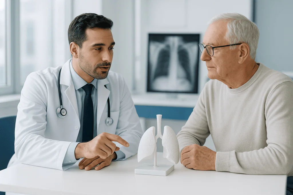
Most people arrive holding a scan, not a diagnosis
A nodule, a shadow, fluid, a mass in the mediastinum, found on a scan done for something else entirely. A large part of this clinic consists of explaining that the finding does not need an operation, and that conversation is as much the job as the operating is.
What was found on the scan
The findings people arrive with, and what each of them actually means before anyone mentions surgery.
Surgery
Keyhole, robotic and open routes into the chest, and the resections and drainage procedures performed through them.
Chest and lung conditions
The diseases behind the findings, several of which are treated jointly with pulmonology and oncology.
The most useful answer is often that no operation is needed
The great majority of lung nodules are benign, and the assessment exists to identify the minority that are not. Deciding which findings need tissue now, which need an interval scan, and which need nothing at all is the substance of this specialty, and getting it wrong in the direction of over-treatment costs someone functioning lung they did not need to lose.
On the cancer side nothing is decided alone. Whether the stage is established, whether the patient is operable as well as the tumour resectable, whether surgery goes first or after systemic treatment, and whether radiotherapy is the better option, all of it goes to a tumour board with the imaging and the pathology on the screen.
What we will not do
- Remove a nodule to settle anxiety. Surveillance is a decision, not a delay, and lung once removed does not come back.
- Resect an anterior mediastinal mass that could be lymphoma. That needs a generous biopsy and chemotherapy, not an operation.
- Choose keyhole access at the cost of the cancer operation. The same lobe, the same margin and the same lymph node dissection, or we convert.
- Offer sympathectomy before the treatments above it have been tried, or without putting compensatory sweating in front of you first.
- Promise a discharge date. It depends on when the air leak seals, and no surgeon knows that in advance.
Thoracic surgeons who lead this work
What actually happens, in order
Send the scans, and the old ones
CT in its original digital form, plus any previous chest imaging however old. For a nodule the old scan is the most valuable thing in the file, one unchanged for four years frequently ends the investigation on the spot.
Review, and the tumour board
Anything with a cancer question goes to the board before a recommendation exists. A proportion of reviews conclude that no operation is needed: that the finding warrants surveillance, or that the right treatment is not surgical.
Assessment on arrival
Lung function and exercise testing, cardiac assessment, and the staging procedures where nodal status is unresolved. Findings here change plans, including in the direction of not operating.
Surgery, then the drain decides
Breathing exercises and walking from the first day. Discharge is governed by the chest drain and the drain by the air leak, so the date genuinely cannot be fixed in advance: this is the part of thoracic surgery that resists scheduling.
Before the flight home
Air left in the pleural space expands at cabin pressure, so an interval and usually a confirmatory chest X-ray precede travel. You leave with the operative note, pathology, staging and the follow-up imaging schedule.
Six things worth knowing first
A nodule is usually not cancer
CT finds them constantly on scans done for unrelated reasons, and most are old infection scars or benign growths. Being told you have a nodule is not being told you have cancer.
Waiting is not inaction
Growth over time is the most reliable discriminator there is, and no single scan can substitute for it. A stable nodule at the end of surveillance is discharged, not followed forever.
Resectable and operable are different questions
One asks whether the tumour can be removed, the other whether this person can spare the lung. Lung function testing answers the second, and for some the better treatment is radiotherapy.
Keyhole matters because of the ribs
Spreading the ribs is what makes chest surgery painful: the nerves run under them. Avoiding that single manoeuvre is where most of the benefit of VATS comes from.
The drain decides the discharge date
Cut lung leaks air until it seals, and a prolonged leak is the commonest complication of a lung resection. It is not dangerous and nothing was done wrong, but it is why no firm date is given.
Sympathectomy has a permanent trade
A majority sweat more elsewhere afterwards, and in a minority severely enough to regret the operation. It is not predictable and not reversible, which is why it sits last in the ladder.
Jump to what you came for
Quick answer
The Thoracic Surgery Department diagnoses and treats diseases of the lungs, chest wall, mediastinum, pleura, diaphragm, and esophagus using surgical and minimally invasive techniques. At Acibadem in Turkey, care is typically planned by a multidisciplinary team and may include advanced imaging, endoscopic evaluation, video-assisted or robotic surgery, and follow-up tailored to the patient’s condition.
What our thoracic surgery unit covers, and who it is for
Thoracic surgery is the surgery of the chest: the lungs, the pleura around them, the mediastinum between them, the chest wall that encloses them, the airway and the oesophagus. It is not lung medicine, asthma, COPD, pneumonia and the long-term management of lung disease belong to pulmonologyand it is not heart surgery, which sits in the same cavity and is a separate specialty entirely.
Most people arrive here in one of two ways, and they need different things from this page. The first group has a scan report with a finding on it, a nodule, a shadow, fluid, a mass in the mediastinum, and no idea what it means. The second has a diagnosis and is being offered an operation. What follows is written for both, and the sections are ordered accordingly: what findings mean first, what the operations involve second.
At Acıbadem International the work is organised into five strands.
- Lung cancer surgeryfrom the assessment of a nodule through staging to resection, by keyhole, robotic or open routes, as part of a multidisciplinary plan rather than in isolation.
- Pleural diseasepneumothorax, effusion, empyema and the drainage, decortication and pleurodesis procedures that treat them.
- Mediastinal surgerythymoma and other masses between the lungs, including thymectomy for myasthenia gravis.
- Chest wall surgerypectus deformities in adolescents and adults, chest wall tumours and reconstruction, and selected rib fractures.
- Airway, diaphragm and oesophageal surgerytracheal resection, diaphragm procedures and oesophageal resection, the last shared with oncology and gastroenterology.
What a thoracic surgeon actually does
A thoracic surgeon is what a general surgeon is not: someone who operates inside the chest daily and whose judgement about which nodules need removing, which need watching and which need only a repeat scan is the substance of the specialty. A large part of this clinic consists of telling people that the thing found on their scan does not need an operation, and that conversation is as much the job as the operating is.
Lung nodule: what a nodule on a scan actually means
A lung nodulea pulmonary noduleis a rounded opacity in the lung measuring up to three centimetres. Anything larger is called a mass, and that distinction matters because the two are approached differently from the first moment.
Here is the fact that should be the first thing anyone with a new nodule reads: the great majority of lung nodules are not cancer. CT scanning is sensitive enough that nodules are found constantly on scans done for entirely unrelated reasons: a chest scan after a car accident, a CT of the abdomen that catches the lung bases, a scan for chest pain that turns out to be muscular. Most are scars from infections people never knew they had, small collections of inflammatory tissue, or benign growths. Being told you have a nodule is not being told you have cancer, and the interval between the report and the explanation is where most of the fear in this specialty lives.
What separates a nodule that needs attention from one that does not is not a single feature but a combination, and this section is the only place on this page where that logic is set out in full; later sections refer back to it.
- Size. Very small nodules carry a very low probability of malignancy; the probability rises with diameter, and the thresholds for acting are built around that.
- Growth over time. The single most informative feature there is. A nodule unchanged over an adequate interval of surveillance is behaving benignly, whatever it looks like; one that grows is investigated regardless of how reassuring its appearance was.
- Density and edge. Solid, part-solid and ground-glass nodules behave differently, see below. Smooth round edges are reassuring; a spiculated, star-shaped edge is not. Certain patterns of calcification are essentially diagnostic of a benign process.
- The person. Age, smoking history, previous cancer, occupational exposure and family history shift the probability considerably before the scan is even looked at. The same nodule in a thirty-year-old lifelong non-smoker and a sixty-eight-year-old with forty pack-years is not the same problem.
Those inputs produce one of three answers: no further imaging, interval surveillance CT at a defined schedule, or tissue and staging now. The schedules themselves are set by published guidelines and by the individual’s risk, and they are followed by the team rather than chosen by the patient, but understanding why an interval scan is the recommendation, rather than a biopsy, prevents the common and understandable feeling that nothing is being done.
Ground glass nodule
A ground glass nodule is a hazy area on CT through which the underlying lung markings remain visible, as opposed to a solid nodule that obscures them. It behaves quite differently from a solid nodule and is frequently misunderstood in both directions.
Pure ground-glass nodules that persist across scans can represent the very earliest end of the lung adenocarcinoma spectrum, but at that end the biology is extremely indolent, growing over years rather than months, and many never progress at all. That is why they are followed with far longer surveillance intervals than a solid nodule of the same size, and why immediate surgery for a small pure ground-glass lesion is usually the wrong answer. Many resolve entirely, because inflammation and infection look the same way.
What changes the plan is the appearance of a solid component within the hazy area, a part-solid nodule, and growth of that solid component in particular. Part-solid nodules carry the highest probability of malignancy of any nodule type, and the solid part is the piece that is measured and watched. This is one of the clearest examples in the specialty of a finding whose appearance frightens patients more than it worries clinicians, and whose evolution matters more than its existence.
Solitary pulmonary nodule and how surveillance works
A solitary pulmonary nodule is a single nodule surrounded by normal lung, with no enlarged lymph nodes and no other lesions: the classical presentation and the one the assessment logic above was built for. Multiple nodules are a different problem, usually infective or metastatic, and are approached differently.
Surveillance means repeat CT at defined intervals to see whether the nodule changes. Two things about it are worth stating plainly, because both cause distress that could be avoided. Waiting is not inaction: growth over time is the most reliable discriminator available, and it is information that no single scan, however detailed, can provide. And a stable nodule at the end of an adequate surveillance period is genuinely reassuring, surveillance is discharged, not extended indefinitely.
Where the probability is high enough, or the nodule grows, the options are a PET-CT to assess metabolic activity and look for spread, a biopsy, or proceeding directly to a diagnostic and therapeutic resection. Which is chosen depends on the size, the position within the lung, and whether the person is fit for an operation, and it is a decision taken with the tumour board rather than by one clinician.
Lung biopsy
A lung biopsy obtains tissue for diagnosis, and there are three routes, chosen by where the lesion sits.
A CT-guided needle biopsy through the chest wall suits lesions near the periphery. It is quick and done under local anaesthetic, and it carries a recognised risk of causing a pneumothorax, which is why the patient is observed and imaged afterwards. A bronchoscopic biopsy reaches lesions near the airways from inside, with navigational and radial ultrasound techniques extending that reach further into the periphery. And a surgical biopsyusually a wedge of lung taken by VATSis the definitive answer where the others have failed or are unsuitable, and has the advantage that a malignant frozen section can be converted into the full cancer operation in the same anaesthetic.
One thing patients are rarely told and should be: a non-diagnostic biopsy is common and does not mean the lesion is benign. A needle that misses, or returns inflammatory tissue only, has answered nothing, and the plan continues rather than stopping there.
Lung cancer surgery
Lung cancer surgery offers the best chance of cure in early-stage non-small cell lung cancer, and the whole of the assessment described above exists to identify who is in that group. Two questions decide whether an operation is the right treatment, and they are separate: is the cancer resectable, anatomically removable with a clear margin, and is the patient operable, meaning able to tolerate the loss of lung and the operation itself.
The second question is answered by lung function testing, cardiac assessment and an appraisal of fitness, because the operation removes functioning lung permanently. Someone whose breathing is already marginal may not tolerate a lobectomy even where the tumour is small and ideally placed, and for that group stereotactic radiotherapy is a genuine alternative rather than a consolation: that comparison is made with radiation oncology at the tumour board, not by a surgeon alone.
Small cell lung cancer is largely a medical rather than a surgical disease and is treated by medical oncology; surgery has a narrow role in a small minority of very early cases.
Lung cancer staging, EBUS and mediastinoscopy
Lung cancer staging establishes how far the disease has spread, and its most consequential question is whether the lymph nodes in the mediastinum, the space between the lungs, are involved. That single answer frequently decides between an operation and chemotherapy or radiotherapy first, which is why it is established with tissue rather than assumed from imaging.
PET-CT identifies metabolically active disease and is the standard imaging step, along with brain imaging where indicated. But PET is not proof: inflammation and infection light up, and small deposits do not, so a positive node on PET is sampled rather than accepted, and in higher-risk cases a negative one may be sampled too.
EBUSendobronchial ultrasound, is how that sampling is now done in most cases. A bronchoscope carrying an ultrasound probe is passed into the airways, the nodes are visualised through the airway wall, and a needle is passed into them under direct vision. It is done under sedation, needs no incision, and has largely displaced the older surgical approach. Mediastinoscopya small incision above the breastbone and direct inspection and sampling of the nodes, remains for the situations EBUS cannot reach or where its result conflicts with the clinical picture.
Lobectomy
A lobectomy removes one lobe of the lung, the right lung has three, the left has two, together with the lymph nodes that drain it. It has been the standard operation for early-stage lung cancer for decades, and the reason is anatomical: a lobe is a self-contained unit with its own artery, vein and bronchus, so removing it takes the tumour with a clear margin and with the lymphatic territory that would spread it.
What people most want to know is how they will breathe afterwards. Removing a lobe removes a proportion of lung capacity, and the remaining lung expands to fill the space over the following months. Most people with reasonable baseline function return to normal daily activity, with reduced reserve that shows on hard exertion rather than at rest. Someone with poor baseline function experiences the loss much more, which is why the operability assessment is done before the decision rather than after.
The lymph node dissection that accompanies it is not an optional extra. It provides the staging that determines whether chemotherapy is offered afterwards, and an operation that removes the tumour without sampling the nodes has left the most important question unanswered.
Segmentectomy and wedge resection
A segmentectomy removes one anatomical segment of a lobe with its own vessels and airway, and a wedge resection removes a non-anatomical piece of lung around a lesion. Both preserve more lung than a lobectomy, and the difference between them is significant: a segmentectomy respects the anatomy and takes the draining lymph nodes with it, while a wedge does neither.
The position has changed in recent years and it is worth stating what changed. For small peripheral early-stage cancers, segmentectomy has been shown in randomised trials to give results comparable to lobectomy while preserving lung function, which has made it a legitimate first choice in selected patients rather than a compromise for those too frail for more. The selection is specific: small tumour, peripheral location, adequate margin achievable, and node sampling still performed.
A wedge resection remains the right operation for a diagnostic biopsy, for removing metastases from another primary cancer, and for patients whose lung function will not tolerate anything larger. What it is not is an equivalent cancer operation for a fit patient, and it should not be presented as one.
Pneumonectomy
A pneumonectomy removes an entire lung. It is required where a tumour crosses between lobes, involves the main bronchus or the central vessels, and cannot be cleared by anything less, and it has become considerably less common as sleeve resections, which remove a segment of bronchus and rejoin it while preserving the rest of the lung, have taken over many of those cases.
It is a substantially bigger undertaking than a lobectomy and is treated as such. Living with one lung is entirely possible and many people do it well, with a permanently reduced exercise ceiling. The assessment beforehand is correspondingly more searching, particularly of cardiac and pulmonary reserve, because there is no margin left afterwards. Where a sleeve resection can achieve the same clearance, it is preferred, and asking whether one has been considered is reasonable.
Lung cancer surgery recovery, and the air leak after lung surgery
Recovery after a keyhole resection is faster than most people expect and slower than the leaflet suggests. The hospital stay is typically a few days, governed almost entirely by one thing: when the chest drain can come out.
That is worth explaining, because it is the commonest reason a discharge date moves. Cut lung surface leaks air until it seals, and a prolonged air leak after lung surgery is the single most frequent complication of a lung resection. It is not dangerous in itself and it is not a sign that anything was done wrong, it is a property of lung tissue, more likely in emphysematous lungs, but the drain stays until it stops. Some patients go home with a portable drainage device rather than waiting in hospital.
Beyond that: pain around the incisions and where the drain sat, which is managed actively because shallow breathing causes the other common complication, which is chest infection. Breathing exercises and early walking are the treatment for both. Fatigue lasts weeks and breathlessness on exertion improves over months as the remaining lung expands. Most people return to office work within a few weeks and to full activity over two to three months, and smoking cessation before surgery measurably reduces every one of these complications.
VATS: video assisted thoracoscopic surgery and thoracoscopy
VATSvideo assisted thoracoscopic surgeryperforms an operation inside the chest through small incisions between the ribs, with a camera and long instruments, without spreading the ribs apart. It is the standard approach for most thoracic operations now, and the reason is specific to this part of the body.
Rib spreading is what makes traditional chest surgery painful. The ribs are levered apart, the intercostal nerves running under them are stretched or crushed, and the resulting pain is both severe in the short term and, in a proportion of patients, persistent for months or longer. Avoiding that single manoeuvre changes the whole recovery: less pain, better breathing afterwards because breathing does not hurt as much, fewer chest infections, shorter stay.
Thoracoscopy is the same access used diagnostically, inspecting the pleural cavity, taking biopsies, draining and treating fluid, and is often done under sedation with local anaesthetic rather than general anaesthesia. Uniportal VATS performs the whole operation through a single small incision; it is a technical refinement rather than a different operation, and the evidence for it over multi-port VATS is modest.
What matters more than the number of ports is that the operation inside is the same one. A lobectomy done by VATS must remove the same lobe with the same margin and the same lymph node dissection as an open one. Where that cannot be achieved: a large central tumour, dense adhesions from previous infection or surgery, involvement of the chest wall, conversion to an open approach is correct rather than a failure, and a surgeon who states in advance under what circumstances they would convert is describing good practice.
Thoracotomy
A thoracotomy is the open approach: an incision along the line of a rib, with the ribs spread to give direct access to the chest cavity. It remains the right operation in a substantial minority of cases, large or central tumours, chest wall involvement requiring resection and reconstruction, complex airway work, dense adhesions, and emergencies.
The muscle-sparing thoracotomy used in modern practice divides fewer of the chest wall muscles than the classical incision, and pain control has improved considerably with regional blocks placed at the time of surgery. It is still the more painful route and the one with the longer recovery, and that is stated plainly rather than minimised. Persistent pain at the incision for months afterwards, post-thoracotomy pain, is a recognised outcome in a minority and is a large part of why the keyhole approach displaced it wherever it can.
Robotic lung surgery
Robotic lung surgery performs the same keyhole operations with instruments held by a robotic platform under the surgeon’s direct control, adding wristed movement, three-dimensional vision and tremor elimination. Its advantages are most apparent in the fiddliest parts of the operation: dissecting lymph nodes out of the mediastinum, and the fine work of a segmentectomy or a sleeve resection.
Compared with VATS, the recovery is broadly similar, both avoid rib spreading, which is where most of the benefit comes from. Robotic surgery reliably produces a more thorough lymph node dissection in several series, which matters for staging. It costs more and takes longer to set up. The honest position is that robotic and VATS are two good routes and the open approach is the third, and the surgeon’s experience with the specific operation matters more than which platform is used.
Pneumothorax
A pneumothorax is air in the pleural space, between the lung and the chest wall, where there should be none. The air occupies space the lung needs, and the lung collapses to the degree the air allows. It presents with sudden sharp chest pain on one side, worse on breathing in, and breathlessness proportional to how much lung is down.
It occurs in three settings. Primary spontaneous, in young, tall, thin people, typically men in their late teens and twenties, with no known lung disease, from the rupture of small blebs at the lung apex. Secondary, on a background of existing lung disease such as COPD, where the same volume of air is far more dangerous because the reserve is already gone. And traumatic or iatrogenic, after chest injury or after a procedure, a lung biopsy or a central line, which is why those procedures are followed by imaging.
Treatment scales to size and to symptoms. A small pneumothorax in a well person with normal lungs is frequently observed and reabsorbs on its own. A larger one, or a breathless patient, needs the air removed, by aspiration with a needle, or by a chest drain. Tension pneumothorax, where air enters the space and cannot escape and pressure builds until the circulation is compromised, is the emergency form and is decompressed immediately wherever the patient is.
Spontaneous pneumothorax, bullectomy and when surgery is offered
Spontaneous pneumothorax recurs. That is the single most useful thing to know about it: after a first episode a substantial proportion of people have another, and after a second the probability of a third is higher again. Surgery is what changes that.
The accepted indications for operating are consistent across guidelines: a second episode on the same side, a first episode on the opposite side, a persistent air leak that will not seal, failure of the lung to re-expand, both sides simultaneously, and a first episode in someone whose occupation makes recurrence dangerous, pilots and divers in particular, for whom the pressure changes are the specific hazard.
The operation is done by VATS and has two components performed together, and both are necessary. Bullectomy removes or staples off the blebs at the apex that ruptured. And a pleural procedure, abrading the pleural surface or removing a strip of it, makes the lung adhere to the chest wall so that there is no space for air to collect again. Doing only the first leaves a considerably higher recurrence rate, which is worth knowing if surgery is being described as simply removing the blebs.
Chest tube and chest drain
A chest tubea chest drain, is a tube placed through the chest wall into the pleural space to remove air or fluid and allow the lung to re-expand. It is the workhorse of this specialty and it is the thing patients ask about most after an operation, because it is uncomfortable, it tethers them to a drainage system, and it is what decides when they go home.
Modern practice has changed it in ways worth knowing. Smaller-bore tubes are used for air and for simple fluid, and they are considerably more comfortable than the large tubes of the past; large-bore drains are reserved for blood, pus and thick fluid. Digital drainage systems measure the air leak numerically instead of relying on watching for bubbles, which removes a good deal of guesswork from deciding when to remove it. And ambulatory devices allow selected patients to go home with the drain in place and return for removal.
Removal itself is quick and is done at a specific point in the breathing cycle to stop air being drawn in. Most people describe a strange pulling sensation rather than pain. The site is closed with a suture and heals over days.
Pleural effusion
A pleural effusion is fluid in the pleural space. It is a finding rather than a diagnosis, and the entire clinical question is what produced it, because the same appearance on a chest X-ray can be heart failure, infection, cancer, kidney or liver disease, a pulmonary embolus or an autoimmune condition.
The first step is therefore a sample rather than a drain. Fluid is aspirated under ultrasound guidance and analysed, and the single most important division it produces is between a transudatefluid pushed out by pressure, as in heart failure, where the treatment is of the underlying condition and not of the chest, and an exudate, which means the pleura itself is involved by infection, inflammation or malignancy and needs investigating on its own account.
Draining a large effusion relieves breathlessness quickly, and it is done in stages rather than all at once, because removing a very large volume rapidly can cause the lung to re-expand faster than it tolerates. Where the cause is not established by fluid analysis, thoracoscopy with direct pleural biopsy is the next step and gives the highest diagnostic yield of any approach.
Malignant pleural effusion and the PleurX catheter
A malignant pleural effusion is fluid caused by cancer involving the pleura, most often from lung or breast primaries, and it recurs as long as the underlying disease is active. The aim of treatment is therefore not cure but breathlessness, and that reframing matters, because it changes what a good result looks like.
Two approaches dominate and they suit different people. Pleurodesis aims to obliterate the space so fluid cannot reaccumulate, and it requires the lung to be able to re-expand fully to the chest wall. An indwelling pleural catheter: the PleurX catheter is the widely used version, is a soft tube tunnelled under the skin and left in place, through which the patient or a carer drains fluid at home as often as needed. It works where the lung cannot fully re-expand, which is the situation in which pleurodesis fails, and it avoids an inpatient stay.
The honest comparison is that both control symptoms well and the choice turns on the person: prognosis, whether the lung re-expands, whether they would rather have a single procedure or an ongoing device, and whether home drainage is practical. In a meaningful proportion of patients with a catheter the pleura seals spontaneously over weeks and the catheter is removed.
Pleurodesis
Pleurodesis deliberately creates adhesion between the two layers of pleura so that the space between them is obliterated and fluid or air can no longer collect. It is used for recurrent malignant effusion and, in a different form, as part of the operation for recurrent pneumothorax.
It is done two ways. Chemically, by instilling sterile talc, as a slurry through a chest drain or insufflated as powder at thoracoscopy, which provokes an inflammatory reaction that sticks the surfaces together. Or mechanically, by abrading the pleural surface at surgery, which is what is used in the pneumothorax operation in younger patients, since it avoids leaving talc in the chest of someone with decades ahead of them.
Two conditions govern whether it works, and they are worth understanding before consenting to it. The lung must re-expand to meet the chest wall, because surfaces that do not touch cannot adhere, which is why a trapped lung is a contraindication and an indwelling catheter is the answer instead. And the procedure is uncomfortable: the inflammation it provokes causes pain and often fever for a day or two, and that is the mechanism working rather than a complication.
Empyema
An empyema is pus in the pleural space, usually following pneumonia, sometimes after chest surgery or trauma. It is the pleural infection that antibiotics alone will not cure, because antibiotics penetrate a collection of pus poorly and the fluid is walled off from the circulation.
It progresses through three recognised stages and the stage determines the treatment, which is why delay matters so much here. In the first, the fluid is thin and free-flowing and a drain plus antibiotics is usually sufficient. In the second, fibrin strands divide the collection into loculated pockets that a single drain cannot reach; drainage plus fibrinolytic agents instilled through the drain, or surgery, is needed. In the third, a thick fibrous peel forms over the lung and traps it, and no drain will resolve it: the peel must be removed surgically, an operation called decortication, to let the lung re-expand.
The practical consequence: an empyema treated promptly is a drain and antibiotics, and the same empyema treated late is a major operation on a trapped lung. Referral for surgical assessment when a pleural infection is not resolving is not escalation for its own sake, and units that involve a thoracic surgeon early in pleural infection have better outcomes than those that do so once everything else has failed.
Haemothorax (hemothorax) and chylothorax
Hemothorax is blood in the pleural space, most often after chest trauma or as a complication of a procedure or operation. Small volumes are drained and observed; ongoing bleeding, or a large clotted collection that will not drain, requires surgery, and a retained clot left in the chest becomes infected or organises into a trapped lung, which is why it is evacuated rather than watched.
A chylothorax is lymphatic fluid, chyle, in the pleural space, from injury to or obstruction of the thoracic duct. It appears after oesophageal and mediastinal surgery, after trauma, and with lymphoma and other malignancy. The fluid is characteristically milky when the patient has been eating fat, which is the clue that gives the diagnosis.
Its treatment is unusual and worth describing, because it is one of the few places where diet is the treatment. Reducing dietary fat, or feeding intravenously so the gut is bypassed entirely, cuts the flow through the duct and lets many leaks seal on their own. Where they do not, the duct is ligated surgically or embolised radiologically. Losing chyle continuously costs protein, fat-soluble vitamins and immune cells, so a chylothorax that persists is treated actively rather than simply drained.
Mediastinal mass
A mediastinal mass is a lesion in the space between the lungs, containing the heart, great vessels, trachea, oesophagus, thymus, lymph nodes and nerves. The first question is not what it is but where in that space it sits, because the compartment predicts the diagnosis better than the appearance does.
Anterior compartment masses, the commonest to reach a thoracic surgeon, are dominated by four possibilities remembered as the four Ts: thymoma, teratoma and other germ cell tumours, thyroid extending down from the neck, and terrible lymphoma. Middle compartment masses are more often lymph nodes, cysts and vascular. Posterior compartment masses are predominantly neurogenic, arising from the nerve roots and sympathetic chain.
That matters for one practical reason above all: lymphoma is treated with chemotherapy, not surgery, and it needs a generous tissue sample rather than an excision. Operating on a lymphoma in the belief that it is a thymoma is the classic error in this territory, and it is why anterior mediastinal masses are worked up with imaging, tumour markers and, where lymphoma is plausible, a biopsy first, rather than proceeding straight to resection.
Thymoma
A thymoma is a tumour of the thymus gland, the organ in the front of the chest that is active in childhood and largely replaced by fat in adults. It is the commonest anterior mediastinal tumour in adults and it behaves unlike most tumours: growth is typically slow, and its behaviour is defined less by how the cells look than by whether it has breached its own capsule and invaded surrounding structures.
Many are found incidentally on a scan done for another reason. Others present through pressure, cough, chest discomfort, breathlessness, or through an associated autoimmune condition, of which myasthenia gravis is by far the most important and is present in a substantial minority.
Complete surgical removal is the treatment and the strongest determinant of the outcome, which is why the operation is planned to take the whole thymus and surrounding fat rather than just the visible tumour. Encapsulated tumours completely excised do well; invasive ones may need radiotherapy afterwards, and the follow-up is long because late recurrence is a recognised feature of this tumour. The operation is done by sternotomy for large or invasive tumours, and increasingly by VATS or robotic access for smaller ones.
Thymectomy and myasthenia gravis
Thymectomy is removal of the thymus, and it is performed for two distinct reasons: to remove a thymoma, and to treat myasthenia gravis in patients who have no thymoma at all.
The second is the less intuitive one. Myasthenia gravis is an autoimmune disease in which antibodies block transmission at the junction between nerve and muscle, producing fatigable weakness, drooping eyelids, double vision, difficulty swallowing, weakness that worsens through the day. The thymus is implicated in generating that autoimmunity, and randomised evidence has shown that removing it in appropriately selected patients with generalised disease and antibodies to the acetylcholine receptor improves symptoms and reduces the requirement for steroids over the following years.
Three points matter practically. Selection is made by a neurologist rather than a surgeon, age, antibody status and disease pattern all enter it, and the diagnosis and medical treatment belong with neurology. The benefit accrues over years, not weeks, and expecting an immediate change leads to disappointment. And the disease is optimised medically before the operation, because myasthenic weakness affects the breathing muscles and the anaesthetic and recovery are planned around that.
Pectus excavatum
Pectus excavatum is a chest wall in which the breastbone and adjacent ribs are sunken inward, producing the appearance usually described as a funnel chest. It is the commonest congenital chest wall deformity, it is far more frequent in males, and it typically becomes noticeable or worsens sharply during the adolescent growth spurt.
Two questions govern how it is treated, and they are usually asked in the wrong order. The first is physiological: does the depression compress the heart and reduce exercise capacity? In deep deformities it can displace and compress the heart, and patients report exercise intolerance, breathlessness and palpitations out of proportion to their fitness. This is assessed with CT or MRI to measure the severity index, echocardiography, and lung function and exercise testing.
The second is psychological, and it is the one that brings most people to clinic and is most often dismissed. Adolescents with a visible chest deformity avoid swimming, changing rooms, beaches and relationships; the effect on self-image at that age is substantial and well documented. Treating that as vanity is a misreading, in most series it is the dominant reason patients seek surgery, and it is a legitimate one.
Not everyone needs an operation. Mild deformity without physiological effect is managed with observation and posture and exercise programmes. A vacuum bell device, which lifts the sternum by suction applied over months to years, is an option in younger patients with flexible chest walls and mild to moderate depression, and it avoids surgery in a proportion of them.
The Nuss procedure
The Nuss procedure corrects pectus excavatum without removing any cartilage or bone. A curved metal bar is passed behind the sternum through two small incisions at the sides of the chest, under thoracoscopic guidance, and then rotated so that its convexity pushes the sunken sternum forward. The bar stays in place for a period of years while the chest wall remodels around the new shape, and is then removed in a second, much smaller operation.
It works best in the adolescent chest, where the cartilage is still flexible, which is why timing matters and why referral around or shortly after the growth spurt is common. It can be done in adults, in whom the chest is stiffer, often requiring more than one bar.
The honest part is the pain. The first days after a Nuss procedure are genuinely painful, because the chest wall has been forcibly reshaped and is held under tension, and pain control is planned deliberately, epidural or regional blocks, and a structured plan afterwards. That is the trade for a scarless-looking chest and a correction that holds, and it should be described before rather than discovered after. The alternative operation, the Ravitch procedure, removes the deformed cartilage and repositions the sternum directly; it remains the choice for complex, asymmetric and some adult deformities.
Pectus carinatum
Pectus carinatum is the opposite deformity: the sternum protrudes outward, giving the appearance described as pigeon chest. It is less common than pectus excavatum and, importantly, it rarely causes any physiological problem: the heart and lungs are not compressed, and the reason for treating it is appearance.
The treatment is also different, and it is good news for most patients. Because the deformity is a protrusion of flexible cartilage, external compression bracing works: a custom brace worn for a defined number of hours daily over months to a couple of years corrects the majority of cases in growing patients, without surgery. The younger and more flexible the chest wall, the better it works, which makes early referral genuinely valuable here rather than merely tidy. Surgery is reserved for rigid deformities, for patients past skeletal maturity and for those in whom bracing has failed.
Chest wall tumours (chest wall tumor) and rib fractures
A chest wall tumor arises from the ribs, sternum, cartilage or the soft tissue between them, and may be benign, primary malignant, chondrosarcoma being the commonest, or a metastasis from elsewhere. The principle of treatment for a primary malignant chest wall tumour is wide resection with clear margins, which frequently means removing a segment of chest wall and reconstructing it with mesh and, where the defect is large, a muscle flap with plastic surgery. Planning that reconstruction before the resection, rather than after, is what distinguishes a well-run case.
Rib fractures are common after chest trauma and the great majority are managed with pain relief, breathing exercises and time: the pain matters because shallow breathing leads to collapse and infection, so analgesia is treatment rather than comfort. Surgical fixation with plates has a defined role in a minority: flail chest, where a segment moves paradoxically with breathing, severely displaced fractures, and fractures that fail to unite and remain painful. Fixing every fracture is not the standard and offering to is over-treatment.
Hyperhidrosis surgery and sympathectomy
Hyperhidrosis is excessive sweating beyond what thermoregulation requires. In its primary focal form it affects the palms, armpits, soles and face, begins in adolescence, and is genuinely disabling in a way that is easy to underestimate, hands too wet to shake, to write, to use a phone or a keyboard, and a constant low-grade social dread.
Hyperhidrosis surgery is the last step in a ladder, and everything above it should have been tried: strong aluminium chloride antiperspirants, iontophoresis, botulinum toxin injections, which are highly effective for the armpits and useful for the palms, and oral anticholinergic medication prescribed by the treating doctor. Surgery is for severe palmar and axillary disease that has not responded.
Sympathectomyendoscopic thoracic sympathectomy, interrupts the sympathetic chain running down inside the back of the chest, at the level that supplies the affected area. It is done by thoracoscopy through two tiny incisions, takes a short time, and is usually a day case. For palmar sweating it is highly effective and the effect is immediate; the hands are dry when the patient wakes.
And then the part that must be said before consent rather than after: compensatory sweating. A majority of patients sweat more elsewhere afterwards, typically the back, chest and abdomen, and in a minority that compensatory sweating is severe enough that they regret the operation. It is not reliably predictable, it does not resolve, and reversal is not realistically possible. Limiting the level divided reduces but does not eliminate it. Any account of this operation that does not put that trade in front of the patient plainly is not informed consent, and it is the single reason this operation is offered late rather than early.
Airway surgery: bronchoscopy, tracheal resection and tracheostomy
The airway is shared territory between thoracic surgery, pulmonology and ENT, and the surgical part of it is small in volume and high in consequence.
Bronchoscopy inspects the airways from inside with a flexible or rigid instrument. Flexible bronchoscopy under sedation is the diagnostic workhorse, visual inspection, washings, brushings and biopsies, and with EBUS the sampling of mediastinal nodes. Rigid bronchoscopy, under general anaesthesia, is the therapeutic tool: it maintains a wide airway while working, and it is what is used to remove a foreign body, to control bleeding, to core out a tumour obstructing a main airway and to place a stent.
A tracheal resection removes a diseased or narrowed segment of trachea and joins the healthy ends directly. The commonest indication is stenosis following prolonged intubation or a previous tracheostomy, and the others are tumours of the trachea and injury. It is a demanding operation with a specific and unusual aftercare instruction: the anastomosis is protected by keeping the neck flexed forward for a period afterwards, sometimes with a stitch from chin to chest, so that the join is not put under tension. It is done in relatively few centres, and volume matters here more than in almost any other operation in the specialty.
A tracheostomy creates an opening into the trachea in the neck, either surgically or percutaneously at the bedside in intensive care. It is done for prolonged ventilation, for airway obstruction above that level, and to allow secretions to be cleared in patients who cannot do so. It is frequently temporary and is closed once its purpose has passed. The most common question from families is whether speech is lost, and the answer is that it is usually preserved or restored with a speaking valve once the patient is stable.
Lung metastases, diaphragm, emphysema and the oesophagus
Four further areas complete the specialty, each briefly, because each carries a decision people search for.
Lung metastasis and metastasectomy
Lung metastasis and metastasectomy. Deposits in the lung from a cancer elsewhere are usually treated systemically, but in selected patients removing them surgically is worthwhile: the accepted conditions being that the primary is controlled, that there is no disease anywhere else, that all the lung deposits can be removed with adequate function remaining, and that no better systemic option exists. It is most established in colorectal cancer and in sarcoma. Wedge resections are used to preserve as much lung as possible, since further metastases and further operations are common. The selection is made at the tumour board with medical oncology.
Diaphragm paralysis and plication
Diaphragm paralysis follows injury to the phrenic nerve, after cardiac surgery, from a tumour, or without identified cause, and produces breathlessness on lying flat and on exertion as the paralysed half of the diaphragm rises and is pushed up by the abdomen. Many people compensate and need nothing. Where symptoms are significant and the paralysis is established, plication, folding and stitching the floppy diaphragm flat so it stops moving paradoxically, reliably improves lung volumes and breathlessness.
Lung volume reduction surgery and endobronchial valves
Lung volume reduction surgery removes the most destroyed, non-functioning areas of an emphysematous lung so that the remaining better lung and the diaphragm can work more efficiently. It sounds counterintuitive and it works, in a narrowly defined group: upper-lobe predominant emphysema, poor exercise capacity after rehabilitation, and specific physiological criteria. Endobronchial valves achieve a similar effect bronchoscopically in selected patients, without an operation. Selection is made jointly with pulmonology and outside those criteria the risks exceed the benefit.
Oesophagectomy (esophagectomy)
Removal of the oesophagus for cancer is among the largest operations in general and thoracic surgery, usually combining an abdominal and a chest approach, with the stomach reconstructed into a tube and brought up to replace it. It is increasingly performed minimally invasively. What matters most for a patient facing it is that outcomes for oesophageal cancer surgery correlate strongly with the volume of the unit performing it, and that it is planned alongside chemotherapy or chemoradiotherapy with medical oncology and gastroenterology rather than as a standalone procedure.
Mesothelioma and lung transplant: two things this unit does not do alone
Mesothelioma is a cancer of the pleura, strongly associated with previous asbestos exposure and typically presenting decades after it, most often as a pleural effusion or chest pain. Surgery has a contested and narrow role: the aggressive operations once offered have not consistently improved survival, and current practice concentrates on establishing the diagnosis with a proper thoracoscopic biopsy, controlling the effusion, and systemic treatment, chemotherapy and, increasingly, immunotherapy, with medical oncology. Anyone offered radical surgery for mesothelioma should ask what evidence supports it in their specific situation, because the honest answer is that it is debated rather than settled.
Lung transplant is the end-stage treatment for irreversible lung disease, pulmonary fibrosis, severe COPD, cystic fibrosis, pulmonary hypertension, and it is performed in a small number of designated transplant centres rather than by general thoracic units. The relevance here is referral timing: patients are assessed for candidacy well before they become too unwell to survive the operation, and the commonest failure in this pathway is referral that comes too late. Assessment is shared with pulmonology.
The tumour board, and what it decides
Nothing on the cancer side of this specialty is decided by a surgeon alone. A lung cancer case is discussed by a board including thoracic surgery, medical oncology, radiation oncology, radiology, nuclear medicine and pathology, with the imaging and the pathology on the screen, before a treatment sequence is fixed.
What the board actually decides is worth spelling out, because it is more than a formality. Whether the stage is established or needs more tissue. Whether the patient is operable as well as the tumour resectable. Whether surgery goes first or after chemotherapy or immunotherapy: a question that has changed substantially in recent years as neoadjuvant treatment has improved. Whether stereotactic radiotherapy is the better option for a patient whose lung function will not tolerate resection. And what happens afterwards, based on the final pathology rather than the pre-operative stage.
For a patient, the useful consequence is that a recommendation should be traceable to that discussion, and that asking what alternatives were considered is reasonable in every case.
Your multidisciplinary team
The thoracic surgeon assesses the finding, decides whether an operation is indicated and performs it. The pulmonologist investigates the lung disease that produced the finding, performs the diagnostic bronchoscopy and EBUS in many units, and manages the lung function that determines operability. The thoracic anaesthetist carries an unusually specific skill: ventilating one lung while the other is deflated for the surgeon to work on, and managing the physiology that produces. The chest physiotherapist does the breathing and mobilisation work that prevents the two commonest complications after any chest operation. And the specialist nurse manages drains, wounds and the long tail of questions after discharge.
Around them: medical oncology and radiation oncology for the treatment sequences that surround an operation, radiology for the imaging and image-guided biopsy, pulmonology for the airway and the lung disease, neurology for myasthenia gravis, and plastic surgery for chest wall reconstruction. The tumour board is where these views meet before a plan is fixed.
The international patient journey
The pathway for a patient travelling from abroad follows the same clinical sequence as for a local patient, with the imaging reviewed before travel and the tissue work done on arrival.
Send the scans, not the reports
CT of the chest in its original digital form, any previous chest imaging however old, and PET-CT if done. Previous scans are the single most valuable thing in the file for a nodule, because they establish whether it is new or has been stable for years, and a nodule unchanged on a scan from four years ago frequently ends the investigation on the spot. Lung function results and the pathology from any previous biopsy complete it.
Review and board discussion
The imaging is reviewed and, for anything with a cancer question, taken to the tumour board before a recommendation is given. A proportion of reviews conclude that no operation is needed: that the finding warrants surveillance, or that the right treatment is not surgical, and that outcome is stated as readily as the other.
Assessment on arrival
What cannot be done remotely: repeat or dedicated imaging where the outside study is inadequate for planning, lung function and exercise testing, cardiac assessment, and the staging procedures, EBUS or mediastinoscopy, where nodal status is unresolved. Findings here change plans, including in the direction of not operating, and that is stated in advance rather than treated as a schedule problem.
Surgery and the days after
The operation, then a ward with a chest drain, breathing exercises and walking from the first day. Discharge is governed by the drain, and the drain is governed by the air leak, so the date is genuinely not fixed in advance: this is the one part of thoracic surgery that resists scheduling.
Before flying home
Flying after chest surgery is timed by the operating surgeon, and the specific concern here is not general, residual air in the pleural space expands at cabin pressure, so an interval and often a confirmatory chest X-ray precede travel. People leave with the operative note, the pathology, the staging and the oncology plan in a form the treating team at home can act on, together with the follow-up imaging schedule, which for a resected cancer continues for years.
Our Specialists Explain
 Lung Cancer Treatment at Acibadem | Prof. Dr. Erkan Kaba
Frequently Asked Questions
Does a lung nodule mean I have cancer?
Usually not. Nodules are found constantly on CT scans done for entirely unrelated reasons, and the great majority are benign, old infection scars, small inflammatory collections or benign growths. What determines the level of concern is a combination rather than any single feature: size, whether it grows over time, its density and edge, and the person’s age, smoking history and previous cancer. Being told you have a nodule is not being told you have cancer, and in many cases the whole assessment ends with a repeat scan showing no change.
How often will my nodule be scanned?
At intervals set by the size and character of the nodule and by your own risk, following published surveillance schedules, commonly a few months for the first repeat in a nodule of moderate size, and considerably longer intervals for very small or pure ground-glass ones. Surveillance is not open-ended: a nodule that has remained stable across an adequate period is discharged rather than followed forever. The interval feels like inaction and is not, growth over time is the single most reliable discriminator available, and no single scan can substitute for it.
What does a ground glass nodule mean?
It is a hazy area through which the lung markings remain visible, and it behaves very differently from a solid nodule. Many are inflammatory and disappear. Those that persist can represent the earliest and most indolent end of the adenocarcinoma spectrum, growing over years rather than months, which is why they are watched at much longer intervals rather than removed promptly. What changes the plan is the appearance or growth of a solid component within the hazy area, a part-solid nodule, because that is the pattern carrying the highest probability of malignancy.
Can a lung nodule disappear?
Yes, and a meaningful proportion do. Nodules caused by infection or inflammation resolve as the process settles, which is precisely why a short-interval repeat scan is often the first step rather than a biopsy: a nodule that has vanished needs nothing further, and one that has shrunk is behaving benignly. This is the single most common reason a frightening initial report ends without any intervention at all.
What is the difference between a nodule and a mass?
Size. A rounded lung opacity up to three centimetres is a nodule; larger than that it is called a mass. The distinction matters because the probability of malignancy rises with diameter, and a mass is generally investigated with tissue and staging promptly rather than entering a surveillance schedule. The words are descriptive rather than diagnostic, neither tells you what the lesion is.
Is a lung biopsy dangerous?
It is a low-risk procedure with one common and manageable complication. A needle biopsy through the chest wall causes a pneumothorax in a proportion of cases, which is why patients are observed and imaged afterwards; most need nothing and a minority need a small drain. Bleeding occurs and is usually minor. The other honest caveat is not a risk but a limitation: a non-diagnostic result is common and does not mean the lesion is benign: it means the plan continues rather than stopping.
Why do I need a PET scan?
Because it measures metabolic activity rather than shape, which adds information a CT cannot. It helps assess how likely a nodule is to be malignant, it identifies lymph nodes that warrant sampling, and it looks for disease elsewhere in the body that would change the treatment entirely. It is not proof in either direction: infection and inflammation are metabolically active and light up, and very small or slow-growing lesions can fail to. That is why a positive node on PET is sampled rather than accepted.
How much lung can be safely removed?
It depends entirely on how well the remaining lung works, which is why lung function and exercise testing are done before the decision rather than after. Someone with normal lungs tolerates removal of a lobe with a reduction in reserve that shows only on hard exertion. Someone whose breathing is already limited may not tolerate the same operation, and for that person a smaller resection, or stereotactic radiotherapy instead, may be the better treatment. The calculation is about predicted function after the operation, not about what looks removable on the scan.
Will I be short of breath after a lobectomy?
Most people with reasonable baseline lung function return to normal daily activity, with reduced reserve that becomes apparent on stairs, hills and hard exercise rather than at rest. Breathlessness is worst in the first weeks and improves over several months as the remaining lung expands into the space and the chest wall recovers. How much permanent reduction remains is predicted before surgery from lung function testing, and it is a fair question to ask for your own numbers rather than a general answer.
Can I live with one lung?
Yes. People live full lives after a pneumonectomy, with a permanently reduced exercise ceiling, normal activity, walking and light exercise are generally fine, while sustained hard exertion is not. Because there is no reserve left afterwards, the assessment before the operation is more searching than for a lobectomy, and where a sleeve resection can achieve the same cancer clearance while preserving lung, it is preferred.
What is the difference between VATS and open surgery?
Whether the ribs are spread apart. VATS works through small incisions between the ribs with a camera and long instruments; a thoracotomy opens the chest and levers the ribs apart, which stretches the nerves running under them. That single difference accounts for most of what patients experience, less pain, better breathing afterwards, fewer chest infections and a shorter stay with the keyhole approach. What must not differ is the operation inside: the same lobe, the same margin, the same lymph node dissection.
Is robotic surgery better than keyhole surgery?
Both avoid spreading the ribs, which is where most of the benefit comes from, and recovery is broadly similar between them. Robotic instruments have wristed movement and three-dimensional vision, which help most in the fiddliest parts, dissecting mediastinal lymph nodes and performing a segmentectomy or sleeve resection, and several series show a more thorough node dissection. It costs more and takes longer to set up. The surgeon’s experience with your specific operation matters more than the platform used.
How long is the hospital stay after lung surgery?
Typically a few days after a keyhole resection, and longer after an open operation. The stay is governed almost entirely by the chest drain, and the drain is governed by whether the cut lung surface is still leaking air. That is why a firm discharge date cannot honestly be given in advance, and why some patients go home earlier with a portable drainage device rather than waiting in hospital for the leak to seal.
Why is my chest drain still in?
Almost always because of a persistent air leak, which is the commonest complication after any lung resection. Cut lung leaks air until the surface seals, and until it does, removing the drain would allow air to collect and the lung to collapse. It is not dangerous and it does not mean anything was done wrong: it is more likely in emphysematous lungs, and it resolves with time. Modern digital drainage systems measure the leak numerically, which takes the guesswork out of deciding when the drain can come out.
Does it hurt when the chest drain is removed?
Less than people expect. Most describe a brief strange pulling sensation rather than pain. It is removed at a specific point in the breathing cycle so that air is not drawn in as it comes out, the site is closed with a stitch, and it heals over days. Pain relief is given beforehand and it is worth asking for it rather than assuming it is not offered.
When can I fly after chest surgery?
Only when the operating surgeon clears it, and the reason is specific to this surgery: any air remaining in the pleural space expands at cabin pressure. A confirmatory chest X-ray showing the lung fully expanded usually precedes travel, alongside the general considerations of clot risk and being far from the team that operated. For an international patient this is the item not to fix in advance: a return booking that cannot move is a clinical liability.
When can I drive after chest surgery?
When you can perform an emergency stop and turn to check blind spots without the pain limiting you, and when you are off any medication that impairs concentration, usually a matter of weeks rather than days, and confirmed by the operating team. Local licensing rules also apply and differ by country and by licence type.
Do I really have to stop smoking before lung surgery?
Yes, and this is one of the few pieces of preoperative advice with a measurable effect within weeks rather than years. Stopping reduces the rate of chest infection, of prolonged air leak and of wound problems after chest surgery. Even a short period of abstinence before the operation improves the ciliary clearance that protects against infection afterwards. Support to stop is offered rather than the instruction being left as an instruction, and it is worth taking.
Will I need chemotherapy after lung surgery?
It depends on the final pathology, which is frequently different from the stage predicted before the operation: that is one of the reasons the lymph nodes are removed and examined. Where the tumour is larger or nodes are involved, adjuvant treatment is usually recommended, and targeted or immune therapy may apply depending on the molecular testing done on the resected tissue. The decision is made by the tumour board with medical oncology once the pathology is complete, not on the day of surgery.
Is a collapsed lung an emergency?
It depends on how much lung is down and on the state of the lungs underneath. A small pneumothorax in a young person with otherwise normal lungs is frequently observed and reabsorbs without any intervention. The same volume of air in someone with severe COPD is far more serious because there is no reserve. The dangerous form is tension pneumothorax, in which air keeps entering the space and cannot escape until the pressure compromises the circulation: that is decompressed immediately wherever the patient is, and it is a recognised emergency rather than a routine presentation.
Will my pneumothorax happen again?
A substantial proportion of spontaneous pneumothoraces recur after a first episode, and the probability rises again after a second. That pattern is precisely why surgery is offered: it is the intervention that changes the recurrence rate rather than treating the current episode. The accepted triggers for operating are a second episode on the same side, a first on the opposite side, a leak that will not seal, a lung that will not re-expand, both sides at once, and a first episode in someone whose occupation makes a recurrence dangerous.
Can I fly or dive after a pneumothorax?
Flying is generally allowed once the lung has fully re-expanded and the surgeon has confirmed it, with the interval set individually. Diving is a different matter and the restriction is far stricter: the pressure changes involved make a recurrence at depth potentially fatal, and diving is usually considered permanently unsuitable after a spontaneous pneumothorax unless definitive surgery has been performed, and even then it is assessed by a diving medicine specialist rather than assumed. Anyone who dives should raise it before decisions are made, because it changes whether surgery is offered after a first episode.
Why does the fluid around my lung keep coming back?
Because draining fluid treats the accumulation, not the cause. Where the pleura itself is involved by cancer, fluid re-forms as long as the underlying disease is active, and the aim of treatment shifts from cure to controlling breathlessness. The two approaches that do that are pleurodesis, which obliterates the space so fluid cannot collect, and an indwelling catheter drained at home. Which suits you depends mainly on whether the lung can fully re-expand, if it cannot, pleurodesis will not work and the catheter is the answer.
Is talc pleurodesis safe, and why is it so uncomfortable?
Graded sterile talc is used specifically because it is effective and its safety in the pleural space is well established. The discomfort is the mechanism rather than a complication: talc provokes an inflammatory reaction between the two pleural layers so that they stick together, and inflammation causes pain and often a fever for a day or two. Pain relief is planned in advance for that reason. In younger patients having surgery for pneumothorax, mechanical abrasion of the pleura is usually preferred over talc, to avoid leaving a permanent foreign material in the chest of someone with decades ahead.
Why would a chest infection need surgery?
Because pus in the pleural space is walled off from the bloodstream and antibiotics penetrate it poorly. An empyema progresses through stages: thin free fluid that a drain and antibiotics resolve, then loculated pockets divided by fibrin that a single drain cannot reach, then a thick fibrous peel that traps the lung and cannot be drained at all. The stage determines whether a drain, drainage with clot-dissolving agents, or an operation to strip the peel off the lung is needed, which is why delay converts a simple problem into a major one, and why surgical assessment early in a non-resolving pleural infection is not over-escalation.
Is a thymoma cancer?
It occupies an unusual middle ground, and that is the honest answer rather than an evasion. Thymomas grow slowly and behave far less aggressively than most tumours, but they are not simply benign: their behaviour is defined by whether they have breached their own capsule and invaded surrounding structures, rather than by how the cells look under the microscope. Complete surgical removal is the treatment and the strongest determinant of outcome. Encapsulated tumours completely removed do well; invasive ones may need radiotherapy, and follow-up is long because late recurrence is a recognised feature.
Will thymectomy cure my myasthenia gravis?
It is not a cure and it should not be described as one. In appropriately selected patients, generalised disease with antibodies to the acetylcholine receptor, randomised evidence shows that removing the thymus improves symptoms and reduces the steroid requirement over the years that follow. The benefit accrues gradually rather than immediately, which is the most common source of disappointment when expectations were not set. Selection is made by the neurologist managing the disease, and medication continues afterwards under their direction.
Is pectus excavatum dangerous, or just cosmetic?
Both questions are legitimate and they are assessed separately. In deep deformities the sunken sternum can displace and compress the heart, producing genuine exercise intolerance and breathlessness, and that is measured with cross-sectional imaging, echocardiography and exercise testing rather than assumed. In milder deformities there is no physiological effect. The psychological impact is separate and is the reason most patients present, avoidance of swimming, changing rooms and beaches during adolescence is well documented, and treating that as vanity is a misreading of what it costs people.
Am I too old for pectus surgery?
No, but the operation is different. The Nuss procedure works best in the adolescent chest, where cartilage is still flexible enough to be reshaped by a bar behind the sternum, and it is why referral around or after the growth spurt is common. In adults the chest wall is stiffer, often requiring more than one bar, and the open Ravitch procedure, removing the deformed cartilage and repositioning the sternum directly, is more frequently chosen. Correction in adults is entirely achievable; the route and the recovery differ.
What is compensatory sweating after sympathectomy?
Increased sweating elsewhere on the body, typically the back, chest and abdomen, after the nerve supply to the hands or armpits has been interrupted. It affects a majority of patients to some degree, and in a minority it is severe enough that they regret having the operation. It is not reliably predictable beforehand, it does not resolve, and reversal is not realistically possible. Limiting the level of the chain divided reduces it without eliminating it. This is the single reason sympathectomy sits at the end of the treatment ladder rather than the start, and any discussion of the operation that does not put it plainly in front of you is incomplete.
Conditions We Treat
- Esophageal Cancer
- Hyperhidrosis
- Lung Cancer
- Lung Nodules
- Mesothelioma
- Pectus Carinatum
- Pectus Excavatum
- Pneumothorax
- Thymoma
9 conditions, listed A–Z
All conditions A–ZMedically reviewed by the Acıbadem International Medical Board September 26, 2026
See our medical review board →
Update history
- PublishedJune 14, 2026
- Medical review approvedSeptember 26, 2026
- Last content updateSeptember 25, 2026
Add us as a Preferred Source to see more of our trusted health content across Google Search, AI Overviews and Discover.
Treatments in Thoracic Surgery Department
Specialists in this Unit









Available at these Hospitals










What patients say about this unit
★★★★★From 2,400+ verified patient reviews“I was nervous about my thoracic procedure, but I felt safe at every single step. Prof. Turhan was calm, precise and deeply reassuring. The facilities were spotless and modern.”
“My thoracic operation was precise and the aftercare in the ward was attentive and kind.”
Speak with our medical team
Share your case and our international patient team will guide you to the right specialist, free of charge.