Brain Aneurysm Treatment (Clipping and Coiling)
Brain aneurysm treatment prevents bleeding from a weak, bulging area in a brain artery. The two main methods are surgical clipping, in which a neurosurgeon places a small clip across the aneurysm…
Quick answer
Brain aneurysm surgery seals off a weak, bulging spot in a brain artery so it cannot bleed. Clipping places a small metal clip across the aneurysm through an opening in the skull; coiling fills it with soft coils delivered through a catheter from an artery in the groin or wrist. Both are used for ruptured and selected unruptured aneurysms.
What is Brain Aneurysm Treatment (Clipping and Coiling)?
A brain aneurysm (also called a cerebral aneurysm) is a weak, bulging spot in the wall of an artery in the brain. Because the wall is thinner than normal, it can leak or burst. When a brain aneurysm ruptures, blood spills into the space around the brain. This is called a subarachnoid hemorrhage (bleeding between the brain and the thin membranes that cover it), and it is a life-threatening emergency.
Brain aneurysm treatment aims to stop blood from flowing into the aneurysm so that it cannot rupture, or cannot bleed again if it already has. The two most widely used approaches are:
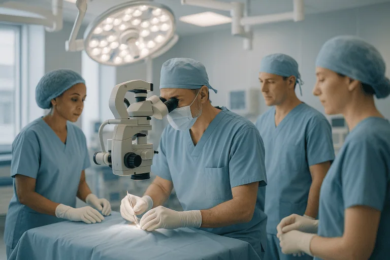
- Aneurysm clipping: a form of open brain aneurysm surgery. A neurosurgeon opens a small section of the skull and places a tiny metal clip across the neck (the base) of the aneurysm, sealing it off from the artery.
- Aneurysm coiling: a minimally invasive procedure done from inside the blood vessels (an endovascular procedure). A specialist threads a thin tube called a catheter through an artery, usually starting at the groin or wrist, up to the aneurysm and fills it with soft platinum coils. The coils cause a blood clot to form inside the aneurysm, which blocks blood flow into it.
Both methods are used for ruptured aneurysms (as emergency treatment) and for unruptured aneurysms that are judged to have a meaningful risk of bleeding in the future. Newer endovascular devices, such as flow diverters (stent-like mesh tubes that redirect blood past the aneurysm) and stent-assisted coiling, are variations of the coiling approach used for certain aneurysm shapes.
Who is a candidate for brain aneurysm surgery
Not every brain aneurysm needs to be treated. Many small aneurysms are found by chance on scans done for other reasons and never cause symptoms. The decision to treat depends on balancing the estimated risk of the aneurysm bleeding against the risks of the procedure itself.
Brain aneurysm treatment is usually considered when:
- The aneurysm has ruptured and caused a subarachnoid hemorrhage. In this situation, securing the aneurysm is typically done as soon as safely possible to prevent a second bleed.
- An unruptured aneurysm is relatively large, has grown on follow-up scans, or has an irregular shape that doctors associate with higher rupture risk.
- The aneurysm is pressing on nearby nerves or brain tissue and causing symptoms such as double vision, a drooping eyelid, or pain behind the eye.
- The person has had a previous rupture of another aneurysm, has a strong family history of aneurysm rupture, or has certain inherited conditions that weaken blood vessel walls.
Treatment may not be suitable, or may be delayed, when:
- The aneurysm is very small and has a low estimated risk of rupture, in which case monitoring with periodic imaging is often recommended instead.
- The person is frail, elderly, or has serious heart, lung, or kidney disease that makes anesthesia and surgery unusually risky.
- A ruptured aneurysm has caused such severe brain injury that treatment is unlikely to change the outcome. This is a difficult decision made together with the family.
- The aneurysm’s location or shape makes both clipping and coiling technically unsafe; in some cases other techniques, such as bypass surgery, are discussed.
The choice between clipping and coiling depends on the aneurysm’s size, location, shape, and neck width, the patient’s age and general health, and whether the aneurysm has ruptured. Treatment decisions are typically made by a multidisciplinary team that includes neurosurgeons, neuroradiologists (doctors who treat blood vessel problems from inside the vessels using imaging), and neurologists.
How the procedure works
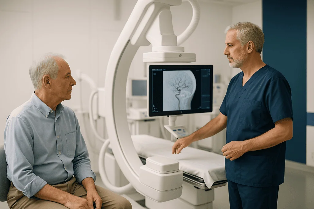
Before the procedure. Detailed imaging is essential. This usually includes a CT scan (computed tomography, a detailed X-ray image), a CT angiogram or MR angiogram (scans that show the blood vessels), and often a catheter angiogram, in which contrast dye is injected directly into the brain arteries to map the aneurysm precisely. The team uses these images to decide whether clipping or coiling is the safer option and to plan the approach.
Aneurysm clipping, step by step.
- You receive general anesthesia, so you are fully asleep and feel nothing.
- A small area of the scalp is shaved and an incision is made. The surgeon removes a section of skull bone (a craniotomy) to reach the brain.
- Using an operating microscope, the surgeon gently moves aside brain tissue and follows the artery to the aneurysm.
- A small titanium clip is placed across the neck of the aneurysm. The clip stays in place permanently. Surgeons often check with a special dye or ultrasound that the parent artery still carries blood normally.
- The bone is replaced and secured with small plates, and the scalp is closed with stitches or staples.
- The operation typically takes several hours.
Aneurysm coiling, step by step.
- Coiling is usually done under general anesthesia, although some centers use sedation with local anesthesia for selected cases.
- A small puncture is made in an artery in the groin or wrist and a thin catheter is guided, using live X-ray imaging, up through the body and into the brain arteries.
- A smaller catheter is advanced into the aneurysm itself. Soft platinum coils are released one at a time until the aneurysm is packed.
- If the aneurysm has a wide neck, a stent (a small mesh tube) or a balloon may be used to hold the coils in place, or a flow diverter may be placed across the aneurysm opening.
- The catheters are removed and pressure or a closure device is applied to the puncture site. There is no incision in the skull.
- The procedure often takes one to three hours.
After the procedure. Most people are monitored in an intensive care or neurological care unit for at least the first night. After a ruptured aneurysm, monitoring is longer and more intensive because of the risk of complications from the bleed itself, such as vasospasm (narrowing of brain arteries) and hydrocephalus (a build-up of fluid in the brain).
Preparation for brain aneurysm treatment
Preparation depends heavily on whether the treatment is planned or an emergency. After a rupture there is little time to prepare, and the medical team manages everything. For a planned (elective) procedure, the following are commonly involved:
- A full medical review, blood tests, and often a heart tracing (ECG) and chest X-ray to confirm fitness for anesthesia.
- A review of all medicines. Blood-thinning medicines and some supplements may need to be paused before clipping. In contrast, before coiling with a stent or flow diverter, your doctor may ask you to start antiplatelet medicines (such as aspirin and a second drug) several days beforehand to reduce the risk of clots forming on the device.
- Not eating or drinking for a set number of hours before general anesthesia, as instructed.
- Stopping smoking as early as possible. Smoking is linked to aneurysm growth and rupture and slows healing.
- Controlling blood pressure, since high blood pressure increases the risk of rupture.
- Arranging practical support at home, because you will not be able to drive for a period afterward and may need help with daily tasks.
It also helps to write down your questions in advance. Reasonable topics include why one method is recommended over the other, what the team expects your recovery to look like, and what follow-up imaging you will need.
Recovery and aftercare
Brain aneurysm surgery recovery varies widely. The main factors are whether the aneurysm had ruptured, which technique was used, and your overall health.
After coiling of an unruptured aneurysm, many patients stay in the hospital for one to two nights. Bruising or soreness at the groin or wrist puncture site is common and usually settles within a week or two. Headache and tiredness are common in the first days. Many people return to light daily activities within one to two weeks, though lifting and strenuous exercise are typically restricted for a short period.
After clipping of an unruptured aneurysm, the hospital stay is often several days to a week. Recovery from a craniotomy usually takes longer: headaches, fatigue, jaw stiffness, and difficulty concentrating are common for several weeks. Many patients return to most normal activities within four to eight weeks, and return to work varies with the type of job.
After a ruptured aneurysm, the recovery is mainly recovery from the hemorrhage itself rather than from the procedure. Hospital stays of two to three weeks or longer are typical, and many people need a period of rehabilitation afterward. Fatigue, memory and concentration problems, mood changes, and headaches can persist for months. Some people recover fully; others have lasting effects. Rehabilitation with physical, occupational, and speech therapists is often part of this phase.
General aftercare advice commonly includes:
- Take pain relief and other medicines exactly as prescribed. If you were given antiplatelet drugs after a stent or flow diverter, do not stop them without speaking to your specialist.
- Keep the incision or puncture site clean and dry as instructed, and watch for redness, swelling, or discharge.
- Avoid driving until your doctor confirms it is safe. Rules vary by country and by whether you had a bleed or seizure.
- Increase activity gradually. Rest when you are tired; fatigue is one of the most common complaints.
- Attend all follow-up imaging. Coiled aneurysms in particular are checked with angiography or MR angiography at set intervals because coils can compact over time and the aneurysm can partly refill.
- Manage blood pressure and avoid smoking, both to protect the treated site and because people who have had one aneurysm are somewhat more likely to develop another.
The Neurosurgery department is typically the unit that coordinates both clipping and endovascular treatment along with the follow-up plan; at Acibadem this care is organized within that department together with interventional neuroradiology.
Risks and side effects
Both procedures carry serious risks, which is why treatment is only recommended when the risk of leaving the aneurysm alone is considered higher. Your team should discuss your individual risk, which depends on the aneurysm’s size and position and on your health.
Risks shared by clipping and coiling include:
- Stroke, caused by a clot or by blockage of a small artery during the procedure, which can lead to weakness, speech problems, or vision loss.
- Bleeding, including rupture of the aneurysm during the procedure.
- Seizures.
- Reactions to anesthesia or contrast dye, and kidney strain from contrast in people with kidney disease.
- Infection.
- Death, which is uncommon in planned treatment of unruptured aneurysms but is a real risk, and is more common when treatment follows a rupture.
Risks more specific to clipping include wound infection, cerebrospinal fluid leak (leakage of the fluid that surrounds the brain), swelling of the brain, temporary or permanent injury to nearby brain tissue, and cosmetic effects such as a visible scar or slight indentation.
Risks more specific to coiling include damage to the artery at the puncture site, bleeding in the groin, coils moving out of position, and incomplete filling of the aneurysm. Because coiling does not always seal an aneurysm as durably as clipping, there is a higher chance that the aneurysm will need retreatment later. Stents and flow diverters require antiplatelet medicines, which increase bleeding risk elsewhere in the body.
After a ruptured aneurysm, additional complications relate to the bleed itself: vasospasm leading to delayed stroke, hydrocephalus that may require a drain or a permanent shunt, salt imbalances, and heart or lung problems. These occur regardless of which treatment is used.
Results and outlook
The purpose of treatment is prevention: a well-clipped or well-coiled aneurysm is very unlikely to bleed. For unruptured aneurysms treated electively, most people who do not have a complication return to their previous level of function, and long-term studies generally show that clipping provides a very durable seal, while coiling has a somewhat higher rate of the aneurysm partially reopening and needing a second procedure.
Large randomized trials comparing the two approaches in ruptured aneurysms have generally found that, for aneurysms suitable for either method, patients treated with coiling were somewhat more likely to be living independently at one year, at the cost of a higher retreatment rate. This is one reason coiling has become more common. However, many aneurysms are still better treated with clipping because of their shape or position, and both methods remain standard care.
The brain aneurysm survival rate depends far more on whether and how severely the aneurysm bled than on the treatment chosen. A ruptured aneurysm is fatal in a substantial proportion of people, some before they reach a hospital, and a significant share of survivors have lasting disability. In contrast, an unruptured aneurysm treated in a planned way has a much more favorable outlook. Because published figures vary between studies and populations, your specialist is the best source for how these general findings apply to your situation.
Cost considerations
The cost of brain aneurysm treatment varies widely between countries, hospitals, and individual cases. The main factors that drive the total include:
- Type of procedure. Coiling typically involves expensive single-use devices, including coils, stents, or flow diverters, and specialized imaging suites. Clipping involves a longer operation, an operating theater, and a neurosurgical team, but fewer implanted devices.
- Length and intensity of hospital stay. Intensive care days are the largest driver, and a ruptured aneurysm usually means a much longer stay than a planned procedure.
- Imaging. Pre-treatment angiography and repeated follow-up scans over the years add to the overall cost, particularly after coiling.
- Complications and rehabilitation. Treatment of vasospasm, placement of a shunt, or inpatient rehabilitation substantially increases costs.
- Medicines and follow-up visits. Long-term antiplatelet therapy after stent-assisted procedures and outpatient specialist reviews are ongoing costs.
Insurance coverage, national health systems, and whether treatment is emergency or elective all affect what a patient actually pays. A written estimate from the treating hospital that lists what is and is not included is the only reliable guide.
Frequently asked questions
Is brain aneurysm surgery always necessary if an aneurysm is found?
No. Many small unruptured aneurysms have a low estimated risk of bleeding, and the risks of treatment may outweigh the benefits. In these cases doctors often recommend regular imaging to check for growth, along with controlling blood pressure and stopping smoking. Treatment is usually advised when the aneurysm is larger, growing, symptomatic, or has features linked to higher rupture risk.
Which is better, aneurysm clipping or aneurysm coiling?
Neither is better in every situation. Coiling avoids opening the skull and often allows a quicker recovery, but it carries a higher chance that the aneurysm will need retreatment. Clipping is more invasive but usually provides a more permanent seal. The decision depends on the aneurysm’s size, shape, and location, your age and health, and the experience of the treating team, and is ideally made by a multidisciplinary group.
How long does brain aneurysm surgery recovery take?
It depends mainly on whether the aneurysm had ruptured. After coiling of an unruptured aneurysm, many people are home in a day or two and back to light activities within one to two weeks. After clipping, recovery often takes four to eight weeks. After a rupture, recovery is measured in months and may involve rehabilitation, and some effects on memory, energy, or mood can be long-lasting.
What is the brain aneurysm survival rate?
Survival depends heavily on whether the aneurysm has bled. Unruptured aneurysms treated in a planned way have a favorable outlook, with most people returning to normal life. A ruptured aneurysm is a life-threatening event that causes death in a substantial proportion of cases and leaves many survivors with some disability. Figures vary between studies, so it is best to discuss your own outlook with your specialist rather than rely on a single number.
Can a brain aneurysm come back after treatment?
A treated aneurysm can partly reopen, especially after coiling, because the coils can compact over time. This is why follow-up scans are scheduled for months and sometimes years afterward. Recurrence after clipping is uncommon. Separately, people who have had one aneurysm have a somewhat higher chance of developing a new one elsewhere, so long-term monitoring may be recommended.
Will I need to take medicines long term after brain aneurysm treatment?
After simple coiling or clipping, long-term medicines are usually limited to those for blood pressure or other existing conditions. If a stent or flow diverter was placed, you will typically need antiplatelet medicines for several months, and sometimes longer, to prevent clots forming on the device. Never stop these without checking with your specialist.
When to see a doctor
You should be assessed by a specialist, usually through a neurology or neurosurgery service, if:
- A brain aneurysm has been found on a scan, even if it was discovered by chance and you have no symptoms.
- Two or more close relatives have had a brain aneurysm or subarachnoid hemorrhage, or you have an inherited condition such as polycystic kidney disease or a connective tissue disorder linked to aneurysms.
- You develop persistent pain behind or above one eye, a drooping eyelid, a dilated pupil, or new double vision.
Call emergency services immediately, whether or not you have ever been diagnosed with an aneurysm, if you or someone near you experiences:
- A sudden, extremely severe headache, often described as the worst headache of your life or like being struck on the head.
- Sudden headache with a stiff neck, vomiting, sensitivity to light, or loss of consciousness.
- Sudden weakness or numbness on one side of the body, trouble speaking, confusion, or a seizure.
After brain aneurysm treatment, seek urgent medical attention if you notice any of the following, as they may indicate a complication:
- A new severe headache, or a headache that is rapidly worsening rather than improving.
- New weakness, numbness, difficulty speaking, vision loss, or confusion.
- Fever, chills, or redness, swelling, or discharge from the head incision or the groin or wrist puncture site.
- Clear fluid leaking from the wound, nose, or ear after clipping.
- A rapidly growing, painful lump at the puncture site, or a cold, pale, or numb leg or hand after coiling.
- Increasing drowsiness, repeated vomiting, or a seizure.
Follow-up appointments and scheduled imaging remain important even when you feel well, because some changes at the treated site cause no symptoms until they become serious.
Preparation
- For a planned procedure, you will have detailed imaging of the brain arteries and a medical review to confirm fitness for anesthesia. Your doctor may ask you to pause blood-thinning medicines before clipping or to start antiplatelet medicines several days before stent-assisted coiling. Do not eat or drink for the set period before anesthesia, stop smoking as early as possible, and arrange help at home since you will not be able to drive for a while.
Aftercare
- Take prescribed medicines exactly as directed and do not stop antiplatelet drugs without specialist advice. Keep the incision or puncture site clean and watch for redness, swelling, or discharge. Increase activity gradually, avoid driving until cleared, and attend all follow-up imaging, since coiled aneurysms can partly reopen without causing symptoms.
Medically reviewed by the Acıbadem International Medical Board September 8, 2026
See our medical review board →
Update history
- PublishedSeptember 8, 2026
- Medical review approvedSeptember 8, 2026
- Last content updateSeptember 8, 2026
References2
From the Acibadem Blog

When Is an Unruptured Brain Aneurysm Monitored and When Is Treatment Recommended?
An unruptured brain aneurysm is usually monitored with repeat scans when it is small, smooth, stable and in a lower-risk location, and treatment is…
How Brain Aneurysm Imaging Works: CT Angiography, MRA and the Catheter Angiogram
CT angiography (CTA) is usually the first test for a suspected brain aneurysm because it is fast, widely available and detects most aneurysms larger…

How Does Recovery Differ After Brain Aneurysm Coiling and Clipping? Timelines Side by Side
After an unruptured brain aneurysm is treated, coiling recovery is usually shorter: most people go home within one to two days and ease back…