Cerebrovascular Disease Treatment
Cerebrovascular disease affects blood flow to the brain, increasing the risk of stroke and neurological disability. Care focuses on diagnosis, prevention, medication, endovascular therapy or surgery when needed.
Quick answer
Cerebrovascular disease covers disorders of the blood vessels supplying the brain, including stroke, transient ischaemic attack, carotid artery narrowing, aneurysms and vascular malformations. Treatment combines medication and risk-factor control with, in selected cases, catheter-based procedures such as thrombectomy, stenting or coiling, or open surgery such as carotid endarterectomy or aneurysm clipping, followed by rehabilitation and long-term prevention.
Stroke and Cerebrovascular Disease: What the Diagnosis Means
Cerebrovascular disease is the group of conditions that affect the blood vessels supplying your brain. It includes stroke, transient ischaemic attack (TIA), narrowing of the carotid arteries in the neck, brain aneurysms, arteriovenous malformations and chronic small vessel disease. Treatment ranges from medication and risk-factor control to catheter-based procedures and open surgery, and the right choice depends on the exact cause, where it sits in the circulation, and how urgently it threatens brain tissue.
For most patients and families, the word that matters is stroke. A stroke can change speech, movement, vision, memory and independence within minutes, and the first questions are usually practical ones: what happened, why it happened, and what can be done to stop it happening again. Other patients arrive at this diagnosis by a quieter route, transient symptoms that resolved, an abnormal scan, dizziness that would not settle, carotid narrowing found during a check-up, an aneurysm discovered incidentally, or a family history of vascular disease.
Cerebrovascular care is not one-size-fits-all. Some patients need rapid emergency treatment. Others need careful risk assessment, detailed imaging, medication management and close follow-up, with no procedure at all. In selected cases, endovascular therapy or neurosurgery can reduce the risk of a future stroke or treat an existing vascular problem. The goal throughout is the same: protect brain function, prevent future events where possible, and preserve your ability to think, speak, move and live independently.
At Acibadem, cerebrovascular disease is evaluated and treated through coordinated neurological, neurosurgical, interventional neuroradiology, cardiology, vascular medicine, rehabilitation and intensive care expertise as the case requires. That structure matters because the safest and most effective treatment depends on the precise cause, location, severity, timing and your overall health, and those factors rarely fall neatly within one specialty.
What is cerebrovascular disease?
Cerebrovascular disease is any disorder of the arteries or veins that carry blood to, through or away from the brain. Doctors divide it into two broad groups. Ischaemic disease means blood flow is reduced or blocked, by narrowed arteries, travelling clots or diseased small vessels deep in the brain. Haemorrhagic disease means a vessel has bled, either into the brain tissue itself or into the spaces around it. Both groups share the same underlying problem: the brain depends on a constant blood supply, and it tolerates interruptions poorly. Some cerebrovascular disease is dramatic and sudden. Some is silent for years, showing up only as gradual changes on an MRI scan or subtle shifts in walking, memory or mood.
Is a stroke a cerebrovascular disease?
Yes. A stroke is the most common and most serious acute expression of cerebrovascular disease. The term cerebrovascular disease is wider, though: it also covers conditions that have not yet caused a stroke but could, such as carotid artery narrowing, intracranial stenosis, unruptured aneurysms and vascular malformations, as well as chronic small vessel disease that damages the brain gradually rather than in a single event. In practice, much of cerebrovascular medicine is about treating strokes that have happened and preventing strokes that have not.
What is a stroke?
A stroke is the sudden loss of brain function that occurs when part of the brain stops receiving enough blood. In an ischaemic stroke, an artery is blocked, usually by a clot, and the brain tissue beyond the blockage is starved of oxygen. In a haemorrhagic stroke, a vessel ruptures and blood escapes into or around the brain, damaging tissue directly and raising pressure inside the skull. The symptoms depend on which region of the brain is affected: one area controls speech, another movement of the arm and leg, another vision, another balance. That is why one person’s stroke looks nothing like another’s.
Why is a stroke called a cerebrovascular accident?
A cerebrovascular accident, usually shortened to CVA, is the formal medical name for a stroke. You will still see it in hospital records, discharge summaries and insurance documents. Many clinicians now prefer the plain word stroke, partly because “accident” suggests something random, when in fact most strokes have identifiable causes and many of those causes can be treated. If your paperwork says CVA, it means stroke, nothing more mysterious than that.
What does “TIA of CVA” mean in medical records?
If a referral letter reads “TIA of CVA”, the word “of” is simply Dutch for “or”: the phrase means a TIA or a stroke. This kind of terminology turns up regularly when medical records cross language borders. A TIA, transient ischaemic attack, is a temporary blockage whose symptoms resolve, usually within minutes to hours. A CVA is a completed stroke with lasting tissue injury. The two are investigated in very similar ways, because a TIA is frequently a warning that the mechanism behind it is still active.
What Cerebrovascular Disease Treatment Means
Treatment for cerebrovascular disease covers the medical, endovascular, surgical and rehabilitation strategies used to diagnose, manage and prevent disorders of blood flow in the brain. It includes care for ischaemic disease, where flow is reduced or blocked, and haemorrhagic disease, where bleeding has occurred in or around the brain. Because the causes differ so widely, treatment is built in layers rather than delivered as a single procedure.
Medication is often the foundation. Drugs can reduce clotting risk, control blood pressure, treat cholesterol, regulate heart rhythm and manage diabetes and other contributors. For many patients, particularly those with small vessel disease or moderate artery narrowing, well-chosen medication and disciplined risk-factor control are the treatment, and no operation is needed.
Endovascular therapy works from inside the blood vessels. A specialist threads small catheters through the arteries under real-time imaging to remove clots, open narrowed segments, place stents, treat aneurysms from within, or block abnormal vessels with embolic materials. Surgical treatment is considered for selected patients: severe carotid narrowing, certain aneurysms, vascular malformations, some brain haemorrhages, and conditions that call for bypass or decompression.
Modern cerebrovascular care also includes prevention and recovery. Prevention means identifying why a vascular event occurred and reducing the chance of another one. Recovery may involve early neurological rehabilitation, speech and swallowing support, cognitive assessment, physical and occupational therapy, and long-term control of the risk factors that caused the problem. Cerebrovascular treatment is therefore both immediate and long-range: it addresses today’s danger while planning for future brain health.
What Causes Cerebrovascular Disease?
Cerebrovascular disease develops when the vessels supplying the brain are narrowed, blocked, weakened or malformed. The most common driver is atherosclerosis, the gradual build-up of fatty plaque inside artery walls, accelerated by high blood pressure, smoking, diabetes, high cholesterol, obesity, sleep apnoea and inactivity. Atherosclerosis is a whole-body process: the same disease that narrows brain and neck arteries also underlies coronary artery disease and peripheral vascular disease, which is why a cerebrovascular diagnosis often prompts a wider vascular review.
Ischaemic problems have several possible sources. Plaque in the carotid arteries or inside the skull can restrict flow or shed fragments downstream. Clots can form in the heart, most often with atrial fibrillation, but also with heart valve disease or after a heart attack, and travel to the brain. Small vessel disease damages the fine, deep arteries of the brain over years. Less common causes include arterial dissection (a tear in the vessel wall, sometimes after minor neck trauma), vasculitis, genetic disorders, clotting abnormalities and plaque in the aortic arch, which links this field to aortic disease.
Haemorrhagic problems have their own list of causes: long-standing high blood pressure that weakens deep brain vessels, aneurysms that rupture, arteriovenous malformations, cavernous malformations, trauma, blood-thinning medication and fragile abnormal vessels, including those affected by amyloid deposits in older patients. Identifying the specific cause is not academic: it determines the treatment.
What causes a stroke?
A stroke is caused by either a blocked artery or a burst vessel. In the blocked-artery form, ischaemic stroke, the more common type, a clot or plaque fragment cuts off blood flow to part of the brain. The clot may form locally on diseased artery wall, or travel from the heart or the carotid arteries in the neck. In the burst-vessel form, haemorrhagic stroke, blood escapes into or around brain tissue, most often because of high blood pressure, an aneurysm or a vascular malformation. When people ask what a stroke is caused by, the honest answer is that there are many mechanisms, and finding yours is the central task of the diagnostic work-up, because prevention differs for each one.
Symptoms and Warning Signs: Who Needs Cerebrovascular Evaluation?
Some patients need evaluation after sudden neurological symptoms. Others need it because of abnormal imaging, a known vascular diagnosis, or a cluster of risk factors that raises stroke risk. And some seek a second opinion before deciding whether an aneurysm, carotid stenosis or vascular malformation should be treated at all.
Sudden symptoms that suggest a stroke or TIA include weakness or numbness on one side of the body, facial drooping, difficulty speaking or understanding speech, sudden loss of vision or double vision, severe dizziness with imbalance, confusion, trouble walking, and an abrupt severe headache unlike any previous headache. Emergency teams treat these symptoms as a stroke until proven otherwise, even when they improve quickly, because a TIA that resolves within minutes can still signal a high near-term risk of a completed stroke.
Other findings are quieter. Carotid narrowing discovered at a check-up. Silent infarcts seen on an MRI performed for another reason. Chronic small vessel changes. Recurrent unexplained headaches, pulsatile tinnitus, or a family history of aneurysm. Patients with atrial fibrillation, uncontrolled hypertension, diabetes, a smoking history, high cholesterol, obesity, sleep apnoea, kidney disease or a previous stroke may also need structured risk assessment, even without symptoms.
What are 5 warning signs of a stroke?
The five classic warning signs of a stroke all begin suddenly:
- Sudden weakness or numbness of the face, arm or leg, especially on one side of the body.
- Sudden confusion, trouble speaking, or difficulty understanding what others are saying.
- Sudden loss of vision in one or both eyes, or double vision.
- Sudden trouble walking, dizziness, loss of balance or coordination.
- Sudden severe headache with no known cause.
The word “sudden” carries the weight in every line. Symptoms that build gradually over weeks usually have other explanations; symptoms that switch on in seconds or minutes point to a vascular event.
What are the signs of a mild stroke?
The signs of a mild stroke or TIA are the same as those of a major stroke, one-sided weakness, slurred speech, facial drooping, visual loss, imbalance, but briefer or less severe. They may last only minutes and then vanish completely, which is exactly why they are so often dismissed. The practical difference between a mini-stroke and stroke is duration and tissue damage: in a TIA the blockage clears before the brain is permanently injured, while in a stroke injury remains. The underlying cause, however, may be identical in both, and it may still be active after the symptoms fade. That is why a resolved episode still warrants the same vascular work-up as a lasting one.
What type of stroke causes leg weakness?
Any stroke that involves the brain’s motor pathways can weaken the leg, but the pattern gives clues to the location. A stroke in the anterior cerebral artery territory characteristically causes weakness that is greater in the leg than in the arm or face, because the leg’s control area sits high on the inner surface of the brain, where that artery supplies blood. A lacunar stroke, a small deep infarct from small vessel disease, often causes equal weakness of the face, arm and leg on one side. Weakness in both legs together is less typical of stroke and prompts doctors to consider the spinal cord and other causes. The pattern matters diagnostically, but no pattern is reliable enough to identify the mechanism without imaging.
Conditions Treated Within Cerebrovascular Care
Cerebrovascular medicine covers a wide spectrum. The most urgent condition is acute ischaemic stroke, in which a blocked artery prevents oxygen-rich blood from reaching part of the brain. Treatment may include intravenous clot-dissolving medication in carefully selected patients within an appropriate time window, mechanical thrombectomy for certain large-vessel blockages, intensive monitoring, and immediate work on secondary prevention.
Transient ischaemic attack is treated with equal seriousness even though the symptoms disappear. The underlying cause may still be active, and identifying carotid stenosis, atrial fibrillation, unstable plaque or another mechanism gives doctors the chance to intervene before a completed stroke occurs.
Carotid artery disease, narrowing of the major neck arteries that feed the brain, is one of the most common cerebrovascular conditions. Depending on the degree of narrowing, symptoms, plaque characteristics, age and medical risk, care may involve medication and surveillance, carotid endarterectomy, or carotid artery stenting. Intracranial arterial stenosis, narrowing of the arteries inside the skull, can cause recurrent strokes or transient symptoms; treatment usually begins with intensive medical therapy and risk-factor control, with endovascular treatment considered in selected cases after careful review.
Brain aneurysms are weakened, bulging areas of a vessel wall. Some are discovered before rupture, often incidentally on a scan performed for another reason. Others present after bleeding into the space around the brain, subarachnoid haemorrhage, a medical emergency. Decisions rest on aneurysm size, shape, location, rupture status, patient age, family history and procedural risk. Options include observation with periodic imaging, endovascular coiling or flow-diversion techniques, and surgical clipping.
Arteriovenous malformations and dural arteriovenous fistulas are abnormal connections between arteries and veins. They may cause bleeding, seizures, headaches or focal neurological symptoms, or may be found incidentally. Treatment can involve endovascular embolisation, microsurgery, radiosurgery or a staged combination, chosen according to the anatomy and the risk each approach carries in that particular location.
Chronic cerebral small vessel disease deserves its own mention, because it rarely announces itself with a dramatic event. It develops slowly as the fine, deep arteries of the brain stiffen and narrow, and it can show itself as gradual changes in walking, thinking speed, mood, bladder control or memory: a pattern sometimes described as vascular cognitive impairment. There is no operation for small vessel disease; management centres on sustained blood pressure control, treatment of diabetes and cholesterol, stopping smoking, physical activity and careful review of medications that affect bleeding and clotting. Slowing its progression is one of the quiet successes of long-term cerebrovascular care.
Haemorrhagic stroke requires rapid identification of the bleeding source, blood pressure control, reversal of certain blood-thinning medications where clinically appropriate, intensive care monitoring, and sometimes surgery or endovascular treatment. Beyond these headline conditions, cerebrovascular teams also manage cerebral venous sinus thrombosis, moyamoya disease, arterial dissections, cavernous malformations, vasculitis affecting brain vessels, and stroke related to cardiac or systemic disease.
How Cerebrovascular Disease Is Diagnosed
Diagnosis begins with history and examination. Doctors want to know when symptoms started, how long they lasted, which functions were affected, what medications you take, and whether heart rhythm disorders or vascular risk factors are present. The examination maps neurological function in detail, strength, sensation, speech, vision, coordination, because the pattern of deficits points to the territory involved.
Imaging is central. Brain MRI and CT show stroke, bleeding, tissue at risk, older vascular injury and structural abnormalities. CT angiography and MR angiography map the arteries; carotid ultrasound and transcranial Doppler measure flow in the neck and, in some cases, within the brain itself. Perfusion imaging can estimate how much brain tissue is receiving reduced blood flow, information that helps determine whether urgent intervention may still help. Catheter angiography is reserved for situations where the vascular anatomy must be defined precisely before endovascular or surgical treatment; although invasive, it gives the most detailed picture of aneurysms, malformations, narrowing and collateral circulation.
Cardiac testing is a routine part of stroke work-up, because the heart is a frequent source of clots that travel to the brain. This may include electrocardiography, extended rhythm monitoring to detect intermittent atrial fibrillation, and echocardiography to look for clots, valve disease or structural abnormalities. Laboratory testing assesses cholesterol, diabetes control, inflammation, clotting disorders, kidney function and medication safety.
Part of the diagnostic task is also ruling out stroke mimics. Migraine with aura, focal seizures, low blood sugar, inner-ear disorders, certain infections and functional neurological symptoms can all imitate a vascular event, and each has a completely different treatment. Careful history-taking combined with modern imaging usually separates a true cerebrovascular problem from its imitators, one more reason why a thorough work-up is worth the time it takes.
The pathway is tailored to the situation. A patient with sudden stroke symptoms needs emergency imaging within minutes, because treatments for blocked flow and for bleeding are entirely different and the wrong one is dangerous. A patient seeking a second opinion for an unruptured aneurysm needs something else: an unhurried, expert review of existing scans, sometimes supplemented by new imaging, and a candid discussion of whether treating or monitoring carries the lower lifetime risk.
Treatment Step by Step
The pathway depends on whether you are experiencing an emergency or planning elective evaluation. In acute stroke, triage is time-sensitive: the team establishes when symptoms began, assesses neurological function, checks vital signs and blood glucose, and performs emergency brain imaging to distinguish blocked flow from bleeding. For planned care, the sequence typically runs like this:
- Record review. Previous imaging, medication lists and symptom history are collected. Having earlier scans reviewed in advance, where feasible, lets the team identify whether further imaging or urgent planning is needed.
- Specialist assessment. Neurologists and related specialists examine you and design a diagnostic plan matched to the suspected condition.
- Definitive imaging. MRI, CT, angiographic studies, ultrasound and, where needed, catheter angiography define the anatomy and the problem.
- Multidisciplinary decision. Where more than one reasonable treatment exists, the options are weighed together: medication alone, endovascular treatment, surgery, or structured monitoring.
- Preparation. Blood tests, kidney function, allergy history, anaesthesia assessment and a review of current medication, including blood thinners, which the treating team manages around any procedure. Contrast-dye procedures take kidney health and allergy history into account.
- Treatment. The chosen intervention is carried out with anaesthesia and neurological monitoring appropriate to its complexity.
- Recovery and monitoring. Neurological status, blood pressure, access sites, swallowing and mobility are watched closely, in an intensive or specialised unit where required.
- Prevention and follow-up. Medication, rehabilitation, follow-up imaging and risk-factor control are set out before discharge.
Medical treatment
Medication is often where treatment starts and where it continues for life. Depending on the diagnosis, the plan may include antiplatelet therapy, anticoagulation for selected heart rhythm or clotting conditions, cholesterol-lowering treatment, blood pressure management, diabetes care, and, in specific situations, therapies to reduce swelling or prevent seizures. All medication decisions belong to the treating doctor, who balances stroke prevention against bleeding risk for your individual case. Lifestyle counselling runs alongside: smoking, diet, activity, sleep apnoea, weight and long-term metabolic control all shape how vascular disease progresses. Consistency matters as much as the prescription itself, vascular protection works cumulatively, over years, and a plan you understand and can sustain is worth more than an ambitious one you abandon.
Endovascular procedures
Endovascular treatment works through small vascular access points, commonly in the groin or wrist. A specialist guides microcatheters through the vessels using real-time imaging. In acute large-vessel ischaemic stroke, a clot can be retrieved mechanically to restore flow. For aneurysms, devices are placed inside or across the aneurysm to reduce rupture or rebleeding risk. For certain narrowed segments, balloon angioplasty or stenting may be appropriate. For vascular malformations, embolic materials reduce or close abnormal flow. These procedures typically take from one to several hours depending on complexity, and are performed with close anaesthesia and neurological monitoring. Many patients recover from elective endovascular treatment relatively quickly, though this varies with the condition being treated.
Surgical treatment
Open surgery is recommended when the anatomy or the disease favours it. Carotid endarterectomy removes plaque directly from the carotid artery in selected patients with significant narrowing. Aneurysm clipping uses microsurgical techniques to isolate an aneurysm from the circulation. Surgery for vascular malformations removes abnormal vessels when this can be done safely. In moyamoya disease and selected complex occlusive disorders, bypass procedures can improve blood supply to vulnerable brain regions. Operative duration varies widely by procedure, and postoperative care usually takes place in a specialised monitoring unit.
The role of technology
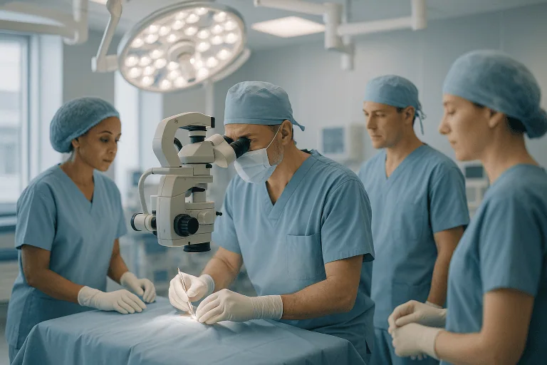
Advanced imaging identifies the location, cause and severity of disease. Digital angiography maps vessels in detail during procedures. Operating microscopes, navigation systems, intraoperative imaging where appropriate, neurophysiological monitoring and intensive care systems help physicians work on delicate brain and vessel structures with greater precision. The point of the technology is not sophistication for its own sake; it is matching the intervention to your anatomy and reducing avoidable uncertainty.
Why Acting Early Matters
Brain tissue is highly sensitive to interruptions in blood flow. During an ischaemic stroke, every minute affects how much tissue remains at risk, and early treatment may reduce disability in eligible patients. This is why doctors treat sudden weakness, speech trouble, facial drooping, visual loss, severe imbalance and abrupt severe headache as emergencies rather than symptoms to watch at home.
Early action matters outside the emergency setting too. A TIA may be brief, but it can signal an unstable vascular condition. Symptomatic carotid narrowing carries a different risk profile from silent narrowing. An aneurysm with certain high-risk features deserves discussion before rupture, not after. A vascular malformation may be safer to treat before it bleeds, depending on its anatomy and natural history. Equally, some findings are best monitored rather than treated, and timely specialist review is what distinguishes one situation from the other.
Delayed evaluation lets risk factors run uncontrolled. High blood pressure keeps damaging small vessels. Undetected atrial fibrillation can send further clots to the brain. Diabetes, cholesterol abnormalities, smoking and sleep apnoea accelerate vascular disease over years. In some patients, recurrent small strokes accumulate quietly, gradually eroding walking, memory, mood and independence: a decline that is easier to slow than to reverse.
There are practical reasons too. Many cerebrovascular treatments need planning: medication review, pre-procedure imaging, anaesthesia evaluation, rehabilitation arrangements and coordination across specialties. An early, structured assessment lets the medical team rank urgency honestly, and avoid both dangerous delay and unnecessary haste.
Potential Benefits of Cerebrovascular Disease Treatment
What treatment can achieve depends on the diagnosis, the timing and your overall condition. The central aim is always to protect brain function and reduce preventable neurological harm.
| Benefit | What It Means for You |
|---|---|
| Reduced risk of future stroke | Identifying the cause of reduced blood flow or clot formation allows physicians to recommend medication, procedures, or surgery aimed at lowering future risk. |
| Rapid treatment of urgent events | In selected acute stroke cases, time-sensitive therapies may restore blood flow and help limit neurological injury. |
| More accurate diagnosis | Detailed brain, vessel, and heart evaluation can clarify whether symptoms are due to carotid disease, cardiac embolism, aneurysm, malformation, small vessel disease, or another cause. |
| Personalised treatment planning | Care can be adapted to your anatomy, symptoms, age, medication needs, and long-term health priorities. |
| Support for recovery and independence | Early rehabilitation, swallowing evaluation, speech therapy, mobility support, and cognitive care may improve day-to-day function after stroke or brain bleeding. |
| Long-term vascular health management | Ongoing control of blood pressure, cholesterol, diabetes, heart rhythm, and lifestyle factors helps protect both brain and cardiovascular health. |
Recovery Timeline After Cerebrovascular Treatment
Recovery varies widely with the condition and the treatment. The timeline below gives a general sense of what many patients experience after evaluation, endovascular treatment, surgery or stroke-related care, treat it as orientation, not prediction.
| Time Period | What Patients Can Expect |
|---|---|
| Day 1 | Patients are monitored closely for neurological status, blood pressure, heart rhythm, breathing, swallowing, and procedure access sites. Emergency stroke patients may be cared for in an intensive or specialised stroke unit. |
| First Week | Further imaging, medication adjustment, rehabilitation assessment, and specialist consultations may take place. Some elective endovascular patients may leave the hospital sooner, while stroke or surgery patients may need longer observation. |
| First Month | Many patients focus on medication adherence, blood pressure and risk-factor control, wound or access-site healing, and rehabilitation. Follow-up visits or imaging may be scheduled depending on the diagnosis. |
| Three to Six Months | Neurological recovery often continues, particularly after stroke. Therapy may target strength, balance, speech, swallowing, cognition, and daily activities. Some patients undergo repeat vascular imaging. |
| Longer Term | Prevention becomes the central focus: maintaining vascular risk control, monitoring treated aneurysms or stenoses when needed, and continuing follow-up with neurology, cardiology, or neurosurgery as appropriate. |
Behind these timeframes sits a biological process worth understanding: neuroplasticity, the brain’s capacity to reorganise and let undamaged regions take over some of the work of injured ones. Rehabilitation is how that capacity is trained. Physiotherapists work on strength, balance and walking; occupational therapists rebuild everyday skills such as dressing, cooking and using a keyboard; speech and language therapists address speaking, understanding and safe swallowing; neuropsychologists assess memory, attention and mood. Progress after a stroke is rarely a straight line, plateaus and setbacks are part of the pattern, but repetition, gradually increasing challenge and realistic goal-setting give the recovering brain the practice it needs.
What Influences Outcomes and a Good Result?
Outcomes in cerebrovascular disease depend on timing, diagnosis, anatomy, treatment selection and overall health, in combination, not in isolation. In acute ischaemic stroke, one of the most important factors is how quickly the blocked artery is evaluated and whether you meet the criteria for reperfusion treatment. The location and size of the stroke, the presence of collateral blood flow around the blockage, and your baseline health all play major roles.
For aneurysms, outcomes hinge on whether the aneurysm has ruptured, its size, shape and location, and whether endovascular or surgical treatment offers the better risk-benefit profile in your specific anatomy. Ruptured aneurysms are medical emergencies and may require intensive care for complications such as rebleeding, vasospasm, hydrocephalus and brain swelling. Unruptured aneurysms demand careful judgement: the risk of treatment has to be weighed honestly against the estimated risk of future rupture, and for some aneurysms monitoring is the better answer.
For carotid disease, good outcomes rest on proper selection between medical therapy, endarterectomy and stenting. The degree of narrowing matters, but it is not the only factor. Symptoms, plaque stability, age, heart and lung health, prior neck surgery or radiation, and vessel anatomy all influence which approach is safest for you. Careful blood pressure control and medication management around the procedure matter as well.
For vascular malformations, the pattern of blood flow, the location relative to critical brain functions, any prior bleeding and associated aneurysms shape the plan. Some malformations are observed, some are treated in stages, and some need combined methods. A thoughtful plan avoids overtreatment while still addressing meaningful risk.
Rehabilitation is a major determinant of recovery in its own right. After stroke or haemorrhage, improvement may continue for months, and some patients keep progressing for longer with structured therapy. Early mobilisation when medically safe, swallowing protection, prevention of complications, mood and sleep support, and family education all contribute. Patients who understand their medications and their warning signs are also better prepared for long-term prevention.
A good result is not defined only by an imaging report. It may mean preventing a first stroke, avoiding another one, safely treating an aneurysm, improving blood flow, reducing seizure or bleeding risk, regaining speech or mobility, or going home with a prevention plan you can actually sustain. The right goal for your treatment should be stated plainly before treatment begins, and if it has not been, that is a fair question to raise with your doctors.
How Acibadem Organises Cerebrovascular Care
Patients facing a cerebrovascular diagnosis need more than a procedure. They need a reliable diagnosis, a clear explanation of the options, coordinated decision-making, and support before, during and after treatment. These conditions are technically complex and emotionally demanding, so the organisation of care matters as much as any single intervention.
At Acibadem, cerebrovascular conditions are managed by physicians across the related specialties. Depending on the case, care may involve neurologists, neurosurgeons, interventional neuroradiologists, vascular surgeons, cardiologists, anaesthesiologists, intensive care physicians, radiologists, rehabilitation specialists, speech and swallowing therapists, and specialised nursing teams. This multidisciplinary structure is particularly valuable when your condition could reasonably be treated in more than one way, which, in cerebrovascular medicine, is often.
Complex cases may be reviewed through specialist boards or multidisciplinary discussions that integrate imaging findings, procedural feasibility, medical risk and your own priorities. A patient with carotid stenosis may need medical treatment, stenting and endarterectomy compared side by side. A patient with an aneurysm may need observation, endovascular treatment and microsurgery weighed against each other. A patient recovering from stroke may need acute care and rehabilitation planned as a single pathway. Evidence-based international protocols guide the process, while the plan itself is adapted to the individual diagnosis.
Technology supports this decision-making rather than replacing it: advanced MRI and CT, vascular ultrasound, angiographic imaging, neurophysiological monitoring, intensive care systems and minimally invasive endovascular tools are used according to medical need, to confirm the diagnosis, define the anatomy, guide treatment precisely and monitor recovery.
Second opinions have a legitimate place in this field. They tend to be most useful for unruptured aneurysms, carotid stenosis, recurrent transient ischaemic attacks, unusual stroke causes, vascular malformations, and cases where previous imaging does not fully explain the symptoms. The purpose is not to repeat testing for its own sake, but to clarify the diagnosis and establish whether the proposed plan is appropriate, urgent, or safe to monitor.
Moving Forward With Clarity
Cerebrovascular disease can feel overwhelming because it involves the brain, the possibility of stroke, and decisions that sometimes have to be made quickly. Yet most patients benefit from something quite ordinary: a structured, expert evaluation that identifies the cause of the problem and lays out the realistic options, medication, monitoring, endovascular therapy, surgery, rehabilitation, or some combination. Clarity is the first form of care.
Personalised care also means recognising your wider context. Some patients are still working and need a plan built around returning to activity. Some are older, with several medical conditions that shape what is safe. Some are caring for others and need a realistic picture of how much support they will need at home during recovery. Wherever you are evaluated, the process moves faster and more accurately when previous imaging, medication lists, medical history and a clear symptom timeline are available from the start, and a careful cerebrovascular plan accounts for these realities from the beginning, not as an afterthought.
Preparation
- Bring previous brain imaging, vascular reports, medication lists and details of any stroke-like symptoms. Your doctor may request MRI or CT angiography, carotid Doppler ultrasound, blood tests and cardiac evaluation. Do not stop blood thinners, blood pressure or diabetes medicines unless your care team instructs you.
Aftercare
- Aftercare may include antiplatelet or anticoagulant medication, blood pressure and cholesterol control, diabetes management and smoking cessation support. Neurological rehabilitation may be recommended to improve movement, speech or daily function. Attend follow-up visits and imaging, and seek emergency care for sudden weakness, speech difficulty, facial drooping or vision loss.
Frequently Asked Questions
What affects the cost of cerebrovascular disease treatment?
Cost depends on the diagnosis, urgency, imaging needs, specialist team, hospital stay, need for intensive care, procedure type, rehabilitation and follow-up plan. A personalised quote requires review of medical records and imaging.
How can I get a quote from Acibadem International?
You can request a free consultation by sharing your medical reports, imaging files, medication list and a summary of symptoms. The team can then coordinate specialist review and prepare an individual care plan and quote.
Is travel suitable for someone with suspected stroke symptoms?
Sudden weakness, speech difficulty, facial drooping, severe headache, vision loss, confusion or loss of balance may be signs of stroke and require immediate emergency care locally. International travel should only be considered after a specialist confirms it is safe.
What is usually included in an international patient package?
When clinically appropriate, a package may include specialist consultations, diagnostic tests, hospital coordination, interpreter support, accommodation guidance, airport support and treatment planning. Inclusions vary by case and should be confirmed in the written quote.
Will I need surgery or an endovascular procedure?
Not always. Many patients are managed with medication, risk factor control and monitoring. Procedures are considered only when imaging, symptoms and overall risk profile suggest a potential benefit.
Is this information medical or financial advice?
No. This is general educational information. Diagnosis, treatment choice and cost can only be confirmed after specialist evaluation, so a free consultation is recommended for a personalised plan.
Medically reviewed by the Acıbadem International Medical Board September 13, 2026
See our medical review board →
Update history
- PublishedJune 8, 2026
- Medical review approvedSeptember 13, 2026
- Last content updateSeptember 13, 2026
References2
- Stroke medlineplus.gov
- Cerebrovascular Disease my.clevelandclinic.org
Trusted care for international patients
From the Acibadem Blog

How Is Cerebrovascular Disease Managed Day to Day? Blood Pressure, Cholesterol and Habits
Cerebrovascular disease is managed day to day by keeping blood pressure in the range your clinician sets, lowering LDL cholesterol (usually with a statin),…