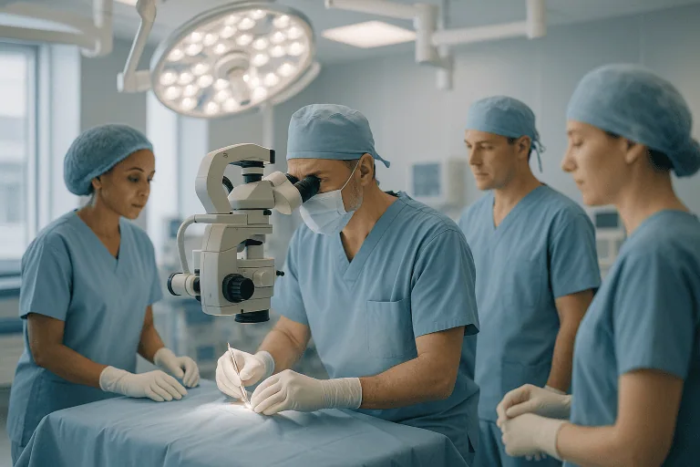
Disc Operations
Disc operations are spine surgeries that remove or relieve pressure from a damaged or herniated disc to reduce pain, numbness, and nerve compression.

Quick answer
Disc operations are surgical procedures that treat damaged spinal discs pressing on nerves or the spinal cord. Depending on the problem, the surgeon may remove a herniated fragment (discectomy or microdiscectomy), decompress the nerve endoscopically, or remove the disc and reconstruct the space with fusion or an artificial disc. They are considered when nerve pain, numbness or weakness persists despite non-surgical treatment.
Disc Operations: What They Are and Who They Help
Disc operations are surgical procedures that relieve pressure on compressed or irritated nerves in the spine when a damaged intervertebral disc is the cause. The goal is not simply to “remove a disc” but to correct the specific structural problem behind your pain or neurological symptoms. For some people, that means taking out a small herniated fragment pressing on a nerve root. For others, it means a wider decompression, stabilisation of the spine, or replacement of the disc itself when the disc and surrounding structures are significantly damaged. Disc operations are considered when nerve-related pain, numbness or weakness has not improved with non-surgical care, or when the neurological findings make earlier treatment sensible.
If you are reading this page, you probably already know that disc pain is rarely a purely physical problem. It affects sleep, work, travel, walking, sitting, and the confidence to move normally. Many people describe a sharp, electric pain travelling into the arm or leg, numbness in the fingers or toes, weakness, or the unsettling feeling that the body no longer responds as it should. When symptoms persist despite medication, physical therapy, injections and lifestyle changes, the possibility of surgery can feel both hopeful and daunting at the same time. That mix of feelings is normal, and it is exactly why the decision deserves careful, unhurried information.
It helps to start with an honest baseline: most disc problems do not require surgery. Many herniations shrink or settle on their own, and non-surgical care resolves symptoms for a large share of people. Disc operations earn their place when nerve compression causes persistent pain, progressive weakness, loss of function, or serious neurological warning signs, situations in which continuing to wait carries its own cost. A well-planned operation can reduce nerve irritation, improve mobility, and help you return to daily life with less pain and more confidence in your own body.
Deciding whether to have spine surgery requires three things working together: an accurate diagnosis, realistic expectations, and an experienced team. This page walks you through each of them, what the different types of disc operations involve, who genuinely needs one, how the procedures are performed, what recovery looks like, and which factors most influence a good result.
How Spinal Discs Work, and How They Fail
Intervertebral discs are the soft cushioning structures between the bones of your spine. Each disc has a tougher outer layer, the annulus, and a gel-like inner centre, the nucleus. Together they absorb load and allow the spine to bend and rotate. With ageing, repetitive strain, injury, genetic factors or degeneration, a disc may bulge, tear, collapse or herniate. When disc material moves out of its normal space, it can narrow the passage available for spinal nerves. The result is the familiar pattern of disc-related symptoms: pain that radiates along the path of the affected nerve, numbness, tingling, or weakness in the muscles that nerve supplies. Certain levels bear the most load and fail most often: in the lower back, the discs between the fourth and fifth lumbar vertebrae and between the fifth lumbar vertebra and the sacrum account for the majority of symptomatic herniations, while in the neck the most commonly involved discs sit in the lower cervical spine, where movement and mechanical stress concentrate.
One point matters more than almost any other in this field: the same MRI finding can mean very different things in different people. A small disc herniation can be disabling if it compresses a sensitive nerve root in a tight canal, while a larger bulge may cause few symptoms if there is enough space around it. Disc bulges and age-related degeneration are common even in people with no pain at all. This is why experienced spine teams never make surgical decisions from a scan alone. They interpret the imaging alongside your physical examination, your symptom pattern, and how you have responded to non-surgical care. When all three point to the same spinal level and the same mechanism of compression, the case for surgery, and the likelihood that it will help, becomes much clearer.
Types of Disc Operations
There is no single “disc operation”. The right procedure depends on where the problem sits, what the disc and the structures around it look like, and what your symptoms and examination show. The main options are outlined below; in practice, your surgeon may combine elements of several.
Discectomy and microdiscectomy
Discectomy is the most common disc operation: the surgeon removes the portion of disc material that is compressing a nerve. A microdiscectomy performs the same task using an operating microscope and smaller instruments through a limited incision, which allows the surgeon to treat many lumbar disc herniations while disturbing as little normal tissue as possible. The herniated fragment is removed; the rest of the disc is usually left in place to keep doing its job. This is the standard operation for a lumbar herniation causing sciatica that has not settled with conservative care. You can read more about the procedure and its indications on our herniated disc surgery page.
Endoscopic discectomy
Endoscopic discectomy treats selected herniations through an even smaller access channel, using a camera and specialised instruments rather than an open corridor. In appropriate cases this approach can reduce tissue disruption, but it is genuinely suitable only for certain herniation patterns. The location of the fragment, the degree of stenosis, your spinal anatomy and the surgeon’s assessment all determine whether it is an option: it is a tool for the right case, not an upgrade for every case. Our page on endoscopic disc surgery explains when it is and is not appropriate.
Cervical disc surgery
In the neck, disc surgery usually means approaching the spine from the front: a procedure often known as anterior cervical discectomy. The damaged disc is removed in its entirety to decompress the nerve root or the spinal cord. Because the disc is fully removed, the space it occupied must be reconstructed. Most commonly this is done with a fusion device and bone graft material, joining the two vertebrae into one solid segment. The front-of-neck approach sounds alarming to many patients, but it uses natural tissue planes and is one of the most established procedures in spine surgery.
Artificial disc replacement
Artificial disc replacement reconstructs the removed disc space with a mobile implant designed to preserve motion at that level, rather than fusing it. In carefully selected patients: the selection depends on age, the state of disc degeneration, arthritis in the small joints of the spine, alignment, the number of levels involved and overall spinal stability: a motion-preserving implant can be a genuine alternative to fusion. It is not suitable for everyone, and an honest surgeon will explain why one option fits your anatomy better than the other. See our dedicated artificial disc replacement page for the details of candidacy and technique.
Decompression, stabilisation and fusion
Some disc operations are combined with other spine procedures. If a disc problem occurs together with spinal stenosis, bony overgrowth, instability, deformity or a recurrent herniation, the operation may include a wider decompression, removal of part of a lamina, or stabilisation with screws, rods, cages or spacers to support alignment and promote fusion. The guiding principle is proportionality: a limited discectomy is ideal for a contained herniation in an otherwise stable spine, while a collapsed, unstable segment may need reconstruction. Doing too little risks leaving the problem unsolved; doing too much exposes you to avoidable risk.
Is disc surgery a major surgery?
It depends on which disc operation you are having. A lumbar microdiscectomy is a contained procedure performed under general anaesthesia, often with a hospital stay of a night or less, and most patients are walking on the day of surgery. A multi-level cervical reconstruction or a lumbar fusion is major surgery by any definition, with a longer operation, a longer stay and a longer recovery. What unites them is that all spine surgery is taken seriously, because the work happens millimetres from nerves. “Minimally invasive” describes the size of the corridor, not the importance of the structures at the end of it.
Who May Need a Disc Operation?
A disc operation may be considered when your symptoms strongly suggest nerve compression and have not improved adequately with conservative treatment. Conservative care typically includes activity modification, anti-inflammatory medication, nerve pain medication, physical therapy, guided exercise, weight management, epidural steroid injections, and, importantly, time. Many disc herniations improve without surgery over weeks to months. Surgery enters the conversation when symptoms persist, worsen, or affect strength and function despite a genuine trial of these measures.
The symptom patterns that most often lead to a surgical evaluation are distinctive. In the lower back, the classic picture is pain radiating from the back into the buttock, thigh, calf or foot, commonly called sciatica. In the neck, a herniation may send pain into the shoulder, arm, hand or fingers. Alongside the pain, you may notice numbness, tingling, muscle weakness, difficulty lifting the foot, reduced grip strength, or pain that worsens with coughing, bending, sitting or standing. Some people find walking itself becomes difficult because nerve compression affects coordination or leg strength. The more precisely your symptoms follow the territory of a single nerve, the more informative they are.
Diagnosis begins with a detailed medical history and a neurological examination, muscle strength, reflexes, sensation, walking pattern, posture and pain distribution. Imaging then confirms the suspected level and cause of compression. Magnetic resonance imaging is the most common test because it shows discs, nerves, the spinal cord and soft tissues in detail. Computed tomography is useful when the bone anatomy needs closer evaluation, particularly after previous surgery or in complex degeneration. Plain X-rays, including bending and extension views, can reveal alignment problems, disc height loss or instability that a static MRI misses. In selected cases, electrodiagnostic testing helps distinguish spinal nerve compression from peripheral nerve conditions such as carpal tunnel syndrome, which can mimic a cervical disc problem.
Some symptoms change the clinical picture entirely. New bladder or bowel dysfunction, numbness in the saddle area, rapidly progressive weakness, severe balance problems or signs of spinal cord compression point to conditions that spine teams treat as emergencies, because the window for protecting nerve function can be short. These presentations follow a different, much faster pathway than the planned evaluation described on this page.
Conditions Treated With Disc Operations
Disc operations are most often performed for herniations in the lumbar spine, where displaced disc material compresses nerve roots and causes leg pain. They are also common in the cervical spine, where herniation or degeneration may compress nerve roots or, more seriously, the spinal cord itself. Thoracic disc problems in the mid-back are less common, but they can be significant when they cause cord compression or severe pain, and they demand particular surgical expertise because of the anatomy of that region.
The main indications include a lumbar disc herniation causing sciatica, a cervical disc herniation causing arm pain or weakness, and degenerative disc disease associated with nerve compression or instability. Surgery may also be considered for a recurrent disc herniation after a previous operation, when symptoms return and imaging confirms renewed compression at the same level. In some patients, disc degeneration contributes to spinal stenosis, a narrowing of the passages the nerves travel through; when disc collapse and bony changes occur together, decompression alone may not be enough, and stabilisation may enter the discussion.
Disc operations can also form part of the treatment for cervical myelopathyspinal cord compression in the neck that causes hand clumsiness, gait imbalance, weakness or changes in fine motor control, such as difficulty with buttons or handwriting. Because spinal cord symptoms can progress, evaluation of suspected myelopathy is typically more urgent than for uncomplicated nerve root pain, and the goal of surgery is often to stop the condition worsening as much as to improve existing symptoms.
It bears repeating that not every disc abnormality on MRI requires surgery. A good surgical indication requires a close match between your symptoms, your examination findings and your imaging. That careful matching, deciding who should have a disc operation as much as how to do one, is among the most important factors in achieving a good outcome, and it is a fair test of any spine team you are evaluating.
How a Disc Operation Is Performed
The process begins well before the day of surgery. Your imaging, medical history, medications, previous treatments and general health are reviewed in full. A complete set of records: the MRI images themselves rather than reports alone, an up-to-date medication list and any previous surgical notes, makes this review more accurate and often prevents repeat testing.
Preoperative preparation may include blood tests, cardiac or anaesthesia assessment, updated imaging, and a structured discussion of your medications. If you take blood thinners, certain diabetes medications or supplements, any adjustment before surgery is decided and directed by your treating team: it is never something to change on your own. Stopping smoking is strongly encouraged, particularly when fusion is being considered, because smoking measurably affects healing and bone union. You will also be told what to expect about fasting, anaesthesia, the likely hospital stay, pain control, wound care and early movement after the operation.
What happens during a lumbar microdiscectomy?
A lumbar microdiscectomy follows a well-defined sequence. Understanding it removes much of the mystery:
- Step 1: Positioning and anaesthesia. You are placed under general anaesthesia and positioned face down on a frame that protects your abdomen and supports your spine.
- Step 2: Confirming the level. The surgeon uses intraoperative imaging to confirm the exact spinal level before making any incision: a formal verification step, not an assumption.
- Step 3: Access. A small incision is made over the affected level and, under magnification, a narrow passage is created through the tissues down to the spine.
- Step 4: Exposure. A limited amount of bone or ligament may be removed if needed to reach the compressed nerve safely.
- Step 5: Decompression. The nerve root is carefully protected and gently moved aside while the herniated disc fragment is removed. The surgeon checks that the nerve is free along its course.
- Step 6: Closure. The small corridor is closed in layers. Because little normal tissue has been disturbed, mobilisation can usually begin the same day.
In an endoscopic disc procedure, the same decompression is achieved through an even smaller channel using a camera and dedicated instruments. The steps are conceptually similar, but the technique and the case selection differ, which is why not every herniation can be treated this way.
How is cervical disc surgery different?
Cervical disc surgery usually approaches the spine from the front of the neck, through natural tissue planes, rather than from behind. The damaged disc is removed completely to decompress the nerve root or spinal cord, and the empty disc space is then reconstructed, most commonly with a fusion device and bone graft, or in selected patients with an artificial disc that preserves motion at that level. The choice between fusion and disc replacement is one of the most important conversations you will have before surgery. It depends on your age, the extent of disc degeneration, arthritis in the facet joints, your spinal alignment, the number of levels involved and the stability of the segment. Neither option is universally better; each fits a different anatomy and situation.
When a disc operation requires stabilisation, the surgeon may use implants such as screws, rods, cages or spacers to support alignment and promote fusion. Depending on the case, the team may also use navigation systems, intraoperative imaging, surgical microscopes, endoscopes, neuromonitoring and high-resolution diagnostic imaging. These technologies help identify anatomy, confirm the operative level, visualise delicate nerve structures and place implants accurately. It is worth keeping perspective, though: technology supports surgical judgement: it does not replace the importance of correct diagnosis, sound planning and experience. A well-chosen operation done with standard tools beats the wrong operation done with impressive ones.
Operating time varies. A straightforward lumbar microdiscectomy takes a relatively short time, while a complex cervical or lumbar reconstruction takes considerably longer. Hospital stay also differs by procedure and by your condition: some patients go home the same day or after one night, while others stay longer for monitoring, pain control or rehabilitation planning. Whatever the scale of the procedure, your team will review you before clearing longer journeys, because prolonged sitting and immobility in the early weeks after spine surgery carry risks of their own that sensible planning avoids.
Why Timing Can Matter
Many disc problems can be observed safely for a period, especially when pain is improving and there is no significant weakness. Watchful waiting is a legitimate strategy, not a failure of nerve. But delay carries costs in certain situations. Persistent nerve compression can prolong pain and limit mobility, which in turn leads to muscle deconditioning, disrupted sleep, anxiety and reduced work capacity: a downward spiral that becomes harder to reverse the longer it runs. When weakness is present, and particularly when it is progressing, earlier decompression may improve the chance that the nerve recovers.
The cervical spine deserves special mention. When the spinal cord itself is compressed, symptoms such as balance difficulty, hand clumsiness, stiffness in the legs or subtle changes in coordination may reflect myelopathy. This condition can worsen over time, sometimes gradually enough that patients adapt without realising how much function they have lost. The purpose of surgery in myelopathy is often to halt progression as much as to improve existing symptoms, and waiting too long can allow neurological changes to become difficult or impossible to reverse.
Certain presentations sit outside the planned-surgery timeline altogether. Loss of bladder or bowel control, numbness around the groin or inner thighs, severe or rapidly worsening weakness, fever accompanying back pain, unexplained weight loss, or pain following major trauma each point to conditions that clinicians assess urgently rather than electively.
Benefits of Disc Operations
When surgery is appropriately selected and carefully performed, a disc operation can offer several meaningful benefits for someone living with nerve compression. The table below summarises what each benefit actually means in daily life.
| Benefit | What It Means for You |
|---|---|
| Relief of nerve compression | Removing the disc material or narrowing that presses on a nerve can reduce radiating leg or arm pain and give the nerve the conditions it needs to begin recovering. |
| Improved mobility | Less pain can make walking, standing, sitting and daily movement easier, allowing a gradual return to normal routines, work and travel. |
| Protection of neurological function | In patients with weakness, spinal cord compression or progressive symptoms, surgery may help prevent further deterioration. |
| Reduced reliance on pain medication | As nerve pain improves, many patients are able to decrease pain medication under the supervision of their treating doctor. |
| More precise treatment planning | Modern imaging and specialist evaluation identify the exact pain generator and match the procedure to your anatomy and goals, rather than applying a standard operation to every back. |
Note what is absent from this list: a promise that all pain disappears. Disc operations are most reliable at relieving the nerve-related arm or leg pain caused by compression. Back or neck pain at the spine itself has more possible causes, and an honest surgeon will tell you which of your symptoms the operation is designed to address.
Recovery After a Disc Operation
Recovery varies with the type of operation, the severity and duration of nerve compression, your general health, and whether stabilisation or fusion was performed. The timeline below describes a typical course; your own plan will be set by your surgical team.
| Time Period | What Patients Can Expect |
|---|---|
| Day 1 | You are monitored after anaesthesia, pain control begins, and you are usually encouraged to stand or walk with assistance when medically appropriate, often on the day of surgery. |
| First week | Incision discomfort is normal. Most patients walk short distances several times a day, avoid bending and heavy lifting, and follow wound care instructions closely. |
| First month | Activity increases gradually. Radiating nerve pain may already be significantly improved, while numbness or weakness recovers more slowly. Follow-up reviews guide rehabilitation. |
| Six to twelve weeks | Many patients return to desk work and light daily activities, depending on the procedure. Physical therapy focuses on posture, core strength and safe movement patterns. |
| Longer term | Strength and endurance continue to rebuild. After fusion, bone healing and activity progression are monitored over a longer period with follow-up imaging. |
How long does it take to recover from a disc operation?
There is no single answer, because “disc operation” covers procedures of very different scale. After a lumbar microdiscectomy, many patients walk on the day of surgery, manage light daily activity within days, and return to desk-based work within a few weeks; physically demanding work takes longer and is cleared step by step. After a fusion, the timeline extends, because the bone itself must heal and that healing is monitored over months. Two patterns are consistent across all procedures: nerve pain tends to improve faster than numbness, and weakness recovers gradually, because nerves heal slowly. Your surgeon’s milestones, not a calendar date, should guide each step up in activity.
How painful is a diskectomy?
A diskectomy causes incision-site soreness that is expected and managed with a structured pain-control plan from your team; it typically eases substantially over the first week or two. What surprises many patients is the other side of the equation: the radiating nerve pain in the leg or arm, the pain that drove the decision to operate, often improves early, sometimes noticeably within the first days, because the pressure on the nerve is gone. Numbness and weakness follow a slower course, and some residual sensory change can persist while the nerve recovers. If pain increases rather than settles after the first days, that is something your team wants to know about at follow-up, not something to endure quietly.
Between discharge and your follow-up appointment, most of recovery happens at home, and it is worth understanding wound care before surgery rather than after. In general, teams ask you to keep the incision clean and dry, follow the dressing instructions you were given, and pay attention to the changes that matter most in the first weeks: increasing redness or swelling around the wound, discharge, fever, pain that escalates rather than settles, new or worsening numbness or weakness, or swelling and tenderness in the calf. These findings matter because they can signal infection, a blood clot or renewed nerve compression, problems that are far easier to treat when identified early.
Rehabilitation deserves its own mention, because much of the final result is built there. Walking is the primary exercise after most disc operations, frequent, short and gradually longer. Structured physiotherapy usually begins once the wound has settled and works on posture, core and hip strength, hamstring flexibility and safe movement patterns for lifting and bending. Just as important is what you avoid early on: prolonged sitting, twisting under load and heavy lifting, each cleared step by step by your team. Patients who treat rehabilitation as part of the operation, rather than an optional extra, tend to regain function more smoothly and protect the rest of their spine in the process.
What Influences a Good Outcome?
The single strongest predictor of a good result is correct diagnosis. Outcomes are generally best when your symptoms, your neurological examination and your imaging all point to the same spinal level and the same mechanism of compression. Classic leg pain from a lumbar herniation that matches the MRI and the examination responds well to decompression. More complex pain patterns, long-standing nerve damage, widespread degeneration or chronic pain syndromes call for more nuanced expectations, and a surgeon who sets them honestly before the operation rather than explaining them afterwards.
The duration and severity of your symptoms also matter. Nerve pain tends to improve faster than numbness, and muscle weakness can recover, but recovery may be incomplete if the compression has been severe or prolonged, which is one reason progressive weakness shifts the timing conversation. Age, diabetes, smoking, obesity, bone quality, overall fitness and previous spine surgery all influence healing and rehabilitation. You are not a passive participant in any of this: patients who walk regularly, respect the early restrictions, attend therapy and avoid tobacco give themselves measurably better conditions to heal in.
Surgical planning is the third pillar. A limited discectomy is ideal for a contained herniation in an otherwise stable spine. Where instability, deformity, severe disc collapse or repeated herniation is present, a more comprehensive procedure may be needed, but the reverse error is just as real: performing a larger operation than necessary exposes you to avoidable risk. The decision should be proportionate, evidence-based and tailored to you, and you are entitled to ask why this operation rather than a smaller or larger one.
Finally, expectations. Disc surgery is most effective for nerve-related arm or leg pain caused by compression. It may also improve back or neck pain when that pain is genuinely linked to the treated disc, but axial spine pain can have multiple contributing causes, and no operation addresses all of them at once. A careful preoperative discussion should leave you clear on the goals of surgery, which of your symptoms are most likely to improve, how long recovery will take, and where the limits of treatment lie.
How serious is spinal disc surgery?
Every spinal operation is serious, because the work takes place next to nerves and, in the neck, next to the spinal cord. The recognised risks include infection, bleeding, a tear in the membrane covering the nerves, nerve injury, recurrence of the herniation at the same level, and, after fusion, the possibility that the bone does not unite as planned. How likely each of these is depends on the specific procedure, your health and your anatomy, which is why generic figures are less useful than a direct conversation about your case. A trustworthy surgeon will explain the risks that apply to you plainly rather than minimising them, and will weigh them openly against the risks of not operating, because ongoing nerve compression carries risks of its own.
Disc Operations at Acibadem
Spine care at Acibadem hospitals is delivered by physicians from the specialties this work requires: neurosurgery, orthopaedics and traumatology, radiology, anaesthesiology, physical medicine and rehabilitation, pain medicine and, where needed, neurology. Complex cases can be reviewed through multidisciplinary boards, where imaging, symptoms, previous treatments and surgical options are discussed collectively before a recommendation is made. This matters most for the situations where a single opinion is least reliable, recurrent herniation, multi-level disease, spinal cord compression, deformity, or a spine that has already been operated on.
Because the decision to operate must rest on more than an MRI report, diagnostic pathways use high-resolution MRI, CT, digital X-ray and functional views where needed to evaluate compression, alignment, instability and prior surgical changes. In theatre, teams draw on surgical microscopes, endoscopic systems, intraoperative imaging, navigation support and neuromonitoring according to the demands of the individual case, tools that help surgeons work through smaller corridors when appropriate, confirm anatomy, and protect neural structures during the operation.
Follow-up after a disc operation is structured rather than left to chance: wound review, staged clearance of activity, physiotherapy planning and, after fusion procedures, imaging at intervals to monitor bone healing. The discharge summary you receive documents the procedure performed, any implants used, the medication plan and the rehabilitation programme, so that every physician involved in your later care has a complete and accurate picture to work from.
Making the Decision on Your Own Terms
Disc pain narrows a person’s world. It complicates ordinary choices, whether to sit through a meeting, take a flight, lift a child, return to sport, or trust yourself to sleep through the night. When non-surgical care is no longer enough, disc operations offer a realistic path toward less nerve pain and better function, provided the diagnosis is right and the procedure fits the problem. The most valuable step you can take is a careful evaluation by a qualified spine specialist who examines you, studies your imaging alongside your symptoms, and explains in plain terms whether surgery is appropriate, which procedure fits your condition, and what recovery will genuinely involve.
A second opinion is a reasonable and common part of this process, particularly if you have been told you need a fusion, if symptoms have returned after a previous operation, or if your imaging findings do not clearly match your pain pattern. Good spine teams welcome the scrutiny, because the same careful matching of symptoms, examination and imaging that produces good surgical outcomes also, sometimes, produces the most useful recommendation of all: that you do not need an operation yet.
Frequently Asked Questions
What is a disc operation and when is it considered?
Disc operations are spine surgeries that remove or relieve pressure from a damaged or herniated disc pressing on nerves or the spinal cord. Depending on the problem, the surgeon may remove a herniated fragment, decompress the nerve endoscopically, or remove the disc and reconstruct the space with fusion or an artificial disc. They are usually considered when nerve pain, numbness or weakness persists despite non-surgical care; your doctor will decide whether surgery is appropriate for you.
What types of disc operations are there?
There is no single disc operation. A discectomy or microdiscectomy removes the portion of disc material compressing a nerve, usually leaving the rest of the disc in place. Endoscopic discectomy treats selected herniations through an even smaller channel. In the neck, the disc is often removed completely and the space reconstructed with fusion or, in selected patients, an artificial disc. Some operations also include wider decompression or stabilisation with screws, rods or cages when the spine is unstable.
Is disc surgery considered a major operation?
It depends on which procedure you are having. A lumbar microdiscectomy is a contained procedure performed under general anaesthesia, often with a hospital stay of a night or less, and most patients are walking on the day of surgery. A multi-level cervical reconstruction or lumbar fusion is major surgery, with a longer operation, stay and recovery. All spine surgery is taken seriously, because the work happens millimetres from nerves.
How long does recovery take after a disc operation?
Recovery varies with the type of operation, the severity of nerve compression, your general health and whether fusion was performed. After a lumbar microdiscectomy, many patients walk on the day of surgery, manage light activity within days and return to desk-based work within a few weeks. After fusion, the timeline extends because the bone must heal. Nerve pain tends to improve faster than numbness or weakness, and your surgeon's milestones, not a calendar date, should guide each step.
What are the risks of spinal disc surgery?
Every spinal operation is serious because the work takes place next to nerves and, in the neck, next to the spinal cord. Recognised risks include infection, bleeding, a tear in the membrane covering the nerves, nerve injury, recurrence of the herniation at the same level and, after fusion, the possibility that the bone does not unite as planned. How likely each is depends on the procedure, your health and your anatomy, which your surgeon will discuss with you.
When should I seek urgent medical advice for disc symptoms?
Some symptoms change the picture entirely. New bladder or bowel dysfunction, numbness in the saddle area around the groin or inner thighs, rapidly progressive weakness, severe balance problems, fever accompanying back pain, unexplained weight loss or pain following major trauma can point to conditions that clinicians assess urgently rather than electively. If you feel unwell, or your symptoms are severe or getting worse, get medical care where you are now rather than waiting or relying on this page.
Preparation
- Patients usually undergo a spine examination, MRI or CT imaging, and anesthesia assessment before surgery. Blood tests and medication review are completed, and blood thinners may need to be stopped with medical approval. Fasting is typically required from midnight before the procedure.
Aftercare
- Early walking is encouraged, but bending, heavy lifting, and twisting should be avoided during initial recovery. Pain control, wound care, and follow-up visits are important. Physical therapy may be recommended to strengthen the back and reduce recurrence risk.
Medically reviewed by the Acıbadem International Medical Board September 13, 2026
See our medical review board →
Update history
- PublishedJune 8, 2026
- Medical review approvedSeptember 13, 2026
- Last content updateSeptember 12, 2026
References1
- Slipped disc nhs.uk
From the Acibadem Blog

When Does a Damaged Disc Need an Operation? Nerve Symptoms That Change the Plan
Disc surgery is usually considered when a damaged disc presses on a nerve and causes progressive weakness, worsening numbness, or leg or arm pain…

Discectomy vs Disc Replacement: Which Disc Operation Fits Which Spine Problem
A discectomy removes the fragment of a herniated disc that is pressing on a nerve and leaves the rest of the disc in place;…