

Valvuloplasty
Valvuloplasty is a catheter-based procedure that widens a narrowed heart valve, often using a balloon. It can improve blood flow and reduce symptoms in selected valve stenosis patients.
Quick answer
Valvuloplasty is a catheter-based procedure that widens a narrowed heart valve. A thin tube is guided through a blood vessel to the heart, and a balloon at its tip is briefly inflated across the valve to stretch the opening and separate fused leaflets. It is used in selected cases of mitral, pulmonary, aortic and tricuspid stenosis. It improves the existing valve; it does not replace it.
What Is Valvuloplasty?
Valvuloplasty is a procedure that widens a narrowed heart valve so blood can move through it more easily. In its most common modern form, a thin flexible tube called a catheter is guided through a blood vessel, usually from the groin, up to the heart, and a small balloon at the catheter tip is briefly inflated across the narrowed valve to stretch the opening. It is considered for selected patients with valve stenosis, which means a valve that no longer opens fully, when the shape and condition of the valve make a catheter-based widening likely to help.
The mechanism is simple to describe and demanding to perform. Many stenotic valves, particularly those damaged by rheumatic fever, have leaflets that have fused together along their edges. When the balloon inflates, it stretches the valve ring and can split those fused areas, called commissures, so the leaflets can separate more fully with each heartbeat. The inflation lasts only seconds and may be repeated. The team measures pressures across the valve before and after each inflation and uses imaging throughout, so decisions are made on live information rather than a single measurement.
The heart has four valves: mitral, aortic, pulmonary and tricuspid. Valvuloplasty is most commonly considered for certain cases of mitral stenosis, especially rheumatic mitral stenosis with favourable valve anatomy. It is also well established for pulmonary valve stenosis, where selected patients often see marked improvement in symptoms and pressure. In aortic stenosis it has a narrower role, usually as a bridge to another therapy, for symptom relief in patients who are not immediate candidates for valve replacement, or for urgent stabilisation. Tricuspid valvuloplasty is uncommon and reserved for specific situations.
One point deserves emphasis from the start: valvuloplasty does not replace the valve. It reshapes and widens the valve you already have. That distinction shapes everything else on this page, because how long the result lasts depends on the cause of the narrowing, the condition of the leaflets, the amount of calcium in the valve, whether the valve also leaks, and how well the heart is pumping. In some patients the improvement is substantial and lasts for years. In others the narrowing returns, or a different treatment becomes the better option later.
What does valvuloplasty mean, and how do you pronounce it?
Valvuloplasty means, literally, the reshaping or repair of a valve, from “valvula” (small valve) and “-plasty” (moulding or forming). It is pronounced VAL-vyoo-loh-plas-tee, with the stress on the first syllable. You may see the word used in two senses. Surgeons sometimes use “valvuloplasty” for the open surgical repair of a valve, most often a leaking one. Cardiologists more often mean the catheter-based balloon procedure for a narrowed valve, which is the subject of this page. If a report or referral letter uses the word, it is worth confirming which of the two is intended, because the procedures, recovery and risks are very different.
What is the difference between a valvuloplasty and a valvotomy?
In everyday clinical use, the two words usually describe the same balloon procedure, and you will see “balloon valvuloplasty” and “balloon valvotomy” used interchangeably in reports. Strictly speaking, valvotomy means cutting into or opening a valve, historically this was a surgical operation in which fused leaflets were separated by hand or with an instrument, either through a beating heart (closed valvotomy) or under direct vision (open valvotomy). Valvuloplasty is the broader term for reshaping the valve. The balloon technique achieved by catheter what the surgical valvotomy achieved by operation, which is why the names have merged in practice.
When a Narrowed Heart Valve Begins to Limit Your Life
Valvular stenosis means that one of the heart valves does not open fully. Instead of letting blood move forward smoothly, the narrowed valve creates resistance, and the heart chamber behind it must generate more pressure to push blood through the smaller opening. Over months and years, that extra workload changes the heart. Depending on which valve is involved, the result can be shortness of breath, fatigue, chest discomfort, dizziness, fainting, palpitations, swelling in the legs or abdomen, or a steady loss of exercise capacity. Some people have few symptoms at first, even when the narrowing has become significant, because the heart adapts before it complains.
Being told that a valve is narrowed raises practical questions. Are the symptoms really coming from the valve? How urgent is the situation? Is an operation unavoidable, or can a catheter-based treatment postpone or replace it? None of these questions can be answered from symptoms alone; they depend on imaging and on a considered review of the whole heart.
Not every narrowed valve can be treated with valvuloplasty, and not every patient is best served by it even when it is technically possible. The most important step is a precise diagnosis and a thoughtful review by a heart team experienced in valve disease. At Acibadem, that review draws on modern cardiac imaging, interventional cardiology and, where needed, input from the cardiovascular surgery team. The purpose is to establish whether valvuloplasty is the right option for your valve, whether a different catheter-based therapy fits better, or whether surgical repair or replacement would give you a more durable result.
Who May Need Valvuloplasty?
Valvuloplasty may be considered for people whose symptoms or heart findings are caused by valve stenosis and whose valve anatomy is suitable for balloon widening. The decision is never based on symptoms alone. It requires detailed assessment of the valve itself, the pressure gradient across it, the size and function of the heart chambers, the heart rhythm, the pressure in the lung circulation, and the rest of your medical picture. Two patients with the same symptom can need entirely different treatments.
The symptoms that most often lead to evaluation are breathlessness with activity, reduced exercise capacity, fatigue, chest pressure, dizziness, fainting episodes, a rapid or irregular heartbeat, and swelling in the legs or abdomen. Some patients notice the change indirectly: stairs that used to be easy, a hill that now forces a stop, difficulty sleeping flat, or a daily routine that has quietly shrunk. In mitral stenosis, symptoms may first become obvious during pregnancy, infection, anaemia or an episode of atrial fibrillation, because each of these situations increases the workload on the heart and unmasks the obstruction.
How is valve stenosis diagnosed before valvuloplasty?
Diagnosis usually begins with a careful history, physical examination and echocardiography. A heart murmur may raise the suspicion of valve disease, but imaging is what defines its severity. Transthoracic echocardiography, an ultrasound scan through the chest wall, is typically the first key test. It shows how the valve leaflets move, the valve area, the pattern of blood flow, the pressure gradient across the valve, the dimensions of the heart chambers and the pumping function of the heart. Transoesophageal echocardiography, performed through the food pipe, may be added when more detailed images are needed, particularly before mitral valvuloplasty, to define the valve anatomy precisely and to check the left atrium for blood clots.
Further tests are chosen according to the individual case: an electrocardiogram to document heart rhythm, chest imaging, blood tests, exercise testing in selected patients whose symptoms and imaging findings do not quite match, cardiac CT to assess calcification and anatomy, and cardiac catheterisation when direct pressure measurements or a coronary artery assessment are required. Previous echocardiograms, angiogram reports, CT or MRI scans, medication lists and physician letters allow any reviewing team to work from your real history rather than starting from zero.
Candidates typically fall into a few groups: people with symptomatic moderate to severe stenosis and favourable valve anatomy; selected patients with severe stenosis whose heart findings justify treatment before major symptoms develop; and people who need temporary stabilisation before a more definitive valve intervention. In some cases valvuloplasty is recommended precisely because it is less invasive than open surgery. In others the heart team will advise against it, for example when the valve is heavily calcified, already leaking significantly, structurally unsuitable, or simply unlikely to respond well to balloon dilation.
Conditions Balloon Valvuloplasty Can Treat
Balloon valvuloplasty is not a single-purpose procedure; its role differs according to the valve involved and the cause of the narrowing. Careful patient selection matters because the expected benefit, and the honest limitation, is different for each valve.
Mitral valve stenosis is one of the most established indications. Mitral stenosis often develops years after rheumatic fever, which leaves the leaflets thickened and fused along their edges. When the anatomy is favourable, balloon mitral valvuloplasty can substantially improve valve opening and reduce pressure in the left atrium and the lung circulation. It is generally most appropriate when there is no significant mitral regurgitation and no clot in the left atrium; both are checked carefully beforehand, because either can change the plan.
Pulmonary valve stenosis is usually congenital, present from birth, and can come to light in children, adolescents or adults. Balloon pulmonary valvuloplasty is frequently the first-line treatment when the narrowing is significant and the valve structure is suitable, because it reduces the pressure the right ventricle must generate to pump blood to the lungs. In children, the procedure sits within the broader field of paediatric cardiac care, where congenital valve disease is planned and followed over years, not just around a single intervention.
Tricuspid valve stenosis is uncommon and usually occurs with rheumatic disease or other structural valve conditions. Valvuloplasty may be considered in selected cases, particularly when the anatomy is favourable and the goal is to relieve the obstruction between the right atrium and right ventricle.
Balloon aortic valvuloplasty: a bridge rather than a destination
Balloon aortic valvuloplasty is the widening of a narrowed aortic valve with a catheter-mounted balloon, and its role in adults differs from valvuloplasty of the other valves. Aortic stenosis in older adults is usually caused by age-related calcification, sometimes on a bicuspid (two-leaflet) valve. Calcified leaflets respond less well to stretching, and the valve tends to narrow again after balloon dilation. For that reason, balloon aortic valvuloplasty in the modern era is mainly used as a bridge, to stabilise a patient before transcatheter or surgical valve replacement, to relieve symptoms in someone who cannot yet undergo definitive treatment, to improve a patient’s condition before another major operation, or as a palliative measure in selected frail patients. It is rarely offered as a stand-alone, long-term solution for degenerative aortic stenosis. In children and young adults with congenital aortic stenosis, where the valve is not calcified, the calculation is different and balloon treatment can play a larger role.
Valvuloplasty is also discussed in particular clinical situations that cut across valve types: pregnancy with severe symptomatic mitral stenosis, high surgical risk, urgent decompensation from severe stenosis, or the need to improve a patient’s condition before other major treatment. In each of these, the expected benefit is weighed against the procedural risk and against how long the improvement is likely to last.
How the Heart Balloon Procedure Is Performed
The heart balloon procedure follows a defined sequence: planning, catheter access, balloon positioning and inflation, immediate assessment, and monitored recovery. Understanding each stage makes the experience far less abstract.
Preparation and planning
The work begins well before the day of the procedure. Your cardiology team reviews your symptoms, medical history, medications, imaging and general health. Echocardiography is central to planning: it confirms the severity of the stenosis, maps the valve anatomy, and identifies factors that could make valvuloplasty less suitable, such as heavy calcification or significant pre-existing leakage of the valve.
Before mitral valvuloplasty, transoesophageal echocardiography is commonly performed to look for clots in the left atrium and to define the valve in fine detail. If a clot is found, the procedure is usually postponed or the treatment plan changed, because manipulating catheters inside the heart in the presence of a clot could dislodge it. In older patients, or in anyone with risk factors for coronary artery disease, the coronary arteries may be assessed before the intervention.
Your team will also review blood thinners, diabetes treatment, kidney function, allergies to contrast dye, previous anaesthesia issues and any implanted cardiac devices; any adjustments are decided by your treating doctors as part of the plan. You will usually be asked not to eat or drink for several hours beforehand. On the day, an intravenous line is placed and monitoring is attached so that heart rhythm, blood pressure and oxygen levels are tracked continuously.
During the procedure
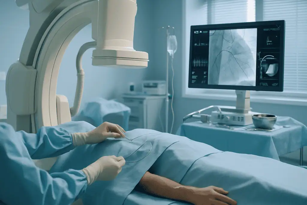
Valvuloplasty is performed in a cardiac catheterisation laboratory or hybrid cardiovascular suite. Most procedures use a vein or artery in the groin, although the access route depends on the valve being treated. The skin is numbed with local anaesthetic. Sedation is often used to help you relax, and general anaesthesia may be recommended in selected cases, particularly when transoesophageal echocardiography is used throughout, or when depth of anaesthesia is safer for the individual patient. Discomfort during and after catheter procedures is actively managed; you can read how in how we control pain after surgery and invasive procedures.
The devices used are sometimes loosely described as balloons for heart surgery, but the phrase is misleading in one important way: the chest is not opened. The cardiac balloon travels folded on the catheter tip through the blood vessels, and everything happens from inside the circulation. Using live X-ray (fluoroscopy) and echocardiographic guidance where appropriate, the interventional cardiologist steers the catheter to the heart. In mitral valvuloplasty, the catheter is usually passed from the right atrium across the wall between the two atria into the left atrium, and then across the mitral valve: a step called transseptal puncture. In pulmonary or aortic valvuloplasty the route differs according to the anatomy and access site.
The core steps, once access is established, run in order:
- Guidewires and catheters are advanced to the target valve under imaging guidance.
- Baseline pressures are measured on both sides of the valve to quantify the obstruction.
- The balloon is positioned precisely across the narrowed valve.
- The balloon is inflated in a controlled way for a matter of seconds, and deflated; inflation may be repeated, sometimes with a stepwise increase in balloon size.
- Pressures are re-measured and the valve is re-imaged to check the new opening and to look for any new or worsened leakage.
- When the result is acceptable, or when further inflation would risk creating leakage, the equipment is withdrawn and the access site is closed.
The technology exists to sharpen judgement, not to replace it. Fluoroscopy guides catheter movement; echocardiography shows valve structure and blood flow in real time; pressure traces show exactly how much obstruction remains; and, in some patients, cross-sectional imaging performed beforehand has already defined the anatomy and the approach. Duration varies with the valve, the anatomy and the complexity of the case. Many valvuloplasty procedures are completed within a few hours, including preparation in the procedure room and the immediate post-procedure assessment; complex cases take longer.
Immediately after the procedure
Once the catheters are removed, pressure or a closure device is used at the access site to prevent bleeding, and you are moved to a recovery area or cardiac unit. Nurses and physicians monitor your heart rhythm, blood pressure, oxygen levels, the access site and your symptoms. You may need to lie flat for a period, especially after groin access, before gradually sitting up and walking with assistance.
An echocardiogram is usually performed after the procedure or before discharge to document the result: the new valve opening, the residual pressure gradient, heart function and the degree of any regurgitation. Some patients notice easier breathing or better effort tolerance quickly; for others the change is more gradual as the heart and lungs adjust to the improved flow.
Length of stay varies. Some patients go home the next day; others need longer observation because of their underlying condition, age, rhythm problems, heart failure or kidney function. Some patients are also advised to stay near the hospital for a follow-up check before undertaking a long journey, particularly after a complex procedure or when medications are being adjusted. General guidance on timing air travel around procedures is set out in flying after surgery: how soon is safe, procedure by procedurethough the final word on your fitness to fly always belongs to your treating team.
How Long Does Balloon Valvuloplasty Last?
How long the result lasts depends mainly on which valve was treated and why it was narrowed; there is no single answer that covers all patients. Broad, honest patterns can still be described. In rheumatic mitral stenosis with favourable anatomy, a good initial result can last for many years, though the valve remains a diseased valve and periodic echocardiography is needed because narrowing can slowly recur. In congenital pulmonary stenosis, the improvement is often long-lasting, which is one reason balloon treatment is so widely used in this condition. In calcific aortic stenosis in adults, the situation is different: re-narrowing after balloon dilation is common and often occurs within months, which is precisely why balloon aortic valvuloplasty is treated as a bridge to valve replacement rather than a destination therapy.
Two practical consequences follow. First, valvuloplasty should be understood as one stage in the long-term management of valve disease, not a one-off fix that closes the file. Second, follow-up imaging is not optional courtesy; it is how recurrence or new leakage is detected before it causes trouble. If the narrowing does return, options may include repeat valvuloplasty in suitable cases, or surgical or transcatheter valve treatment: the possibilities are described in our guide to heart valve surgery.
Why Acting Early Matters
Valve stenosis usually progresses gradually, and the body compensates for a long time. That makes it tempting to wait until symptoms become severe, and it is exactly what cardiologists try to talk patients out of. Delay allows secondary changes to develop. In mitral stenosis, long-standing obstruction raises pressure in the lungs and can lead to pulmonary hypertension, enlargement of the left atrium, atrial fibrillation, blood clot risk and strain on the right side of the heart. In aortic stenosis, advanced disease can bring fainting, chest pain, heart failure or sudden deterioration. In pulmonary stenosis, persistent obstruction loads the right ventricle year after year.
Early evaluation does not mean early intervention. It means knowing the true severity of the disease, identifying the right moment to treat, and avoiding deterioration that could have been prevented. Some patients are best served by careful monitoring with medication and periodic imaging. Others benefit from intervention before the heart muscle weakens or before lung pressures become difficult to reverse. Changing symptoms, new breathlessness, fainting, a racing irregular heartbeat, swelling, or a falling exercise capacity, are among the main reasons cardiologists bring forward the timing of assessment, because they can signal that the balance has shifted.
Potential Benefits of Valvuloplasty
For appropriately selected patients, valvuloplasty can deliver meaningful improvement while avoiding the physical impact of open-heart surgery. The benefits below are potential benefits, whether they apply to you depends on your valve, your anatomy and your overall condition.
| Benefit | What It Means for You |
|---|---|
| Improved valve opening | A wider valve opening lets blood move forward more easily and reduces the pressure burden on the heart chamber behind the valve. |
| Symptom relief | Many well-selected patients experience less breathlessness, better activity tolerance and reduced fatigue after successful widening of the valve. |
| Catheter-based approach | The procedure is performed through the blood vessels, usually without opening the chest, which may mean a shorter recovery than traditional valve surgery. |
| Useful bridge to further treatment | In selected high-risk or urgent cases, valvuloplasty can stabilise symptoms or circulation before a more definitive valve procedure. |
| Personalised timing | For some patients, the procedure postpones the need for valve replacement while keeping future treatment options open. |
Recovery Timeline After Valvuloplasty
Recovery varies with the valve treated, your overall heart condition, the access site, your medications, and whether the procedure was planned or urgent. The pattern below describes a typical, uncomplicated course; your own team’s instructions take precedence over any general timeline.
| Time Period | What Patients Can Expect |
|---|---|
| Day 1 | Monitoring of heart rhythm, blood pressure, the access site and the early valve result. Many patients can sit up and walk with assistance once the required bed-rest period has passed. |
| First week | Mild soreness or bruising at the catheter site is common. Light daily activities usually resume, while heavy lifting and strenuous exercise wait until the care team clears them. |
| First month | Follow-up typically includes examination, medication review, rhythm assessment and echocardiography. Activity increases gradually according to symptoms and heart function. |
| Longer term | Periodic monitoring continues, because narrowing can recur, valve leakage can change, and another valve treatment may become appropriate later. |
Because valvuloplasty avoids a chest incision, day-to-day recovery is usually driven by the access site and by how the heart responds to the improved flow rather than by wound healing. Fatigue in the first days is common and usually settles. What matters most in this period is attending the follow-up echocardiogram, taking prescribed medications exactly as directed by the treating team, and reporting new or returning symptoms so they can be assessed rather than absorbed into daily life.
How Risky Is Valvuloplasty?
Valvuloplasty is generally less physically demanding than open-heart surgery, but it is still a heart procedure and carries real risks that differ from patient to patient. The recognised complications include bleeding or bruising at the access site, injury to a blood vessel, abnormal heart rhythms during or after the procedure, stroke, new or worsened valve leakage, perforation of the heart, allergic reaction to contrast dye, strain on the kidneys from contrast, and, rarely, the need for urgent surgery. The likelihood of each depends on the valve treated, the state of the valve and heart, your age and other conditions, and whether the procedure is planned or performed urgently.
This is why the risk conversation before valvuloplasty should be specific rather than generic. A young patient having balloon dilation of a congenital pulmonary valve and an elderly patient having balloon aortic valvuloplasty as a bridge to valve replacement are not taking the same risk, even though the procedure shares a name. It is also why the procedure belongs in a hospital environment where imaging, anaesthesia, intensive care and cardiac surgery are available, not because complications are expected, but because the safest catheter programmes are the ones prepared for the unexpected.
What Influences Outcomes and a Good Result?
The success of valvuloplasty is decided as much before the procedure as during it. Valve anatomy is one of the strongest predictors. Valves with flexible leaflets and fused commissures, but limited calcification and limited leakage, are the most likely to respond well to balloon dilation. Heavily calcified valves, or valves that already leak significantly, may not be suitable: the balloon may fail to improve the opening, or the stretch may worsen the leak.
The specific valve matters. Balloon mitral valvuloplasty can be highly effective in carefully selected rheumatic mitral stenosis, particularly when the valve score and anatomy are favourable. Pulmonary valvuloplasty often produces strong, lasting pressure reduction when the narrowing sits at the valve itself and the valve is not severely malformed. Aortic valvuloplasty behaves differently: it often improves flow temporarily, but recurrence is common, so it is usually one element of a broader strategy rather than the definitive treatment for degenerative aortic stenosis.
Heart rhythm and chamber function shape both the risk and the recovery. Atrial fibrillation, pulmonary hypertension, an enlarged left atrium, right ventricular dysfunction or advanced heart failure all influence the picture. Some of these improve once the obstruction is relieved; others need ongoing management, which may involve blood thinners, rhythm control, diuretics and treatment of related conditions, all directed by your treating cardiologist as part of the overall plan.
General health completes the picture. Kidney disease, lung disease, frailty, prior stroke, diabetes, active infection, anaemia and coronary artery disease affect planning and recovery. For women who are pregnant or planning pregnancy, timing and imaging choices need particular care, especially in significant mitral stenosis, where the physiological demands of pregnancy can unmask or worsen the obstruction.
Finally, experience and coordination matter. Valvuloplasty demands precise imaging interpretation, careful catheter technique, real-time decision-making, and readiness to manage complications immediately. A good result is not defined by how the valve looks the moment the balloon deflates. It is defined by improved blood flow, an acceptable degree of leakage, genuine symptom improvement, stable rhythm and circulation, a safe recovery, and a sensible long-term plan, because valve disease is a chronic condition even when a catheter procedure brings significant relief.
How Much Does a Balloon Valvuloplasty Cost?
There is no single, honest price for a balloon valvuloplasty, because the cost is built from elements that vary widely between patients. Anyone quoting a fixed figure before reviewing your imaging is guessing. The main cost drivers are:
- The valve being treatedmitral, pulmonary, aortic and tricuspid procedures differ in complexity, equipment and time.
- Diagnostic work-uphow much imaging and testing is needed before the procedure, including transoesophageal echocardiography, cardiac CT or catheter-based measurements.
- Anaesthesia typelocal anaesthetic with sedation versus general anaesthesia.
- Length of hospital stayfrom overnight observation to a longer admission driven by heart failure, rhythm problems or other conditions.
- Case complexityanatomy, prior procedures, urgency, and whether additional treatments are performed in the same admission.
- Follow-uppost-procedure echocardiography, reviews and any medication adjustments.
The practical way to understand cost is to obtain an itemised quotation after your imaging has been reviewed, so that the estimate reflects your valve and your plan rather than an average patient who does not exist.
How Acibadem Approaches Valve Stenosis and Valvuloplasty
Valve disease at Acibadem is evaluated through a multidisciplinary heart-team model. Interventional cardiologists, cardiac imaging specialists, cardiovascular surgeons, anaesthesiology teams and intensive care physicians collaborate where the case requires it. This structure matters because the best treatment for a narrowed valve may be valvuloplasty, but it may equally be surgical repair or replacement, transcatheter valve replacement, medication with monitoring, or a staged combination. A team review guards against choosing a treatment simply because it is less invasive, when another option would be more durable or safer for the specific anatomy. Where surgery is the better path, options range from conventional operations to robotic cardiac surgery in suitable cases.
The diagnostic and procedural pathway follows internationally recognised, evidence-based protocols: echocardiography, transoesophageal imaging where indicated, catheter-based pressure measurement, coronary assessment, CT-based planning when needed, and continuous haemodynamic monitoring during the procedure. The point of the technology is not sophistication for its own sake: it is to make the diagnosis more accurate, the balloon positioning more precise, and the decisions during the procedure better informed.
Planning is individual because the patients are. A young adult with congenital pulmonary stenosis, a woman with rheumatic mitral stenosis who is considering pregnancy, and an older patient with severe calcific aortic stenosis may all hear the word valvuloplasty, yet their goals, risks and expected outcomes are entirely different. The care plan reflects the valve involved, the severity of stenosis, any leakage, rhythm status, surgical risk, future treatment options and the patient’s own priorities. Because valvuloplasty is performed within full-service hospitals, cardiovascular surgery and intensive care resources sit alongside the catheter laboratory: a catheter-based procedure is still a heart procedure, and coordinated hospital capability supports the safe management of complex patients and provides options if the evaluation points to a different treatment path.
Getting a Second Opinion on a Narrowed Valve
A diagnosis of valve stenosis is exactly the kind of finding where a structured second opinion earns its place. The questions it should answer are concrete: how severe is the narrowing on objective measurements; are the symptoms genuinely explained by the valve; is the anatomy suitable for balloon valvuloplasty; what would surgery or transcatheter replacement offer instead; and what does the timeline look like if nothing is done yet. A useful consultation explains the limitations as clearly as the benefits, including the realistic durability of a balloon result for your particular valve.
Any experienced valve team will base that opinion on your actual imaging rather than a written summary alone. The documents that make a review meaningful are the echocardiography images themselves (not only the report), any angiography or cardiac CT, an up-to-date medication list, and previous physician letters. With those in hand, a heart team can usually say with confidence which of three paths fits: valvuloplasty now, a different intervention, or watchful monitoring with a defined review date. For many patients with suitable anatomy, valvuloplasty offers improved blood flow and symptom relief with a shorter recovery than open surgery. For others it is a temporary bridge, and for some it is not the right tool at all. The value of a good evaluation is knowing, before anything is done, which of those patients you are.
Preparation
- Before valvuloplasty, patients usually undergo echocardiography, ECG, blood tests, and imaging to assess valve anatomy and overall heart function. Blood-thinning medications and other drugs may need adjustment under medical supervision. Fasting is typically required for several hours before the procedure.
Aftercare
- After the procedure, patients are monitored for heart rhythm, blood pressure, and the catheter insertion site. Mild soreness or bruising at the groin or arm access point can occur. Follow-up visits and echocardiography help evaluate valve function, and strenuous activity should be avoided until the cardiology team approves.
Frequently Asked Questions
What affects the cost of valvuloplasty?
The main factors are the valve involved, the complexity of the stenosis, required imaging, catheter lab resources, anaesthesia or sedation needs, hospital stay, medicines, and follow-up care. Travel and interpreter services may also affect the overall budget for international patients.
How can I get a personalised quote for valvuloplasty in Turkey?
You can request a free consultation by sharing recent cardiology reports, echocardiography results, angiography or imaging if available, medication list, and a summary of symptoms. The specialist team reviews suitability and prepares a personalised treatment plan and cost estimate.
Is valvuloplasty always included in a fixed package?
Not always. Some packages include consultation, key tests, the catheter-based procedure, hospital stay, routine medicines, interpreter support, and follow-up planning, while other services may be billed separately depending on clinical need.
Why might the quote change after arrival?
The plan may change if new tests show additional valve disease, coronary artery disease, rhythm problems, lung pressure changes, or other medical issues. Any change should be discussed with the patient before proceeding whenever clinically possible.
Is Turkey suitable for international patients seeking valvuloplasty?
Turkey has hospitals that care for international cardiac patients, including JCI-accredited centres with multilingual support and coordinated travel services. The right choice depends on the patient’s medical condition, urgency, and specialist recommendation.
Medically reviewed by the Acıbadem International Medical Board September 13, 2026
See our medical review board →
Update history
- PublishedJune 8, 2026
- Medical review approvedSeptember 13, 2026
- Last content updateSeptember 12, 2026
Trusted care for international patients
From the Acibadem Blog

Who May Be a Candidate for Balloon Valvuloplasty, and When Valve Replacement Is Preferred
Balloon valvuloplasty is most often considered for people with rheumatic mitral stenosis whose valve is pliable and not heavily calcified, for children and young…

Groin Bruising After Valvuloplasty: What Is Normal and When to Call Your Cardiologist
Some groin bruising after balloon valvuloplasty is normal. The catheter sheath used is wider than for a routine angiogram, and blood-thinning medicine given during…

How Balloon Valvuloplasty Widens a Narrowed Heart Valve Through a Catheter
Balloon valvuloplasty treats a narrowed heart valve by threading a flexible tube called a catheter from a blood vessel in the groin to the…



































