Cataracts Treatment
Cataracts cause clouding of the eye’s natural lens, leading to blurred vision and glare. Treatment usually involves cataract surgery with artificial lens implantation to restore clearer sight.
Quick answer
A cataract is a clouding of the eye's natural lens that makes vision blurred, dim or prone to glare. It usually develops gradually with age, though injury, diabetes and some medications can cause it earlier. The only effective treatment for a visually significant cataract is surgery: the cloudy lens is removed through a small incision and replaced with a clear artificial intraocular lens, usually as a day procedure.
What Are Cataracts?
Cataracts are a clouding of the eye’s natural lens, the small transparent structure that sits behind the iris and focuses light onto the retina. As the lens loses its transparency, light scatters instead of passing through cleanly, and vision becomes blurred, dim or washed out. Cataracts are most often a consequence of ageing, although injury, certain medical conditions and some medications can cause them earlier in life. The only effective treatment for a cataract that meaningfully affects vision is surgery to remove the cloudy lens and replace it with a clear artificial one.
To understand why cataracts change vision the way they do, it helps to know what the lens does. In a healthy eye, the lens is clear and slightly flexible, bending light so that images land sharply on the retina. The lens is made largely of water and precisely arranged proteins. With age, and under the influence of factors described below, those proteins can change structure and clump together. The clumps scatter light, and the lens gradually turns from clear to cloudy. Nothing grows over the eye; the change happens inside the lens itself.
Cataracts usually develop slowly, but their effect on daily life can be significant. At first, you may notice that reading requires brighter light, that night driving feels less comfortable, or that colours seem less vivid than they once did. Over time, vision may become blurred, hazy or distorted by glare. For many people, cataracts create a gradual loss of confidence: avoiding evening activities, hesitating to drive, struggling with screens, or feeling less independent in familiar routines. Because the change is so gradual, some people adapt without realising how much vision they have lost until the cataract is treated.
If you are researching cataract treatment, you are probably weighing practical questions alongside medical ones. Is surgery truly necessary yet? How safe is the procedure? What type of artificial lens is best for your eye and your daily life? How long does recovery take, and when can you fly home if you are travelling for treatment? These questions are reasonable. Cataract surgery is one of the most commonly performed eye procedures worldwide, but it is still a delicate microsurgical operation on one of your most important senses, and it deserves careful planning. This page explains what cataracts are, how they are diagnosed, how surgery works, what recovery looks like, and what genuinely influences the result.
Cataract definition in plain terms
The simplest cataract definition is this: a cataract is an opacity of the eye’s natural crystalline lens that interferes with the passage of light. It is not a film over the surface of the eye, not a growth, and not a tumour, three common misunderstandings. It cannot be wiped away, dissolved with drops, or corrected fully with glasses once it is advanced, because the problem lies inside the lens itself rather than in how light is focused. A cataract in one eye does not spread to the other, although both eyes often develop cataracts over a similar period. Understanding this distinction matters, because it explains why the treatment is replacement of the lens rather than any external remedy.
One condition, several names
The same condition appears under different names depending on the language you search in. Cataract in English, catará or catarata in Spanish and Portuguese, and katarata in several other languages all describe the identical clouding of the lens. Similarly, what some patients call a katarata operation is exactly the same procedure described on this page: removal of the cloudy lens and implantation of a clear artificial one. Whichever term you use, the diagnosis, the decision-making and the surgery follow the same principles.
Cataract Symptoms
Cataract symptoms build gradually and often affect specific situations before they affect everything. The most common symptoms that lead a patient to seek evaluation include:
- Blurred, cloudy or misty vision, often described as looking through a dirty window or a fogged lens
- Glare from headlights, low sun or bright indoor lighting
- Halos or starbursts around lights, particularly at night
- Reduced night vision and reluctance to drive after dark
- Fading or yellowing of colours
- Frequent changes in glasses prescription without lasting improvement
- Double or ghosted vision in one eye
- Difficulty reading, working on a computer, cooking, or recognising faces at a distance
None of these symptoms is unique to cataracts. Retinal disease, glaucoma, corneal problems and simple refractive error can produce similar complaints, which is one reason a full eye examination, not just a vision test, is the correct first step.
What are the first signs of a cataract?
The first signs of a cataract are usually subtle: needing brighter light to read, slightly duller colours, more glare from oncoming headlights, and a sense that glasses are never quite right anymore. Some people notice that one eye sees a little more dimly or more yellow than the other when they cover each eye in turn. In nuclear cataracts, an early and curious sign can be a temporary improvement in near vision, sometimes called second sight, as the hardening lens shifts the eye’s focus. This improvement does not last, and the underlying clouding continues to progress.
What do stage 1 cataracts look like?
Stage 1, or early, cataracts are usually invisible to the person who has them and to anyone looking at their eye in a mirror. The lens changes at this stage are subtle opacities that an ophthalmologist can see with a slit lamp during a dilated examination, but the pupil still looks black from the outside. Visually, an early cataract may cause nothing more than mild blur, slightly increased light sensitivity, or a small change in prescription. Only in very advanced, mature cataracts does the pupil sometimes appear whitish or grey to the naked eye: a stage that modern care aims to avoid, because very dense cataracts are harder to remove safely.
What Causes Cataracts?
The most common cause of cataracts is ageing. Over decades, the proteins in the lens change structure and clump together, and the lens gradually loses its clarity. This process is universal to some degree; whether it becomes a visually significant cataract, and when, varies from person to person. Beyond age, several factors can cause cataracts to appear earlier or progress faster: diabetes, blunt or penetrating eye trauma, long-term corticosteroid medication use, previous eye surgery, inflammation inside the eye, radiation exposure, long-term unprotected exposure to ultraviolet light, smoking, and genetic predisposition. In many patients, several of these factors overlap, and it is not possible to point to a single cause.
Rarely, babies are born with cataracts, or develop them in early childhood. Congenital cataracts require specialised paediatric evaluation and, when they threaten visual development, prompt treatment, because a child’s visual system is still forming and a cloudy lens can prevent normal development of sight in that eye.
Types of cataracts
There are different types of cataracts, and each affects vision in a distinct way. Nuclear cataracts form in the centre of the lens; they cause gradual blurring and often a shift towards nearsightedness. Cortical cataracts create spoke-like opacities in the outer lens and are a frequent cause of glare, even when measured visual acuity still looks reasonable on a chart. Posterior subcapsular cataracts form near the back of the lens, directly in the path of focused light; they can cause disproportionate difficulty with reading, bright light and night driving, and they often progress faster than other types. Dense or mature cataracts represent an advanced stage of any type; they can reduce vision severely and may require longer, more complex surgery. Knowing the type of cataract helps explain why two patients with the same visual acuity measurement can have very different experiences of their vision.
Who May Need Cataract Treatment?
Not every cataract needs surgery, and not every cataract needs surgery now. In early stages, stronger glasses, brighter reading light and anti-glare measures may keep vision functional. Surgery is usually considered when the cataract interferes with the activities that matter to you, reading, driving, work, travel, sport, independence, or when it prevents clear examination or treatment of other eye diseases. There is no single visual acuity number at which surgery becomes mandatory; the decision is based on your symptoms, visual testing, eye examination, safety needs and personal goals. A cataract that barely bothers a retired reader may be intolerable for a night-shift driver, and both judgements are legitimate.
Can I live with cataracts without surgery?
Yes, in the early stages, many people live with cataracts for years using updated glasses, better lighting and sensible adjustments, and monitoring with periodic eye examinations is a reasonable plan. A cataract is not, in most cases, an emergency, and delaying surgery for a period does not usually harm the rest of the eye. There are limits, however. As a cataract advances, reduced contrast and glare can make driving unsafe and increase the risk of falls, and a very dense cataract becomes technically harder to remove. In some eyes, a swollen or very mature lens can contribute to inflammation or raised eye pressure. Living with a cataract is a choice you can make deliberately, ideally with an ophthalmologist tracking its progression rather than in the absence of information.
How do you prevent cataracts from getting worse?
Honestly: no eye drop, supplement or medication has been shown to reverse a cataract or reliably stop its progression, and any product claiming to dissolve cataracts should be treated with scepticism. What you can do is protect overall eye health and possibly slow the contributing factors: wear sunglasses with ultraviolet protection outdoors, keep diabetes and other systemic conditions well controlled with your treating doctors, avoid smoking, and attend regular eye examinations so that progression is measured rather than guessed. These steps support the health of the whole eye, which also matters for the eventual surgical result. When a cataract does progress to the point of disrupting daily life, surgery, not prevention, is the effective answer.
How cataracts are diagnosed
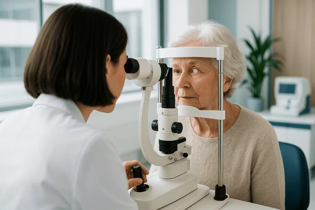
Diagnosis begins with a comprehensive eye examination. This typically includes measurement of visual acuity, refraction to determine whether glasses can still improve vision, slit-lamp examination to assess the cataract itself, pupil dilation to examine the retina and optic nerve, and eye pressure measurement. Additional tests may include corneal measurements, optical biometry for artificial lens calculation, retinal imaging, optical coherence tomography, corneal topography, tear film assessment, or ultrasound in cases where the cataract is too dense to see the back of the eye clearly. The purpose of this breadth is simple: to confirm that the cataract is genuinely the main cause of your symptoms, and to identify anything else that could limit the result of surgery.
If you are travelling for evaluation, it helps to bring previous eye records, glasses prescriptions, details of any prior eye surgeries, a list of current medications, and any known diagnoses such as diabetes, glaucoma, macular degeneration or autoimmune disease. These details allow the care team to plan surgery more accurately and reduce avoidable uncertainty in lens calculations and risk assessment.
Conditions and Indications Cataract Surgery Addresses
Cataract surgery treats vision impairment caused by clouding of the natural lens. The most common indication is age-related cataract, which develops gradually over many years; patients typically seek treatment when vision affects reading, driving, work, travel, sport or daily independence. Other indications include traumatic cataracts, which can develop after blunt or penetrating injury to the eye; diabetic cataracts, which may progress more quickly than average; steroid-related cataracts after long-term use of corticosteroid medications; and cataracts associated with inflammation, prior eye surgery or certain systemic diseases.
Some patients need cataract surgery not primarily for their own vision, but to allow better monitoring or treatment of retinal conditions such as diabetic retinopathy or macular disease, when the cloudy lens prevents a clear view into the eye. In these cases the cataract is an obstacle to other essential care, and removing it serves a double purpose.
Cataract surgery can also be considered when lens opacity creates a troublesome imbalance between the two eyes, reduces quality of life, or increases the risk of falls in older adults. For patients with narrow drainage angles in the eye, lens removal may sometimes help improve the internal anatomy of the eye, although this decision requires careful glaucoma evaluation. In every scenario, the treatment plan should reflect the individual eye and the individual life, not merely the presence of a cataract on an examination report.
How Cataract Surgery Is Performed
Modern cataract surgery removes the cloudy natural lens and replaces it with a clear artificial lens called an intraocular lens, or IOL, which remains permanently inside the eye. Most operations today use a technique called phacoemulsification: the surgeon works through a very small incision, uses ultrasound energy to break the cloudy lens into tiny fragments, removes those fragments, and inserts the folded artificial lens through the same small opening. In selected cases, laser-assisted steps may support incision creation, lens fragmentation or correction of certain astigmatism patterns. The specific technique depends on the structure of the eye, the density of the cataract, the surgeon’s assessment and your visual goals.
Preoperative evaluation and planning
Cataract surgery begins well before the day of the procedure, because the power of the artificial lens must be calculated for your individual eye. Measurements commonly include the length of the eye, the curvature of the cornea, the depth of the front chamber of the eye, and the pattern of any astigmatism. These measurements feed into the calculation of an intraocular lens power that supports the intended focus after surgery. Small errors in measurement translate into refractive surprises afterwards, which is why this stage is treated with the same seriousness as the operation itself.
Your ophthalmologist also evaluates the surface of the eye. Dry eye, blepharitis, allergy or inflammation can distort measurements and affect comfort after surgery; in some patients, treating the ocular surface first improves measurement reliability. The retina and optic nerve are examined to identify conditions that may limit vision even after the cataract is removed: an essential part of setting realistic expectations. If the back of the eye is not healthy, removing the cataract improves the clarity of the window but not the quality of the film behind it, and you deserve to know that before deciding.
You will also discuss your visual priorities. Some patients want the clearest possible distance vision and are entirely comfortable using reading glasses. Others want to reduce dependence on glasses across multiple distances. Professional needs matter: pilots, night drivers, photographers, surgeons and people who spend long hours on screens have different visual requirements, and the right plan for one is the wrong plan for another. The surgeon explains the lens options, their trade-offs, and whether premium lens designs are medically appropriate for your eyes.
Choosing an intraocular lens
Artificial lens selection is a central part of treatment planning. Monofocal lenses are designed mainly to provide clear vision at one chosen distance, usually far, with glasses used for other distances; they remain a reliable, high-quality choice. Toric lenses can be considered for patients with significant astigmatism. Multifocal and extended-depth-of-focus lenses may reduce dependence on glasses for near and intermediate tasks in selected patients, but they involve trade-offs, some patients notice halos around lights or reduced contrast in certain situations, and they are not suitable for every eye. Patients with retinal disease, glaucoma, corneal irregularities, severe dry eye or particular occupational demands may do better with a more conservative choice. The best lens is not the most advanced one on the price list; it is the one that matches your eye’s anatomy, your daily life and sound medical judgement.
Before the procedure
Depending on the clinical protocol, you may be asked to use antibiotic or anti-inflammatory eye drops before surgery. Blood-thinning medications, diabetes medications and systemic conditions are reviewed by the surgical team; in many cases cataract surgery can be performed without stopping essential medications, but this decision is always individualised, and any change to your medication routine belongs to your treating doctors, not to a checklist. On the day of surgery, the eye is cleaned carefully and sterile draping is applied. Anaesthesia is usually local, numbing eye drops, sometimes with an additional local anaesthetic injection, and sedation may be added so you feel calm while remaining responsive. General anaesthesia is uncommon for routine adult cataract surgery, but it may be considered for children, certain complex cases, or patients who cannot comfortably cooperate while lying still.
During the operation
Cataract surgery is typically an outpatient procedure performed under a surgical microscope. In a routine case, the sequence is:
- A very small incision is made at the edge of the cornea; it is designed to be self-sealing.
- A precise circular opening is created in the front of the lens capsule: the thin membrane that holds the lens in place.
- Ultrasound energy breaks the cloudy lens into small fragments (phacoemulsification).
- The fragments are gently removed from the eye while fluid systems keep the eye stable.
- The folded artificial lens is inserted through the small incision and positioned within the remaining capsule whenever possible.
- The incision is checked; it usually seals itself, and a protective shield may be placed over the eye.
Modern cataract surgery relies on magnification, microsurgical instruments, fluidics systems that maintain the stability of the eye, controlled ultrasound energy delivery and precise biometric measurement. Advanced imaging may help evaluate the cornea, retina and lens position. In selected patients, laser-assisted technology can be used for certain steps, such as creating corneal incisions, opening the lens capsule, or softening the cataract before removal. These technologies do not replace surgical judgement; they support precision and planning when the surgeon considers them appropriate for a particular eye.
How long does cataract surgery take?
The operation itself is usually short, routine cataract surgery often takes somewhere in the region of fifteen to thirty minutes per eye, though the full hospital visit is considerably longer because of preparation, anaesthesia checks and recovery observation. Complex situations extend the time: dense cataracts, small pupils, weak lens support, prior eye surgery or corneal disease may require additional manoeuvres and specialised instruments. Plan the day around the visit, not around the minutes of the operation.
Immediately after surgery
After the procedure, you rest in a recovery area while the care team checks your comfort and general condition. Vision is often blurry at first because of dilating drops, mild corneal swelling or protective ointment: this is expected and usually settles. Mild scratchiness, watering, light sensitivity and a feeling of pressure are common early sensations; significant pain, sudden vision loss, increasing redness, or new flashes and floaters are not part of normal healing. Most patients return to their accommodation the same day. Eye drops are used for a period after surgery to reduce infection risk and control inflammation, and it matters that you use them exactly as prescribed. In the early healing phase, you will usually be advised not to rub the eye, swim, use eye makeup or expose the eye to dust. Light walking and normal household activity are often possible soon, while heavy lifting and strenuous exercise are restricted for a short period according to your surgeon’s instructions.
If both eyes need surgery
Many patients have cataracts in both eyes. Surgery is often performed on one eye first, with the second eye treated after the first has begun to heal and the visual result has been assessed: an approach that also lets the surgeon fine-tune the lens plan for the second eye. The interval depends on medical factors, travel plans, surgeon preference and your visual needs. If you are travelling for treatment, planning both eyes in one trip requires careful coordination so that the necessary postoperative checks are completed before you return home, rather than squeezed out by a flight date.
Why Acting Early Matters
Cataracts do not usually require emergency treatment, but waiting too long can make life harder and sometimes make surgery more complex. As the lens becomes denser, it requires more ultrasound energy to remove, which increases stress on the cornea and other delicate structures. Very advanced cataracts can limit the surgeon’s view inside the eye and, in some patients, are associated with inflammation or changes in eye pressure.
Delay also has practical consequences that are easy to underestimate. Reduced contrast sensitivity and glare increase the risk of driving difficulty, falls, medication errors and loss of independence, quiet costs that accumulate long before a cataract looks dramatic on paper. In patients with diabetes, glaucoma, macular degeneration or retinal disease, a cataract may prevent adequate monitoring of the back of the eye, so treating it at an appropriate time also protects the management of those conditions.
Early evaluation does not mean early surgery. It means understanding the cause of your symptoms, tracking progression, and making a decision before vision loss becomes disruptive or the cataract becomes unnecessarily dense. A timely assessment lets you weigh lens options carefully and choose your moment, rather than making decisions under pressure when vision has already failed.
Benefits of Cataract Treatment
The potential benefits of cataract surgery depend on the health of the entire eye, the lens chosen and the individual healing response. No two eyes gain exactly the same result, but the categories of benefit are consistent.
| Benefit | What It Means for You |
|---|---|
| Clearer vision | Removing the cloudy lens can improve sharpness, contrast, and overall visual clarity when the retina and optic nerve are healthy. |
| Reduced glare and halos from cataract clouding | Many patients find bright lights, sunlight, and night driving more comfortable after the cloudy lens is replaced. |
| Improved daily function | Reading, using digital devices, recognising faces, cooking, walking outdoors, and participating in hobbies may become easier. |
| Opportunity to address refractive needs | Artificial lens selection may reduce dependence on glasses for certain distances, depending on eye health and lens suitability. |
| Better view for eye disease monitoring | After cataract removal, doctors may be able to examine and treat retinal or optic nerve conditions more effectively. |
Recovery Timeline After Cataract Surgery
Recovery is usually progressive: most patients notice improvement early, while the eye continues to stabilise over several weeks. The timeline below describes a typical, uncomplicated course; your surgeon’s instructions for your eye take precedence.
| Time Period | What Patients Can Expect |
|---|---|
| Day 1 | Vision may be brighter but still blurry. Mild scratchiness, watering, and light sensitivity are common. A postoperative check is usually performed soon after surgery. |
| First Week | Many daily activities can resume with precautions. Eye drops are continued. Patients should avoid rubbing the eye, swimming, heavy lifting, and dusty environments. |
| First Month | Vision generally becomes more stable as inflammation settles. Glasses may be adjusted after healing, depending on the lens type and visual goal. |
| Longer Term | The artificial lens remains in place permanently. Some patients later develop clouding of the capsule behind the lens, which can often be treated with a brief laser procedure if needed. |
One point in that table deserves expansion. Months or years after surgery, some patients notice a gradual return of blur or glare. This is usually not the cataract returning, a cataract cannot come back once the natural lens is removed, but a clouding of the thin capsule that holds the artificial lens, known as posterior capsule opacification. When it affects vision, it can typically be treated in the clinic with a brief laser procedure called a YAG capsulotomy. It is worth knowing this in advance so a change in vision later does not cause unnecessary alarm.
If you are travelling for treatment, the recovery timeline shapes your itinerary. Your surgeon will want to complete at least the early postoperative checks before you board a flight, and cabin conditions, activity restrictions and drop schedules all need a little planning. Our guides on flying after cataract surgery and how flying rules differ across eye procedures cover the practical details, but the final word always belongs to the surgeon who has examined your eye.
What Influences a Good Cataract Surgery Result
Cataract surgery has a long and well-documented record of safety and effectiveness when performed for appropriate indications, but outcomes are shaped by identifiable factors, and it is better to understand them before surgery than to discover them afterwards. The most important is the health of the rest of the eye. Conditions such as macular degeneration, diabetic retinopathy, glaucoma, corneal scarring, keratoconus, severe dry eye, optic nerve disease or previous retinal detachment can limit how much vision improves, however well the operation goes. Identifying these issues before surgery lets the team plan appropriately and tell you honestly what to expect.
The accuracy of preoperative measurements matters just as much. Artificial lens power calculations are highly refined, but they are still affected by corneal shape, prior laser vision correction, dry eye, very long or very short eyes, and previous eye surgery. If you have had LASIK, PRK, radial keratotomy, a corneal transplant, retinal surgery or glaucoma procedures, tell your ophthalmologist and bring previous records if you can, eyes with a surgical history need adapted calculation methods and more cautious expectations.
Lens selection is a third major factor. A monofocal lens may provide excellent quality of vision at one chosen distance, with glasses for the rest. Toric lenses help patients with significant astigmatism. Multifocal and extended-depth lenses reduce glasses dependence for some patients but can introduce halos or reduced contrast in certain situations. This is a genuine trade-off, not a defect, and it is why the discussion of your lifestyle and priorities before surgery is not a formality.
Surgical complexity affects recovery as well. Dense cataracts, small pupils, weak zonules that support the lens, pseudoexfoliation syndrome, high myopia, previous trauma, corneal endothelial disease and certain medications that affect pupil behaviour may all require additional surgical manoeuvres. In experienced hands, many complex cases can be managed successfully, but they call for careful planning and a frank discussion of risks beforehand.
Finally, your own participation counts. Using prescribed drops correctly, attending follow-up visits, avoiding eye rubbing, protecting the eye during early healing and reporting unusual symptoms promptly all support recovery. If you live far from where you had surgery, the care plan should include clear instructions for travel, the timing of postoperative checks, and arrangements for handover to an eye doctor at home if longer-term follow-up is needed.
Risks and limitations, stated plainly
You should expect an honest discussion of benefits, limitations and risks before agreeing to surgery. Cataract surgery provides meaningful visual improvement for many people, but no responsible medical team promises a specific outcome for a specific eye. Possible risks include infection, bleeding, inflammation, increased eye pressure, corneal swelling, retinal detachment, lens displacement, a persistent refractive error requiring glasses or further correction, glare or halos, a drooping eyelid, dry eye symptoms, or the need for additional procedures. Serious complications are uncommon, but they require prompt attention when they occur, which is one reason the postoperative check schedule exists and should not be skipped. Understanding these risks is not pessimism; it is the basis of an informed decision.
Cataract Care at Acibadem
At Acibadem, cataract care is approached as a personalised process rather than a standard product. Ophthalmologists evaluate not only the cataract itself but the whole visual system, the cornea, retina, optic nerve, eye pressure and any previous eye procedures, because the best outcome depends on understanding the entire eye before surgery is planned. Detailed imaging and measurements guide the treatment plan and the artificial lens calculation. When additional eye disease is present, ophthalmologists may collaborate with retina, glaucoma, cornea or neuro-ophthalmology specialists, and in patients with significant medical conditions such as diabetes or cardiovascular disease, coordination with the relevant medical specialties supports surgical safety.
This structured approach matters particularly for second opinions. Some people are told they have cataracts but are unsure whether surgery is the right next step. Others want to understand whether a premium intraocular lens suits their eyes, why vision remains poor despite new glasses, or whether another condition is contributing to their symptoms. A methodical evaluation can distinguish cataract-related vision loss from retinal, corneal, neurological or refractive causes: a distinction that changes the treatment plan entirely.
Technology supports this work without replacing judgement. Diagnostic devices measure eye length and corneal curvature for lens power calculations, map the corneal surface, image the retina in high detail and assess the optic nerve. Surgical microscopes, microsurgical instruments, ultrasound systems and, where appropriate, laser-assisted platforms help surgeons work precisely through small incisions. The value of these tools lies in how they inform planning and support safe execution, not in the equipment list itself.
For patients coming from abroad, Acibadem International provides coordination services that may include appointment planning, medical record review, multilingual communication, hospital navigation, help with scheduling diagnostic tests and coordination of follow-up timing. Receiving care in another country can feel unfamiliar, and organised logistics are part of good treatment, not an extra. The pathway around a brief operation should be disciplined: infection-prevention practices, sterile operating conditions, appropriate anaesthesia assessment, clear discharge instructions and timely postoperative review, especially if both eyes are being considered within one travel period.
Deciding When to Act
If cataracts are making your vision less clear, treatment can be an important step towards regaining daily function and visual comfort. The sensible time to consider surgery is when the cataract affects your life, your safety, your work or the activities that matter to you, not a fixed point on a chart, and not the moment the word first appears in an eye report. A thorough evaluation clarifies whether cataracts are genuinely the main cause of your symptoms, what the rest of your eye can support, and which type of artificial lens fits your anatomy and your priorities.
What a good decision looks like is consistent everywhere: a complete examination rather than a quick vision test, measurements taken carefully, a lens discussion built around your daily life rather than a catalogue, a realistic account of risks and limits, and a follow-up plan that fits where you actually live. Whether you are seeking first-time advice or a second opinion, understanding your condition clearly is the foundation: the surgery itself is short, but the thinking around it is what determines whether it serves you well.
Preparation
- A detailed eye examination, vision testing, and lens measurements are performed before surgery. Patients may be asked to stop certain medications and use prescribed eye drops. Arrange someone to accompany you on the day of the procedure, as driving is not recommended afterward.
Aftercare
- Use prescribed eye drops as directed and avoid rubbing the operated eye. Heavy lifting, swimming, and dusty environments should be avoided for the first days or weeks. Follow-up visits are important to monitor healing and visual improvement.
Frequently Asked Questions
What affects the cost of cataract surgery in Turkey?
The final cost depends on the lens type, eye measurements, cataract complexity, surgeon and hospital factors, whether other eye conditions are present, and what the international patient package includes. A personalised quote is provided after medical review and consultation.
How can I get a personalised quote for cataract treatment?
You can request a free consultation by sharing recent eye reports if available, your current symptoms, previous eye treatments, and any known conditions such as glaucoma, diabetes-related eye disease, or corneal problems. The medical team can then advise on suitable next steps and package details.
Does the lens choice change the total cost?
Yes. Standard and premium intraocular lens options differ in design and clinical purpose. Toric, multifocal, and extended-depth lenses may require additional measurements and planning, so the specialist will recommend an option based on eye health and lifestyle needs.
What is usually included in an international cataract surgery package?
Package content varies by provider, but it may include ophthalmology consultation, diagnostic measurements, surgery, selected lens, medication guidance, translation support, transfers, and follow-up planning. Patients should confirm exactly what is included before travel.
Is cataract surgery abroad suitable for everyone?
Not always. Suitability depends on the cataract, overall eye health, general medical status, and travel readiness. An ophthalmologist should assess whether surgery abroad is appropriate and whether follow-up can be safely coordinated.
Medically reviewed by the Acıbadem International Medical Board August 31, 2026
See our medical review board →
Update history
- PublishedJune 8, 2026
- Medical review approvedAugust 31, 2026
- Last content updateSeptember 8, 2026
Planning tools for this treatment
Indicative figures for orientation only: your written plan and quote follow a free medical review.
Trusted care for international patients
Compare options, costs and recovery
Cataract surgery costsledger-based guide ranges, or browse the full Turkey Medical Price Index.
From the Acibadem Blog

How Cataracts Cloud the Lens Over Years and What Makes Glare the First Complaint
Cataracts develop when proteins inside the eye's lens slowly break down and clump together over decades, usually beginning after about age 40 and becoming…

Cataracts in Younger Adults: Steroid Use, Injury and Diabetes as Drivers of Early Treatment
Cataracts at a young age most often trace back to a specific cause rather than to aging: long-term corticosteroid use, diabetes, or an eye…

Age-Related, Diabetic and Traumatic Cataracts: How the Cause Changes the Treatment Plan
Cataracts are usually grouped by cause: age-related, diabetic, traumatic, congenital and secondary (linked to medicines, radiation or other eye disease). The cause matters because…