CT And MRI For Urology
CT and MRI for urology are advanced imaging tests used to evaluate kidneys, bladder, prostate and urinary tract conditions, helping doctors plan accurate diagnosis and treatment.
Quick answer
CT and MRI for urology are detailed imaging tests used to examine the kidneys, ureters, bladder, prostate and nearby structures. CT uses X-rays and is often chosen for kidney stones, obstruction and trauma; MRI uses a magnetic field and excels at soft-tissue detail, especially prostate assessment. Neither scan treats disease, both provide the information urologists need to diagnose conditions accurately and plan treatment.
CT and MRI for Urology: What These Scans Are and What They Do
CT and MRI for urology are advanced imaging tests used to examine the kidneys, ureters, bladder, prostate, adrenal glands and the structures around them in fine detail. Computed tomography (CT) builds cross-sectional images from X-rays; magnetic resonance imaging (MRI) does the same with a strong magnetic field and radio waves, without ionising radiation. Neither scan treats disease. Both exist to answer questions, is there a stone, a tumour, a blockage, an infection, a structural abnormality, so that a urologist can diagnose accurately and plan treatment safely.
These scans show what physical examination, blood tests and urine tests cannot: the size and exact position of a kidney stone, the character of a mass, the state of the bladder wall, whether a suspicious area exists within the prostate and where it sits, and whether urine is draining freely from kidney to bladder. Urologists rely on them to confirm a diagnosis, judge the extent of disease, guide biopsies and procedures, plan surgery and monitor how a condition responds to treatment over time. Radiologists rely on them to describe not just what is visible, but what it is likely to mean.
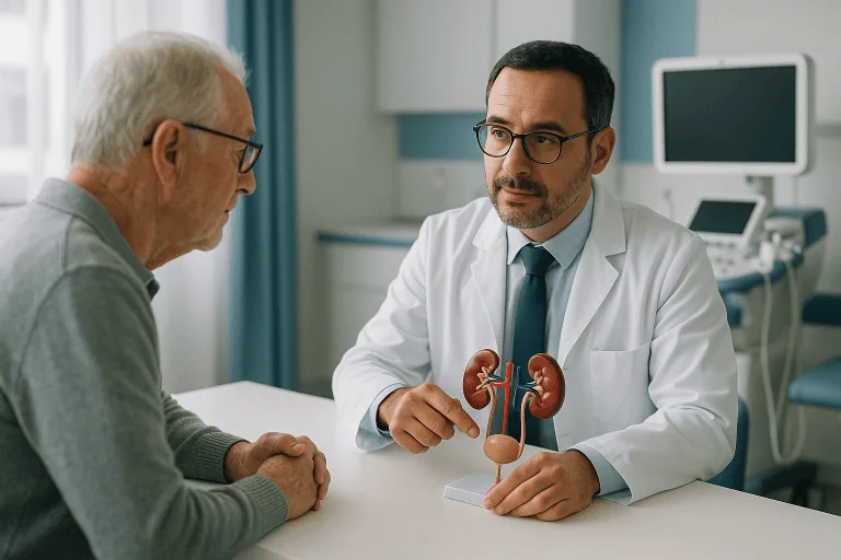
At Acibadem, CT and MRI for urology sit inside a broader diagnostic pathway rather than standing alone. Images are read by radiologists and reviewed together with the urology team, so a finding on a scan is always interpreted next to your symptoms, laboratory results and medical history. The aim is not simply to produce pictures, but to connect precise images with the right clinical interpretation and an appropriate next step.
What is the difference between CT and MRI in urology?
The main difference is how each scan sees the body and what it sees best. CT uses X-rays, is fast, and shows kidney stones, calcification, trauma and the drainage anatomy of the urinary tract with great clarity. MRI uses a magnetic field, involves no ionising radiation, takes longer, and provides superior soft-tissue detail, which is why it dominates prostate assessment and is often chosen to characterise selected kidney, adrenal and pelvic findings. In many complex cases the two are complementary rather than interchangeable: CT maps the stone or the collecting system; MRI characterises the tissue. Choosing between them is a clinical decision, not a matter of one being universally better.
What is CT urography?
CT urography is a specialised CT protocol that examines the entire urinary tract, kidneys, ureters and bladder, usually in several phases. Images may be taken before intravenous contrast is given, again as the kidneys enhance, and later still as the contrast is filtered and excreted into the ureters and bladder. This sequence shows how the kidneys handle contrast and how urine actually travels through the system, which makes CT urography one of the standard investigations for unexplained blood in the urine and for suspected narrowing, blockage or tumours of the upper urinary tract.
What is multiparametric prostate MRI?
Multiparametric prostate MRI is an MRI examination that combines several image types, detailed anatomical sequences, diffusion sequences that assess how water moves through tissue, and contrast-enhanced sequences that show blood flow patterns. Read together, these parameters help radiologists identify areas of the prostate that look suspicious, estimate their location and extent, and assess whether disease may extend beyond the prostate capsule. It is widely used after an elevated PSA result, to support targeted biopsy planning, for staging known prostate cancer and, in selected patients, to monitor low-risk disease under active surveillance.
What is MR urography?
MR urography applies the same whole-urinary-tract logic as CT urography, but uses MRI instead of X-rays. It is considered when ionising radiation should be avoided or when iodinated CT contrast is a concern, for example in some younger patients, in situations assessed case by case around pregnancy, or in people with previous contrast reactions. It can demonstrate dilation, obstruction and certain structural abnormalities of the kidneys and ureters, though it detects stones themselves far less reliably than CT.
When Urologic Symptoms Need Clear Answers
Urinary and reproductive health concerns can be difficult to live with and sometimes uncomfortable to discuss. Blood in the urine, repeated urinary tract infections, kidney or flank pain, changes in urination, an elevated prostate-specific antigen (PSA) result, or an unexpected finding on ultrasound all raise the same understandable questions: Is this serious? Could it be cancer? Is there a blockage, stone, infection or structural problem that has been missed? Do I need surgery, or only observation?
In urology, accurate imaging is often the turning point between uncertainty and a plan. Symptoms overlap heavily in this part of the body. Flank pain can mean a stone, an infection or an obstruction. Blood in the urine can come from a harmless source or from a tumour. A raised PSA can reflect benign enlargement, inflammation or cancer. Physical examination and laboratory tests narrow the possibilities; CT and MRI are usually the tools that resolve them, because they show the actual anatomy and, increasingly, the character of the tissue involved.
Who May Need CT or MRI for Urologic Conditions
A urologist recommends CT or MRI when symptoms, laboratory results, ultrasound findings or medical history suggest that more detailed information is needed before a decision can be made. The scans can help establish whether a symptom comes from a kidney stone, a tumour, an infection, a narrowing of the urinary tract, an enlarged prostate, a congenital abnormality or another condition entirely.
Common reasons for urologic CT or MRI include:
- Blood in the urine (haematuria)whether visible or detected only on urine testing, persistent haematuria usually warrants imaging of the whole urinary tract.
- Flank or back pain suggestive of kidney stones or obstruction.
- Recurrent urinary tract infections, where a structural cause or a complication may be present.
- Unexplained hydronephrosisswelling of the kidney caused by urine backing up behind a blockage.
- Abnormal findings on ultrasound or another scan involving the kidney, bladder, prostate or adrenal gland that need further characterisation.
- Known urologic cancer requiring staging, treatment planning or follow-up.
Why would a urologist order an MRI scan?
Usually because soft-tissue detail is the deciding factor. The most frequent scenario is prostate assessment: after an elevated PSA test, an abnormal digital rectal examination, a previous negative biopsy with persistent concern, or a confirmed prostate cancer that needs staging, MRI shows the gland in a way no other routine test can. MRI is also ordered to characterise indeterminate kidney lesions, to assess the bladder wall in selected cancer cases, to evaluate pelvic organs and pelvic pain, and whenever avoiding radiation exposure matters for the individual patient.
What does a CT scan show in urology?
A urologic CT shows stones, obstruction, masses, trauma, infection complications and the drainage anatomy of the urinary tract. It can reveal the size, number, density and exact position of stones; whether a kidney is swollen behind a blockage; whether a mass is solid or cystic; whether lymph nodes appear enlarged; and how a tumour relates to blood vessels and neighbouring organs, information that directly shapes surgical planning.
Everything begins with a careful review: symptoms, medical history, blood and urine tests, kidney function, current medications, allergies, prior imaging and any previous operations. If you have been scanned before, bringing the actual images on disc or by secure digital transfer, together with the radiology and pathology reports, allows any medical team to compare findings directly. Direct comparison often changes interpretation, and it frequently spares patients unnecessary repeat testing.
Conditions CT and MRI Can Address in Urology
CT and MRI for urology are used across the full range of urinary tract and male reproductive conditions. The most appropriate test depends on the organ involved, the urgency of the situation, kidney function, previous results and the treatment decisions under consideration.
Is CT or MRI better for kidney stones?
CT is the standard test for kidney stones and ureteral stones, and stones are among the most common reasons for CT in urology. A stone-protocol CT shows the size, number, density and precise location of stones, and whether they are blocking the flow of urine. That level of detail lets the urologist choose sensibly between observation, medication, shockwave therapy, ureteroscopy or another procedure, decisions in which a few millimetres of size or position genuinely change the recommendation. MRI can show the consequences of a stone, such as a dilated kidney, but detects the stone itself far less reliably.
Kidney masses and kidney cancer
Kidney masses often require CT or MRI to characterise the lesion: whether it is solid or cystic, how it enhances with contrast, how it is supplied with blood, and whether it appears confined to the kidney. This characterisation drives the treatment discussion, partial nephrectomy that preserves kidney tissue, radical nephrectomy, ablation, surveillance of a small lesion, or further testing. Comparison with older scans is particularly valuable here, because a cyst that has remained stable for years is a very different problem from a lesion that has grown between examinations.
Bladder cancer and haematuria evaluation
Bladder cancer investigation typically pairs imaging with direct inspection. CT urography examines the kidneys and ureters, where tumours can hide beyond the reach of a camera, while cystoscopy remains essential for looking directly inside the bladder, no scan replaces it. In selected bladder cancer cases, MRI is used to assess how deeply a tumour invades the bladder wall and how far it extends locally, information that matters considerably before treatment is planned.
Can MRI detect prostate cancer?
MRI can identify areas of the prostate that look suspicious for cancer, but it does not diagnose cancer on its own: that requires a biopsy and pathological confirmation. What multiparametric MRI does is show where the suspicion lies, estimate tumour location and extent, assess possible spread beyond the prostate capsule, and give the biopsy something specific to target rather than sampling blindly. It also supports multidisciplinary treatment planning once cancer is confirmed, and it is used in selected active surveillance pathways to monitor known low-risk disease without immediate intervention.
Urinary obstruction and strictures
Urinary obstruction, whether from a ureteropelvic junction problem, a ureteral narrowing, a postoperative stricture or compression by a mass, can be mapped with CT or MR urography. Imaging shows where the blockage sits, what is causing it and how it is affecting kidney drainage, which in turn determines whether the sensible answer is endoscopic treatment, reconstructive surgery or careful monitoring.
Infections and inflammatory conditions
Complicated infections are another important indication. Severe, persistent or atypical pyelonephritis, a suspected renal abscess, emphysematous infection or an unusual pelvic infection may all need CT or MRI to detect complications that require drainage or urgent treatment, complications that a routine ultrasound can miss.
Other urologic indications
Advanced imaging also contributes to diagnosis and planning in congenital urinary tract abnormalities: a frequent question in pediatric urologyas well as adrenal masses, trauma to the kidneys or bladder, testicular cancer staging, pelvic pain syndromes evaluated within female urology, and postoperative follow-up after urologic procedures of every kind.
How CT and MRI for Urology Are Performed
The examination begins well before you enter the scanner. Your physician and the radiology team decide the protocol based on the clinical question, because a suspected stone, unexplained haematuria and a kidney mass each call for different image phases, timing and preparation. A prostate MRI, likewise, uses specific sequences and positioning that differ entirely from an MRI of the kidneys or bladder. Ordering the right protocol is half the value of the test.
How do you prepare for a urology CT or MRI?
Preparation is mostly a matter of information and following instructions. Before either scan, you will typically be asked about:
- Kidney functionblood tests may be needed, especially if intravenous contrast is planned. Reduced kidney function rarely rules out imaging, but it may change the protocol to reduce risk.
- Allergies and previous contrast reactions, which influence whether and how contrast is used.
- Current medications and medical conditions, including diabetes and thyroid conditions, which the team reviews when planning contrast. Any adjustment around the scan is decided by your treating doctor, never on your own.
- Implanted devices such as pacemakers, pumps or metal implants, which matter for MRI safety screening.
- Pregnancy status, which affects the choice and timing of imaging.
- Claustrophobia or difficulty lying still, so the team can plan additional support, or sedation in selected cases.
You may also receive instructions about eating, drinking and bladder filling depending on the protocol. Following them matters more than most patients expect: they exist purely to make the images diagnostic.
What happens during the CT scan?
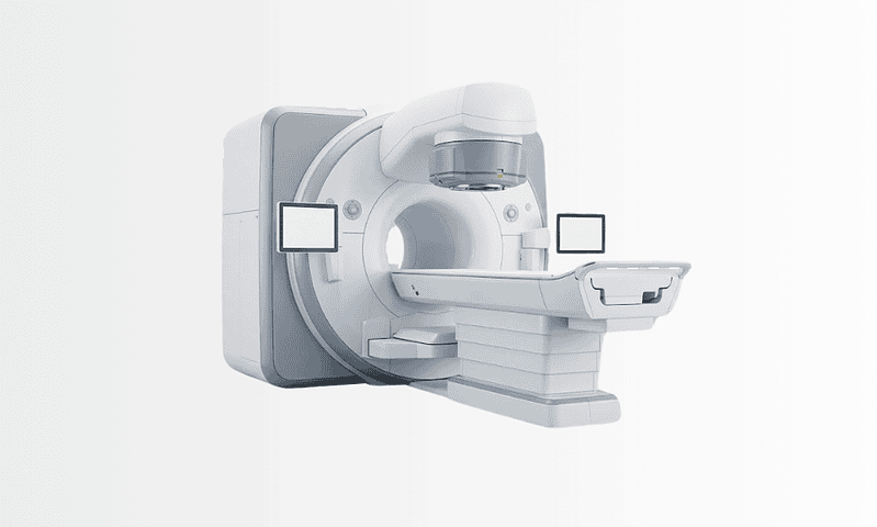
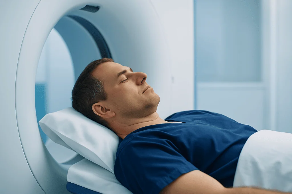
You lie on a table that moves through a scanner shaped like a wide ring, open at both ends rather than a tunnel. The scan itself is usually brief, though preparation and contrast timing can take longer. Some urologic CT protocols involve several passes: images before contrast, during kidney enhancement, and later as contrast is excreted into the ureters and bladder. If contrast is injected, you may feel a brief warm sensation, which is expected. The technologist gives breathing instructions, because holding still and following breath-holds noticeably improves image quality.
What happens during the MRI scan?
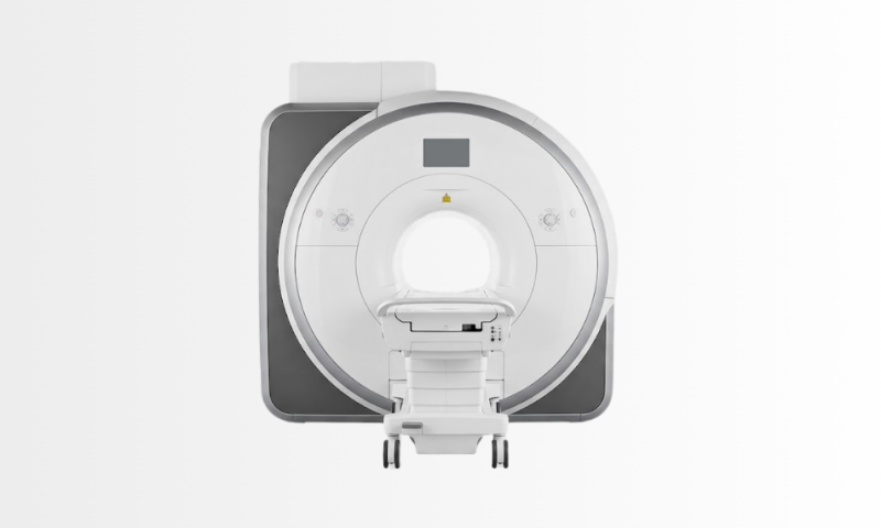
You lie on a table that moves into a tunnel-shaped scanner, and the examination takes longer than CT because MRI acquires multiple separate image sequences. You will hear rhythmic tapping and knocking sounds, this is normal, and ear protection is provided. The team monitors you throughout the examination and you can communicate with them at any point. Some MRI scans use intravenous contrast to highlight blood flow, tissue characteristics or tumour enhancement patterns. In prostate MRI, the protocol combines anatomical images with functional techniques that assess water movement and enhancement within the gland.
How long does a urology CT or MRI take?
It varies with the protocol. A non-contrast CT for stones may take only minutes once preparation is complete. CT urography or multiphase kidney imaging takes longer, because images are captured at several time points after contrast is given. MRI examinations are generally the longest, particularly prostate or complex pelvic studies, which involve many separate sequences. Your care team will explain what to expect for your specific examination, including how much time to allow for preparation and any observation afterwards.
Do you need contrast for CT and MRI in urology?
Not always: it depends on the question. Stone-protocol CT is usually performed without contrast, because stones show clearly against normal tissue. Haematuria workup, mass characterisation, cancer staging and urography protocols typically do use intravenous contrast, because enhancement patterns carry much of the diagnostic information. Contrast decisions are made individually: patients with kidney disease, previous contrast reactions, thyroid conditions or complex medical histories may need modified protocols or additional precautions, and in some situations MRI is chosen precisely because iodinated CT contrast is a concern.
Are CT and MRI scans safe?
Both examinations are considered safe when performed with proper screening, and each has a different safety profile. CT involves ionising radiation, so the dose is kept as low as reasonably achievable and the scan is recommended only when the diagnostic benefit clearly justifies it; modern scanners and dedicated stone protocols are designed to minimise exposure while preserving image quality. MRI involves no ionising radiation, but the strong magnetic field means that pacemakers, implanted pumps, metal fragments and certain other devices must be declared during safety screening before the examination, many implants are compatible, but the team has to know about them in advance. Contrast agents, iodinated for CT, gadolinium-based for MRI, are used selectively, with kidney function and allergy history reviewed beforehand. Reactions are uncommon and the imaging team is prepared to manage them; patients are asked to mention any unusual sensation during or after the injection so it can be assessed straight away.
What technology is behind these scans?
The technology exists to answer specific questions, not to impress. Thin-slice CT can reveal small stones, subtle narrowing of the urinary tract and the detailed anatomy a surgeon needs; advanced reconstruction software builds views in multiple planes, showing exactly how a lesion relates to vessels, the collecting system and nearby organs. MRI uses specialised sequences to separate soft tissues, measure diffusion and track enhancement after contrast, capabilities that matter most in prostate, bladder, kidney and pelvic imaging. This anatomical mapping is also what makes precise operative approaches possible, from laparoscopic urology to robotic urology, where the surgeon plans the operation on the images before making a single move.
After most examinations, you can return to normal activities immediately. If contrast was used, drinking fluids may be recommended unless your doctor has restricted your fluid intake. If sedation was needed, recovery and transport arrangements differ, and the team will explain them beforehand. Radiologists with relevant expertise review the images and report to your treating physician; in complex cases, particularly where cancer care or major surgery is being considered, findings may be discussed in specialist meetings or multidisciplinary boards before a recommendation is made.
Why Acting Early Matters
Many urologic conditions are more manageable when they are identified early and accurately. Delayed imaging can allow an obstruction to keep damaging kidney function, an infection to develop complications, a stone to become more difficult to treat, or a tumour to grow before any plan exists. The cost of waiting is often measured not in symptoms, some of these conditions stay quiet for a long time, but in the treatment options that remain available by the time a diagnosis is finally made.
Blood in the urine is the clearest example. It can be caused by infection, stones, benign prostate enlargement or exercise-related irritation, but it can also be associated with cancers of the kidney, ureter, bladder or prostate. Persistent or unexplained haematuria warrants proper investigation. Imaging, combined with urologic evaluation and, where indicated, cystoscopy, is how the cause is clarified rather than guessed at.
In prostate assessment, timely MRI can reduce uncertainty after abnormal PSA results and help determine whether a biopsy is needed, where it should be targeted, and how treatment should be planned if cancer is confirmed. In kidney masses, early characterisation may preserve more options, including kidney-sparing surgery in selected patients.
Acting early does not automatically mean choosing aggressive treatment. Sometimes early imaging supports careful observation instead of intervention: a small stable lesion followed over time, a low-risk prostate finding monitored under surveillance. The point is that the decision, whatever it is, rests on the condition’s true location, size, behaviour and risk profile rather than on assumption.
Benefits of CT and MRI in Urologic Care
The practical value of these scans is that they turn symptoms and test results into a clearer diagnosis and a more individualised plan. In concrete terms:
| Benefit | What It Means for You |
|---|---|
| More accurate diagnosis | Detailed images help distinguish stones, tumours, obstruction, infection, cysts and structural abnormalities from one another. |
| Better treatment planning | Your urologist can plan surgery, biopsy, medication, surveillance or minimally invasive procedures with clearer anatomical information. |
| Assessment of disease extent | In cancer care, imaging helps show whether disease appears localised or involves nearby tissues, lymph nodes or other organs. |
| Support for targeted procedures | MRI and CT findings may guide biopsy planning, drainage procedures, stone treatment decisions or the surgical approach itself. |
| Monitoring over time | Follow-up scans help evaluate whether a condition is stable, improving, recurring or requiring a change in treatment. |
Recovery and Follow-Up Timeline
CT and MRI are non-surgical diagnostic tests, so recovery is usually brief. What follows the scan depends on the findings and on whether contrast or sedation was used.
| Time Period | What Patients Can Expect |
|---|---|
| Day 1 | Most patients leave shortly after the scan and return to routine activities. If contrast was used, hydration may be recommended. |
| First Week | The radiology report is reviewed by your urologist. Additional tests, cystoscopy, biopsy or treatment planning may be discussed if needed. |
| First Month | Patients with significant findings may move into a treatment pathway, such as stone management, cancer staging, surgery planning or surveillance. |
| Longer Term | Some conditions require repeat imaging to monitor stability, treatment response, recurrence or kidney function over time. |
Can you go home straight after a CT or MRI scan?
In most cases, yes. The scans involve no incisions and no anaesthesia in their standard form, so patients typically resume normal activities immediately afterwards. The exceptions are patients who received sedation, who need a recovery period and accompanied transport, and occasional situations in which the team asks a patient to remain briefly for observation after contrast. Your care team will confirm what applies to you before the scan begins, so there are no surprises on the day.
Factors That Influence Imaging Quality and Results
A good imaging result is not only about the scanner. It depends on choosing the correct test, performing the right protocol and interpreting the images in the context of the patient’s full medical picture. Several factors carry particular weight.
The clinical question matters. A scan ordered simply as “abdomen CT” may not provide the same information as a dedicated kidney mass protocol, CT urography, a stone protocol or a prostate MRI. Urologic imaging is most valuable when the radiology team knows exactly what the physician needs to find out, because the protocol is built around that question.
Patient preparation affects image clarity. Movement during MRI, improper bladder filling for certain pelvic scans, delayed contrast timing or an incomplete medical history can all reduce the diagnostic value of an otherwise well-planned examination. Following the instructions you are given about eating, drinking and arrival time helps the team obtain the best possible study on the first attempt.
Kidney function and contrast decisions are important. Contrast improves diagnostic accuracy in many urologic conditions, but it must be used thoughtfully. Patients with kidney disease, prior contrast reactions, thyroid conditions or complex medical histories may need modified protocols or additional precautions, and those decisions belong to the medical team planning the scan.
Prior imaging is valuable. Comparing current scans with previous studies can show whether a kidney cyst is stable, whether a mass is growing, whether a stone has moved or whether a lymph node has changed. Patients should try to provide the earlier images themselves, not only the written reports, because direct side-by-side comparison often changes the interpretation.
Radiology expertise influences interpretation. Urologic CT and MRI require familiarity with urinary tract anatomy, cancer staging systems, post-treatment appearances, surgical anatomy and the honest limitations of each modality. Prostate MRI is a good example: its interpretation depends on structured assessment and experience with patterns that can mimic cancer, such as inflammation or benign enlargement. The same images can support or mislead a decision depending on who reads them.
Integration with specialist care improves decisions. Imaging findings need to be matched with symptoms, urine tests, PSA levels, biopsy results, kidney function and the patient’s own goals. A small kidney mass may reasonably be managed differently in a young, otherwise healthy patient than in an older patient with several other medical conditions. The value of CT and MRI for urology is highest when the result feeds a personalised treatment plan rather than a one-size-fits-all decision.
How Imaging Fits Into Urologic Care at Acibadem
At Acibadem, urologic CT and MRI are provided in hospitals where radiology, urology, oncology, nuclear medicine, pathology, anaesthesiology and surgical teams can work together when a case requires broader expertise. The advantage for the patient is the connection between imaging and clinical decision-making. A prostate MRI is reviewed alongside PSA history and biopsy considerations. A kidney mass CT can be discussed with urologic surgeons and oncologists to assess whether kidney-sparing surgery is realistic. Bladder cancer imaging is evaluated in relation to cystoscopy, pathology and the available treatment options. In more complex cancer cases, multidisciplinary tumour boards may review the findings and align recommendations with evidence-based international protocols.
The diagnostic environment includes modern CT and MRI systems, urology-focused imaging protocols, digital image review and radiology reporting designed to support treatment planning. Technology is used with a clear purpose: to identify the relevant anatomy, reduce uncertainty, help avoid unnecessary procedures where possible and guide interventions when treatment is genuinely needed, whether that treatment is open surgery or a minimally invasive urology approach. The specific imaging method is always selected according to the patient’s diagnosis, safety profile and the clinical question being answered.
Personalised planning matters especially in urology, because the same imaging finding can lead to different options in different patients: a stone may be observed or treated urgently; a prostate lesion may need biopsy, surveillance or treatment planning; a kidney mass may need follow-up, ablation, partial nephrectomy or more extensive surgery.
For patients seeking a second opinion, careful imaging review is often where the real value lies. Sometimes the sensible next step is not repeating every test, but reviewing the existing studies properly, obtaining the one missing protocol, or clarifying a single unclear finding. When new imaging genuinely is needed, the team can select the examination most likely to answer the unresolved question rather than the widest possible battery of tests.
Understanding Your Results and What Comes Next
If you have been advised to undergo CT or MRI for a urologic concern, the purpose is to obtain the clearest possible understanding of what is happening and what should be done about it. Whether the issue is kidney pain, blood in the urine, an elevated PSA, a suspected stone, a kidney mass, bladder cancer evaluation or follow-up after previous treatment, advanced imaging exists to support safer, more confident decisions.
The radiology report is written for your treating physician, and its language can feel dense: descriptions of enhancement, density, signal characteristics, measurements and comparisons with previous studies. What matters for you is the conclusion your doctor draws from it, whether the finding is benign, suspicious or indeterminate, whether further testing such as cystoscopy or biopsy is warranted, whether treatment is recommended now, and whether follow-up imaging should be scheduled and at what interval. Useful questions to raise with your doctor include which structures the scan actually assessed, how the result compares with previous imaging, and what each realistic next step would involve.
Clear images, expert interpretation and coordinated urologic evaluation together determine how well a diagnosis is understood, and, ultimately, how well the treatment plan fits the person it is written for.
Preparation
- Patients should bring previous imaging, laboratory results and medication lists to the appointment. Inform the team about pregnancy, kidney disease, contrast allergy, implanted devices or claustrophobia. Fasting or a blood creatinine test may be required if contrast material is planned.
Aftercare
- Most patients can return to normal activities immediately after CT or MRI. If contrast was used, drinking water may help clear it from the body unless otherwise advised. Results are reviewed by radiology and urology specialists to guide the next diagnostic or treatment step.
Frequently Asked Questions
What affects the cost of urological CT or MRI?
The final cost depends on the scan type, body area, use of contrast, radiology reporting, specialist consultation, required blood tests and whether additional imaging or follow-up is needed.
How can I get a personalised quote from Acibadem?
You can request a free consultation by sharing your symptoms, referral notes and any previous imaging or laboratory results. The team can then advise which imaging pathway may be appropriate and prepare a personalised quote.
Is contrast always needed for CT or MRI in urology?
No. Contrast is used only when it is clinically helpful and safe. A specialist considers the reason for imaging, kidney function, allergy history and previous test results before recommending contrast.
Will the package include a urologist consultation?
Some pathways may include imaging and a radiology report only, while others may include urology review or further tests. The exact inclusions should be confirmed in your personalised quote before booking.
Can international patients receive the report in English?
International patient teams can usually help coordinate appointments, medical translation and report delivery in English. Availability and format should be confirmed during the consultation process.
Medically reviewed by the Acıbadem International Medical Board September 13, 2026
See our medical review board →
Update history
- PublishedJune 8, 2026
- Medical review approvedSeptember 13, 2026
- Last content updateSeptember 12, 2026
Trusted care for international patients
From the Acibadem Blog

Preparing for a Urology CT or MRI: Creatinine Checks, Contrast Allergies and Implants
Preparing for a urology CT or MRI usually means three things: a recent creatinine blood test to confirm your kidneys can handle contrast dye,…