

Echocardiography
Echocardiography is a non-invasive ultrasound test that shows the heart’s chambers, valves, pumping function and blood flow, helping cardiologists diagnose and monitor many cardiac conditions.
Quick answer
An echocardiogram is an ultrasound scan of the heart. A small probe sends high-frequency sound waves through the chest to create moving images of the heart's chambers, valves and blood flow. Doctors use it to assess pumping strength, valve function and structural problems. A standard transthoracic echocardiogram needs no sedation and usually takes 30 to 60 minutes.
What Is an Echocardiogram?
An echocardiogram is an ultrasound scan of the heart. It uses high-frequency sound waves to create moving images of the heart’s chambers, valves, muscle and blood flow in real time, without needles, incisions or radiation. Cardiologists use an echocardiogram to answer defined questions: how strongly the heart is pumping, whether the valves open and close as they should, and whether symptoms such as breathlessness or chest discomfort are coming from the heart at all.
When a doctor recommends this test, there is usually a specific concern behind it. It may be chest discomfort, palpitations, a heart murmur heard during a routine examination, fainting, high blood pressure, a previous heart attack, or a family history of heart disease. For many patients, especially those arranging care in another country, even a straightforward scan can feel significant. Knowing exactly what the test involves, and what it can and cannot tell you, makes the process easier to navigate.
Echocardiography is one of the most widely used cardiac imaging techniques because it shows the heart in motion without surgery, radiation or a recovery period. It gives cardiologists information that a physical examination or an electrocardiogram cannot provide on its own. Seeing the chambers, valves, muscle movement and blood flow directly helps physicians make more confident decisions about diagnosis, treatment, follow-up, and whether further testing is needed.
At Acibadem, an echocardiogram is rarely an isolated event. It sits inside a broader cardiology pathway, where the imaging findings are interpreted alongside your symptoms, medical history, laboratory tests, electrocardiography, rhythm monitoring, stress testing or coronary imaging when appropriate. The aim is not simply to perform the scan, but to understand what its findings mean for your health and your next steps.
What does echocardiography mean?
Echocardiography means recording images of the heart using sound. The word combines three parts: echo, referring to reflected sound waves; cardio, meaning heart; and -graphy, meaning recording or imaging. In everyday use, the test is often shortened to “echo”. Strictly speaking, echocardiography is the technique, and an echocardiogram is the individual study it produces: the set of recorded images and measurements your cardiologist reviews. In practice, doctors and patients use the terms interchangeably, and both refer to the same ultrasound examination of the heart.
How does the scan create images of the heart?
A small handheld device called a transducer sends pulses of high-frequency sound into the body and listens for the echoes that bounce back from the heart’s structures. Because different tissues reflect sound differently, a computer can convert these returning signals into a moving picture on a monitor, updated many times per second. This is the same principle used in pregnancy ultrasound, adapted for a moving, beating organ.
Several techniques are combined in a single study. Two-dimensional imaging shows the anatomy of the chambers and valves in cross-section. M-mode traces the movement of a single line through the heart over time, which is useful for precise measurements. Doppler ultrasound measures the speed and direction of blood flow, which helps detect valve narrowing, valve leakage and abnormal pressures. Colour flow mapping overlays blood movement onto the anatomical image, making leaks and abnormal connections easier to see. In selected cases, three-dimensional imaging and strain analysis, a way of measuring how the heart muscle deforms with each beat, add further detail.
Types of Echocardiogram
There is no single version of this test. The type of echocardiogram your cardiologist recommends depends on the clinical question being asked, the quality of images that can be obtained from the chest wall, and whether the heart needs to be assessed at rest, under stress, or from a closer vantage point.
Transthoracic echocardiogram (TTE)
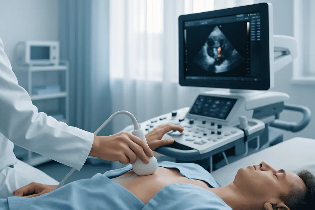
A transthoracic echocardiogram is the standard, most common form of the test. The transducer is placed on the outside of the chest wall while you lie on an examination table, usually on your left side, which brings the heart closer to the chest and improves the images. A water-based gel helps the sound waves pass through the skin. The scan requires no sedation, no anaesthesia and no recovery time. You may feel light pressure from the transducer against your ribs, but nothing enters the body. For most heart questions, a transthoracic echocardiogram is the first-line imaging study, and often the only one needed.
What is transesophageal echocardiography?
Transesophageal echocardiography (TEE) is an echocardiogram performed from inside the oesophagus, the food pipe, which lies directly behind the heart. A specialised ultrasound probe is passed gently down the throat after the area has been numbed, usually with light sedation given through a vein. Because the probe sits so close to the heart, with no ribs or lung tissue in the way, it produces highly detailed images of structures that can be difficult to see from the chest wall: the heart valves, the left atrium, the aorta, and areas where blood clots or infection may be suspected. This test requires fasting for several hours beforehand, your heart rhythm, blood pressure and oxygen levels are monitored throughout, and you should not drive on the same day because of the sedation. It is typically reserved for questions a standard scan cannot answer with enough certainty.
Stress echocardiogram
A stress echocardiogram evaluates how the heart performs when it has to work harder. Images are taken at rest and again after exercise, usually walking on a treadmill or pedalling a stationary bicycle, or after a medication is given under supervision to increase the heart’s workload when exercise is not possible. Comparing the two sets of images can reveal areas of heart muscle that move normally at rest but weaken under stress, which may suggest reduced blood supply from narrowed coronary arteries. It can also assess valve function under load and help explain symptoms that only appear with exertion.
Contrast echocardiography
In selected cases, a contrast agent is injected through a vein to sharpen the images. Contrast can outline the inner border of the left ventricle more clearly, making it easier to assess pumping function and wall motion when standard images are limited by body build, lung disease or chest anatomy. A related technique uses agitated saline, tiny bubbles of sterile salt water, to look for abnormal connections between the heart’s chambers, such as those investigated after certain strokes. Your cardiologist will explain why contrast is being suggested if it applies to you.
Why Would a Cardiologist Order an Echocardiogram?
A cardiologist orders an echocardiogram when symptoms, examination findings or other test results raise a question about the heart’s structure or function that only imaging can answer. The referral is usually specific: is a murmur caused by a valve problem, and how severe is it? Is breathlessness cardiac in origin? Has long-standing high blood pressure changed the heart muscle?
Some patients are referred after a routine check-up reveals a heart murmur: an extra sound made by blood moving through the heart, which may be entirely innocent or may reflect a valve abnormality that deserves assessment. Others seek evaluation because of chest discomfort, fatigue, reduced exercise capacity, swelling in the legs, dizziness or an irregular heartbeat.
Shortness of breath is one of the most common reasons for echocardiography. The scan can help determine whether the symptom relates to heart failure, valve disease, pulmonary hypertension, cardiomyopathy or fluid around the heart. Just as usefully, it can suggest that the heart is unlikely to be the main cause, redirecting the diagnostic effort towards the lungs, the blood or another system. A test that rules things out has real value.
Patients with high blood pressure may need an echocardiogram to see whether the heart muscle has thickened or the chambers have enlarged over time. People with diabetes, kidney disease, a previous heart attack or known coronary artery disease may need their pumping function and wall motion assessed. Those who have had cardiac surgery, valve repair, valve replacement or an interventional heart procedure often need periodic follow-up imaging to confirm the result is holding.
Echocardiography also has a role in the care of people with congenital heart conditions, including adults treated in childhood who now need lifelong monitoring. It may be requested before major non-cardiac surgery to evaluate cardiac risk when there are symptoms or known heart disease. In oncology, echocardiograms monitor heart function before, during or after certain cancer treatments that can affect the heart muscle, so that any change is picked up early.
Whatever the reason, diagnosis starts with a careful history and physical examination. Your cardiology team will ask about symptoms, prior test results, medications, family history and any previous cardiac procedures. Depending on the situation, an electrocardiogram, blood tests, chest imaging, exercise testing, rhythm monitoring, coronary CT or cardiac MRI may also be recommended. The echocardiogram is often central to this pathway because it shows directly how the heart is moving and how blood is flowing through it.
What Can an Echocardiogram Show?
An echocardiogram shows the size and shape of the heart chambers, the thickness and movement of the heart muscle, the structure and function of the four valves, and the direction and speed of blood flow through the heart and the major vessels. From these images, cardiologists can diagnose new problems, grade the severity of known ones, and track change over time.
What abnormalities can be found on an echocardiogram?
The abnormalities most commonly identified on an echocardiogram fall into a handful of groups: valve disease, such as a narrowed or leaking valve; weakened or stiffened pumping function, the hallmark of heart failure; disease of the heart muscle itself, known as cardiomyopathy; fluid collecting around the heart in the pericardial sac; and structural problems present from birth, including holes between the chambers. Beyond these, the scan can detect signs of infection on the valves, raised pressure in the lung circulation, blood clots within the chambers, enlargement of the aorta, and wall-motion changes suggesting a previous heart attack.
Heart valve disease. Echocardiography is the principal tool for evaluating heart valve disease, including aortic stenosis, aortic regurgitation, mitral stenosis, mitral regurgitation, tricuspid valve disease and pulmonary valve abnormalities. By showing valve structure and measuring blood flow across each valve, the test helps determine whether a problem is mild, moderate or severe: a grading that directly shapes whether the right response is observation, medication, a catheter-based intervention or surgery.
Heart failure and pumping function. The scan measures ejection fraction, which describes how much of the blood in the left ventricle is pumped out with each beat. It also evaluates diastolic function, how well the heart relaxes and fills between beats, along with chamber size, wall thickness and estimated pressures within the heart and lungs. These measurements guide medical treatment and provide a baseline against which future scans can be compared.
Coronary artery disease and its consequences. For patients with chest pain or suspected coronary disease, echocardiography can identify segments of heart muscle that are not moving normally, which may reflect prior injury or reduced blood supply. A stress echocardiogram extends this by testing the muscle under exertion. After a heart attack, the scan can assess complications such as reduced pumping function, new valve leakage, aneurysm formation or fluid around the heart.
Cardiomyopathies. These are conditions affecting the heart muscle itself, including dilated cardiomyopathy, hypertrophic cardiomyopathy, restrictive cardiomyopathy and other inherited or acquired disorders. Echocardiography identifies changes in wall thickness, chamber size, obstruction to blood flow and overall function, and it is often the test that first raises the suspicion.
Other findings. The list of indications extends further: suspected pericardial disease, such as pericardial effusion or constrictive pericarditis; infective endocarditis, where infection affects the valves or the inner lining of the heart; congenital heart defects; pulmonary hypertension; unexplained fainting; stroke evaluation when a cardiac source of clot is suspected; and assessment of the aorta in selected cases. Echocardiography can also guide certain cardiac procedures and monitor patients after treatment.
How an Echocardiogram Is Performed, Step by Step
The process begins before the scan itself. Your care team reviews the reason for the test, your symptoms, prior imaging, medications, allergies and any conditions that may influence the examination. This matters, because a sonographer who knows the clinical question can tailor the views and measurements to answer it properly.
For a standard transthoracic echocardiogram, the sequence is straightforward:
- You may be asked to change into a gown so the chest can be reached easily.
- Small adhesive electrodes are placed on your chest to record your heart rhythm throughout the study.
- You lie on an examination table, usually on your left side, which brings the heart closer to the chest wall.
- A water-based gel is applied to the skin to help the ultrasound waves pass through.
- The sonographer or physician moves the transducer over several areas of the chest and upper abdomen to view the heart from different angles. You will feel gentle pressure; at times you may be asked to hold your breath briefly or shift position so a clearer image can be captured.
- The study combines two-dimensional imaging, M-mode measurements, Doppler assessment and colour flow imaging to evaluate anatomy, movement, valve function, blood flow velocity and pressure estimates.
- The gel is wiped away, the electrodes are removed, and you can dress and leave. There is no recovery period after a standard scan.
Modern echocardiography systems add further capability where it is needed: high-resolution imaging, advanced Doppler measurement, three-dimensional views in selected cases, and strain imaging to detect subtle changes in heart muscle function. These tools are particularly useful when monitoring patients at risk of heart muscle changes, evaluating complex valve disease or planning interventions. If images from the chest wall are limited, because of body build, lung disease, chest anatomy or previous surgery, contrast enhancement or a transesophageal study may be considered instead.
What should you not do before an echocardiogram?
For a standard transthoracic echocardiogram, there is usually nothing you need to avoid. You can normally eat, drink and go about your day as usual, unless your treating doctor gives you different instructions. It is sensible not to apply creams, oils or lotions to the chest on the day of the scan, as they can interfere with the gel and the transducer. Wear clothing that is easy to remove from the waist up.
The picture changes for the other types. Before a transesophageal echocardiogram you will be asked not to eat or drink for several hours, because the probe passes into the oesophagus and sedation is usually given. Before a stress echocardiogram, the team may ask you to avoid a heavy meal, and some centres give specific instructions about caffeine; follow whatever guidance you receive for your appointment, and bring comfortable shoes if you will be exercising. Never change any medication on your own initiative before a heart test, whether and how to adjust anything is a decision for the doctor who prescribed it, made ahead of the scan.
How long does an echocardiogram take?
A standard transthoracic echocardiogram is commonly completed within about 30 to 60 minutes, although the time varies with the complexity of the case and the quality of the images. A transesophageal study involves a shorter scanning time, but the appointment as a whole is longer because of preparation, sedation and a period of observation until the sedation wears off. A stress echocardiogram takes longer overall, since images must be captured both at rest and at peak stress, with monitoring in between. Your appointment letter or care coordinator will tell you how much time to allow.
If a transesophageal echocardiogram is recommended, the day runs differently from a standard scan. The throat is numbed, sedating medication may be given through a vein, and the probe is guided gently into the oesophagus to obtain detailed images. Your heart rhythm, blood pressure and oxygen levels are monitored throughout. Afterwards, you are observed until the sedation has worn off, your throat may feel mildly sore for a short time, and you should not drive for the rest of the day.
For a stress echocardiogram, baseline images are recorded first. You then exercise on a treadmill or stationary bicycle while your heart rhythm and blood pressure are monitored, or receive a stress medication under supervision if exercise is not feasible. Images are captured again at peak effort, and the team compares heart movement at rest and under stress to look for changes suggesting reduced blood supply, or stress-related valve or pressure problems.
After the images are acquired, a cardiologist reviews and interprets the study. The report typically includes measurements, descriptions of chamber size and function, valve findings, estimated pressures and any clinically significant abnormalities, along with a comparison to previous studies where these exist. Most patients can return to normal activities immediately after a standard transthoracic echocardiogram; after transesophageal or stress studies, the aftercare instructions depend on the sedation used, the findings and your overall condition.
Why Timely Evaluation Matters
Heart conditions can be quiet in their early stages. A valve may narrow gradually over years. The heart muscle may thicken in response to high blood pressure. Pumping function can decline before symptoms become obvious. An echocardiogram identifies these changes early enough for physicians to monitor them properly and intervene at the right moment rather than the last one.
Delaying evaluation can allow treatable problems to progress. Severe valve disease may lead to heart enlargement, rhythm disturbances, pulmonary hypertension or heart failure. Unrecognised cardiomyopathy may increase the risk of worsening symptoms or sudden complications in selected patients. Fluid around the heart, infection of the heart valves or significant changes after a heart attack all benefit from prompt diagnosis.
Timely echocardiography does not always mean immediate treatment. In many cases, the most valuable outcome is a clear baseline and a structured follow-up plan. If findings are mild, your cardiologist may recommend observation, lifestyle changes, medication or repeat imaging at an appropriate interval. If findings are more serious, early recognition helps direct the right referral, medical therapy, rhythm evaluation, interventional cardiology, cardiac surgery or another specialty, before the situation becomes harder to treat.
Benefits of Echocardiography
The test’s value comes from a simple combination: it shows the heart working, in real time, without an incision and without radiation exposure.
| Benefit | What It Means for You |
|---|---|
| Non-invasive cardiac imaging | A standard transthoracic echo is performed from the outside of the chest and usually requires no needles, no anaesthesia and no recovery time. |
| Detailed assessment of heart function | Your cardiologist can evaluate pumping strength, chamber size, wall motion and signs of strain on the heart. |
| Valve and blood flow evaluation | Doppler imaging identifies valve narrowing, leakage, abnormal flow patterns and pressure changes that guide treatment decisions. |
| Useful for diagnosis and follow-up | The test can establish a diagnosis, monitor known conditions, compare changes over time and assess response to therapy. |
| No ionising radiation | Because ultrasound is used, an echocardiogram can be repeated whenever clinically necessary without radiation exposure. |
| Supports treatment planning | Findings help determine whether medication, further testing, catheter-based intervention, surgery or simple observation is the most appropriate next step. |
Recovery After an Echocardiogram
Aftercare depends on the type of echocardiogram performed. A standard transthoracic study needs essentially none; sedated and stress studies need a little more.
| Time Period | What Patients Can Expect |
|---|---|
| Day 1 | After a standard transthoracic echo, most patients return to normal activities immediately. Mild skin sensitivity from the electrodes or gel is uncommon and temporary. |
| First week | No special restrictions are usually needed after a standard test. After a transesophageal echo, brief throat soreness may occur, and sedation-related precautions apply on the day of the procedure. |
| First month | Your cardiologist may review the findings with you, adjust the treatment plan, recommend additional tests or schedule follow-up based on the results and your symptoms. |
| Longer term | Patients with valve disease, cardiomyopathy, heart failure, congenital heart disease or treatment-related monitoring needs may have repeat echocardiograms at intervals set by their physician. |
What Influences the Accuracy and Value of the Results
An echocardiogram is only as good as its images and the judgement applied to them. Image quality can be affected by lung disease, body build, chest wall structure, previous surgery or difficulty lying in certain positions. In some patients, the ultrasound beam simply cannot capture every structure clearly from the chest wall. When that happens, physicians may turn to contrast enhancement, transesophageal imaging or a different modality, such as cardiac MRI, to answer the clinical question more reliably. A good laboratory says openly when its images are limited, rather than reporting around the gap.
Technical expertise matters at both ends of the study. Acquiring the right views takes training, patience and an understanding of the clinical concern; interpreting them takes experience in cardiac anatomy, physiology, Doppler measurement and disease patterns. A mild valve leak, for example, is a common finding with little clinical impact in many people, while a more significant leak may need surveillance or intervention. The skill lies not just in seeing the finding, but in placing it correctly within the patient’s overall condition. Experienced laboratories follow standardised imaging and measurement protocols, including guidance published by the American Society of Echocardiography (ASE) and its European counterparts, so that studies performed months or years apart can be compared meaningfully.
The most useful report is one connected to a careful clinical assessment. Symptoms, physical findings, blood pressure, heart rhythm, laboratory values and previous imaging all shape what the numbers mean. A patient with breathlessness and preserved pumping function may still have diastolic dysfunction, valve disease, pulmonary hypertension, lung disease, anaemia or another cause entirely. The echocardiogram narrows the possibilities; good care comes from reading the images in context, not in isolation.
For patients monitored over time, consistency is worth a great deal. Comparing current and previous echocardiograms can show whether a valve has worsened, a chamber has enlarged or pumping function has changed, distinctions that drive decisions in valve disease, cardiomyopathy, heart failure, congenital conditions and treatment-related monitoring. A good result is not simply a “normal” or “abnormal” label. It is a clear explanation that supports safe, appropriate decision-making, ideally measured the same way each time so that change is real change and not measurement noise.
Echocardiography at Acibadem
At Acibadem, an echocardiogram is one element of an organised cardiology assessment rather than a stand-alone service. Depending on the clinical question, it may form part of a focused cardiology visit, a broader preventive heart assessment, echocardiography is a standard component of structured check-up and longevity programmes: a preoperative evaluation, or a second opinion after inconclusive results elsewhere. The sequence of tests is personalised, so patients are not routed through examinations that a more focused approach would make unnecessary.
Multidisciplinary review becomes important when the scan identifies a complex finding. Significant valve disease may be discussed among cardiologists, interventional cardiologists, cardiac surgeons, imaging specialists and anaesthesiology teams. Heart failure may involve electrophysiology, nephrology, endocrinology or rehabilitation. Cancer-related cardiac concerns benefit from coordination between oncology and cardiology. In these settings, team-based review helps ensure that the interpretation of an echocardiogram translates into a practical, coherent care plan rather than a report filed away.
Many international patients arrive with prior reports, images or differing opinions. In these cases, a repeat echocardiogram may be recommended to confirm the diagnosis, obtain standardised measurements or match findings against current symptoms. Where previous imaging is available, cardiologists review it alongside the new study, often the most reliable way to judge whether a condition is stable, progressing or in need of prompt attention. Acibadem International supports this process with appointment coordination, medical record transfer, interpreter assistance and follow-up planning, so that the report is not only produced but properly understood.
Physicians at Acibadem work with evidence-based cardiology protocols and consider international guidelines when evaluating heart conditions. Planning is individual: a patient with mild valve disease and no symptoms may need only periodic imaging; a patient with severe, symptomatic valve disease may need prompt referral for intervention; a patient with unclear chest pain may need further ischaemia evaluation. Advanced ultrasound platforms, detailed chamber and valve views, Doppler flow analysis, three-dimensional imaging in selected cases, and functional measurements that detect subtle change, are most meaningful when used by teams who know when they are needed and how the results should influence care.
Making Sense of Your Results
An echocardiogram is often a turning point in cardiac evaluation. It can provide reassurance when the heart is functioning well, identify problems that deserve monitoring, or reveal conditions that should be treated without unnecessary delay. Because it is non-invasive, widely applicable and highly informative, echocardiography remains one of the central tools of modern cardiology.
When your report arrives, three things are worth understanding clearly: what the study showed, in plain terms; whether anything found is stable, new or changed since previous imaging; and what the finding means for the plan, observation, medication, further testing or referral. The most important outcome of the test is exactly that clarity: knowing how your heart is functioning, what your level of risk is, and what should happen next. With well-acquired images, careful interpretation and coordinated follow-up, an echocardiogram gives you and your physicians a solid basis for informed decisions about your heart health.
Preparation
- Most standard transthoracic echocardiography exams require no special preparation. Bring previous cardiac reports, ECGs, medication lists and imaging results if available. Continue regular medicines unless your cardiologist advises otherwise.
Aftercare
- You can usually return to normal daily activities immediately after the test. A cardiologist reviews the images and explains whether further tests, follow-up or treatment is needed. If contrast or a transesophageal approach is planned, specific aftercare instructions may be provided.
Frequently Asked Questions
What affects the cost of echocardiography?
The final cost depends on the type of echo, whether a cardiologist consultation is included, the hospital or clinic setting, reporting requirements, and any additional tests requested by the specialist.
How can I get a personalised quote for echocardiography in Turkey?
You can request a free consultation by sharing your symptoms, previous reports, referral notes, and any recent cardiac test results. The care team can then advise which echo option may be appropriate and provide a personalised quotation.
Is a cardiologist consultation included in the package?
This depends on the package structure. Some packages include the echo, cardiologist interpretation, written report, and discussion of findings, while others may list these services separately.
Will I need extra tests in addition to echocardiography?
Additional tests may be recommended if the cardiologist needs more information about rhythm, blood markers, exercise response, or coronary risk. These tests can affect the final cost and are decided according to clinical need.
Can international patients receive the report in English?
Many international patient programs can support English-language communication and report coordination. It is best to confirm report language, translation support, and follow-up arrangements before booking.
Medically reviewed by the Acıbadem International Medical Board September 1, 2026
See our medical review board →
Update history
- PublishedJune 8, 2026
- Medical review approvedSeptember 1, 2026
- Last content updateSeptember 1, 2026
Trusted care for international patients
From the Acibadem Blog

How an Echocardiogram Is Done, How Long It Takes, and Echo vs ECG
A standard echocardiogram is an ultrasound scan of the heart. You undress from the waist up, wear a gown, lie on your left side,…

What Happens During an Echocardiogram, and How Long Does the Scan Usually Take?
A standard echocardiogram, the ultrasound scan of the heart done through the chest wall, usually takes 15 to 60 minutes according to the NHS,…

What an Echocardiogram Shows (and the Five Abnormalities It Looks for)
An echocardiogram is an ultrasound scan of the heart that shows its size, the thickness of its walls, how strongly it pumps, and whether…



































