Esophageal Varices Treatment
Esophageal varices are enlarged veins in the esophagus, often caused by portal hypertension from liver disease. Treatment aims to prevent or stop bleeding using endoscopic band ligation and medication.
Quick answer
Esophageal varices are enlarged, fragile veins in the lower esophagus, usually caused by raised pressure in the portal vein from liver cirrhosis. Treatment aims to prevent or stop bleeding. It typically combines endoscopic band ligation, which seals the enlarged veins with small elastic bands, medication that lowers portal pressure, and long-term care of the underlying liver disease.
What Are Esophageal Varices?
Esophageal varices are enlarged, swollen veins in the lower part of the esophagus, the muscular tube that carries food from your throat to your stomach. They develop when blood flow through the liver is obstructed and blood is forced to find alternative routes back to the heart. The veins that take on this extra load stretch, thin out and become fragile. Esophageal varices matter for one main reason: they can rupture and bleed, and bleeding from these veins can become a medical emergency.
Many people with esophageal varices feel entirely well. The veins cause no pain, no swallowing difficulty and no sensation at all while they remain intact, which is why they are often discovered during a planned endoscopy in someone with known liver disease rather than because of symptoms. Others learn of the diagnosis only after a frightening episode of vomiting blood or passing black stools. In both situations, the underlying problem is the same: pressure in the portal venous system has risen high enough to reroute blood through veins that were never designed to carry it.
What is the main cause of esophageal varices?
The main cause of esophageal varices is portal hypertension, which means abnormally high pressure in the portal vein: the large vessel that carries blood from the digestive organs to the liver. When the liver becomes scarred or its internal blood flow is blocked, blood backs up in the portal system. The body responds by opening collateral channels, and the veins in the lower esophagus and upper stomach are among the most common escape routes. Under this pressure they enlarge, and varices form.
Cirrhosis is by far the most frequent reason for portal hypertension. Cirrhosis itself has many possible origins: chronic viral hepatitis, alcohol-related liver disease, metabolic dysfunction-associated steatotic liver disease (often still called fatty liver disease), autoimmune liver disease, cholestatic liver diseases and inherited metabolic disorders. Less commonly, varices develop without classic cirrhosis. Portal vein thrombosis, Budd-Chiari syndrome, schistosomiasis in regions where it is endemic, and certain congenital or acquired abnormalities of the portal venous system can all raise portal pressure and produce varices. These non-cirrhotic causes matter because the blood flow problem behind them differs from typical cirrhosis, and the treatment plan may need to differ too.
Are esophageal varices the same as varicose veins in the legs?
No. The comparison is understandable, some people describe the condition as a varicose esophagus, and the veins do share the stretched, tortuous appearance of varicose leg veins. But the mechanism is different. Leg veins enlarge because their valves fail under gravity. In the esophagus varicose enlargement is driven by pressure inside the portal venous system, not by faulty valves. Leg varices are mainly a cosmetic and comfort problem; varices in the esophagus sit millimetres beneath a thin lining, in the path of every swallow, and carry a genuine risk of serious bleeding. That difference is why they are managed by liver and digestive specialists rather than vein clinics.
Symptoms and Warning Signs
Unruptured esophageal varices typically cause no symptoms. You cannot feel them forming, and they do not usually interfere with eating or swallowing. This silence is precisely why doctors recommend screening endoscopy for people with cirrhosis or known portal hypertension: the first sign of a varix should be an endoscopy image, not a bleeding episode.
Some people do have indirect clues that portal pressure is high. An enlarged spleen, fluid accumulation in the abdomen (ascites), visible veins on the abdominal wall, easy bruising and low platelet counts can all point towards portal hypertension, even before any varices have been identified. None of these confirms varices on its own; they are reasons for a specialist to look.
What are red flag signs of esophageal varices?
The red flag signs of esophageal varices are the signs of bleeding. Vomiting fresh blood, vomiting material that looks like coffee grounds, and passing black, tarry, foul-smelling stools all indicate blood in the upper digestive tract. Alongside these, rapid heartbeat, dizziness, fainting, severe weakness, confusion and a sudden fall in blood pressure reflect the volume of blood being lost. In a person with known liver disease, doctors treat this pattern as variceal bleeding until proven otherwise, because bleeding varices in the esophagus can cause rapid, high-volume blood loss.
These signs overlap with other causes of gastrointestinal bleeding, such as ulcers or inflamed stomach lining. Endoscopy distinguishes between them, and it does so while simultaneously providing the tools to treat what it finds. That combination, diagnosis and therapy in a single procedure, is central to how variceal bleeding is managed in hospital.
How Esophageal Varices Are Diagnosed
Diagnosis begins with a detailed medical history and physical examination. The physician asks about liver disease, hepatitis, alcohol use, previous bleeding episodes, medications and family history, and examines for jaundice, fluid in the abdomen, an enlarged spleen and other signs of chronic liver disease.
Blood tests build the next layer of the picture. They assess anaemia, liver enzymes, bilirubin, albumin, clotting function, kidney function and platelet levels. A low platelet count in someone with liver disease often reflects an enlarged spleen trapping platelets, itself a marker of portal hypertension. Abdominal ultrasound, Doppler ultrasound, CT or MRI may then be used to evaluate the liver, spleen and portal vein, to look for thrombosis and to assess the direction and speed of portal blood flow.
Non-invasive assessment increasingly shapes who needs endoscopy and when. Transient elastography measures liver stiffness with a probe placed on the skin, and stiffness readings combined with platelet counts help estimate how likely clinically significant portal hypertension is, sometimes sparing low-risk patients an endoscopy altogether. In specialised settings, physicians can also measure the hepatic venous pressure gradient directly through a small catheter placed in a hepatic vein; this remains the reference method for quantifying portal pressure and is occasionally used to clarify difficult cases or to track the response to pressure-lowering medication.
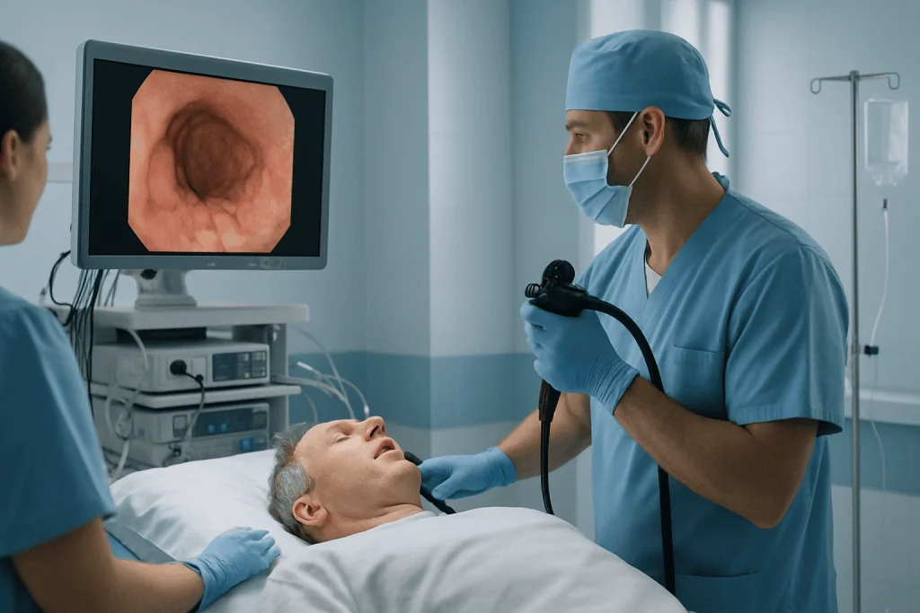
The definitive test is upper gastrointestinal endoscopy. A thin, flexible camera is passed through the mouth into the esophagus and stomach, allowing the physician to see the varices directly, grade their size, and look for high-risk surface features such as red wale markings, thin red streaks on the varix that indicate a weakened wall. Endoscopy also inspects the rest of the upper digestive lining, which means the same examination can identify unrelated conditions such as Barrett esophagus or, rarely, esophageal cancer. Crucially, if high-risk varices are found, band ligation can often be performed during the same session.
Many patients arrive with a stack of prior reports: endoscopy findings from one hospital, imaging from another, laboratory results from a third. A careful review of these records, endoscopy images, variceal grading, liver function trends, medication history and any previous bleeding events, determines whether the existing plan remains appropriate, whether repeat endoscopy is needed, and how urgent further treatment is. This kind of structured second opinion is common in variceal care, because recommendations can differ between centres and the stakes of getting the timing right are high.
Who May Need Treatment for Esophageal Varices
Treatment is considered when varices are large, carry high-risk features, have bled before or are actively bleeding. Not every varix needs intervention. Small varices in a patient with stable, well-compensated liver disease may simply be monitored, sometimes with medication, with endoscopy repeated at intervals to watch for growth.
Risk is assessed during endoscopy by looking at the size of the varices, surface markings such as red wale signs and any evidence of recent bleeding. But the veins are only half of the assessment. Doctors also weigh liver function, platelet count, clotting tests, the presence of ascites, previous bleeding history and the cause of the liver disease. A patient with large varices and more advanced liver disease may be advised to begin preventive treatment even with no symptoms at all, because the combination of fragile veins and limited physiological reserve makes a first bleed particularly dangerous.
The patient’s overall condition shapes the plan as much as the endoscopic findings. Kidney function, infection risk, nutritional status, heart and lung health, use of blood thinners and previous reactions to sedation all influence whether treatment can be done as a scheduled outpatient procedure or requires hospital admission. In active bleeding, none of this is deliberated at leisure, treatment begins immediately with stabilisation and emergency endoscopy. In preventive care, there is time to review records, optimise the patient’s condition and plan the procedure properly.
Conditions and Indications Treated
Variceal treatment sits inside the broader management of portal hypertension, and the indications reflect that. Endoscopic and medical therapy may be recommended for patients with medium or large esophageal varices, varices with high-risk endoscopic features, a previous variceal bleed, active bleeding, or recurrent bleeding despite earlier therapy.
It may also form part of a wider plan for related complications of portal hypertension. Gastric varices, enlarged veins in the stomach, can accompany esophageal varices and sometimes require different techniques, including injection therapies or radiological procedures rather than banding. Portal hypertensive gastropathy, a diffuse congestion of the stomach lining, can cause slower, chronic blood loss. Ascites, an enlarged spleen with low platelet counts, and episodes of confusion caused by hepatic encephalopathy all signal that portal pressure and liver function need attention beyond the varices themselves.
Patients with non-cirrhotic portal hypertension, portal vein thrombosis, Budd-Chiari syndrome, vascular liver disorders, often need more individualised planning. The pressure problem in these conditions may sit before, within or beyond the liver, and that anatomy changes which interventions are feasible and safe. This is one of the situations where coordinated input from hepatology, interventional radiology and, occasionally, liver surgery genuinely changes the plan rather than merely confirming it.
How Esophageal Varices Treatment Works
Treatment for esophageal varices is not a single procedure but a coordinated set of interventions with three aims: prevent a first bleed, stop active bleeding, and reduce the chance of bleeding again, all while managing the liver disease driving the problem. The right combination depends on whether you are being treated preventively or after a bleed, how advanced the liver disease is, and whether varices are present in the stomach as well as the esophagus.
What is endoscopic band ligation?
Endoscopic band ligation is the most commonly used procedure for esophageal varices. A flexible endoscope is passed through the mouth into the esophagus. A special device on the tip of the scope gently suctions a varix into a small chamber, and the physician deploys a tiny elastic band around its base. The band cuts off blood flow within the enlarged vein. Over the following days, the banded tissue sloughs off and the area heals with scar tissue, closing the abnormal channel. Several bands can be placed in one session, depending on the number and size of the varices and the patient’s condition. Most patients need more than one session, spaced weeks apart, until the varices are obliterated.
Are there other endoscopic treatments besides banding?
Yes. Sclerotherapy, injecting a solution into or around the varix that makes it scar and close, was the standard endoscopic treatment before banding and is still used when band placement is technically difficult, for example when scarring from previous sessions makes suction of the varix into the banding device impossible. For gastric varices, injection of a tissue adhesive is often preferred, because bands hold poorly on the wider, deeper veins of the stomach wall. And in the rare situation where esophageal bleeding cannot be controlled endoscopically, a removable covered metal stent can be placed across the lower esophagus to compress the bleeding veins from inside: a temporary bridge that keeps the patient stable while the team arranges definitive therapy. Each of these tools has a specific place; the skill lies in choosing among them, sometimes under considerable time pressure.
What medication is used for esophageal varices?
Nonselective beta blockers are the mainstay of medical therapy. They lower pressure in the portal venous system and can reduce the risk of a first bleed or of rebleeding. They are prescribed and adjusted by the treating physician, because not everyone tolerates them, low blood pressure, asthma, kidney issues and other conditions can make them unsuitable, and the dose has to be balanced against side effects. In an emergency, intravenous vasoactive medications are given to reduce portal pressure quickly, and antibiotics are routinely used, because patients with cirrhosis who bleed into the digestive tract have a markedly increased vulnerability to infection.
What is a TIPS procedure?
TIPS, transjugular intrahepatic portosystemic shunt, is a procedure performed by interventional radiologists that creates a new channel through the liver, connecting the portal vein directly to a hepatic vein. Blood can then bypass the scarred liver tissue, and portal pressure falls. TIPS is considered when bleeding cannot be controlled with endoscopy and medication, or when it keeps recurring despite appropriate therapy. It is not offered casually: candidates are assessed carefully for liver function, heart function and the risk of hepatic encephalopathy, a confusion syndrome that can worsen when blood bypasses the liver’s filtering function. For the right patient, TIPS can be decisive; for the wrong patient, it can trade one problem for another. That judgement is exactly why complex cases benefit from multidisciplinary review.
What Happens During Treatment, Step by Step
For a planned, preventive procedure, the pathway is orderly and predictable:
- Assessment. The team reviews your symptoms, liver disease history, previous endoscopies, medications, allergies and recent laboratory results. Existing records and images from other hospitals are reviewed wherever possible before the appointment.
- Preparation. You are asked not to eat or drink for a set period before the procedure. Blood tests check haemoglobin, platelets, clotting, liver and kidney function and electrolytes. If you take anticoagulants, antiplatelet drugs or certain diabetes medications, the care team gives you individualised instructions: these decisions always sit with the treating doctor.
- Sedation. You receive sedation or anaesthesia matched to your medical condition and the procedure planned, with monitoring throughout.
- Endoscopy. The endoscope is guided through the mouth into the esophagus, stomach and first part of the small intestine. The physician identifies the varices, grades their size and risk features, and checks for signs of recent bleeding.
- Band ligation. If treatment is indicated, bands are placed on the highest-risk varices during the same session.
- Recovery. You are observed until the sedation wears off. The procedure itself is often short; the full hospital visit is longer because of preparation and observation.
- Follow-up planning. Before discharge you receive a written plan covering diet, medication and the timing of the next endoscopy session.
How are bleeding varices treated in an emergency?
Bleeding varices are treated as a hospital emergency in which several things happen at once. Variceal esophageal bleeding can drop blood pressure quickly, so the first priority is stabilisation: intravenous access, fluids, protection of the airway when needed, and blood products when appropriate. Transfusion decisions are made deliberately, because over-transfusing can raise portal pressure and paradoxically worsen bleeding. Intravenous vasoactive medication is started to lower portal pressure, and antibiotics are given to reduce infection risk.
Emergency endoscopy then identifies the bleeding point and treats it, most often with band ligation. If bleeding is difficult to control, temporary measures, such as balloon devices that compress the veins from inside the esophagus, can buy time while the team plans definitive therapy. In selected patients, TIPS is considered at this stage. Throughout, the patient is monitored closely, often in an intensive care or high-dependency setting, tracking blood pressure, oxygen levels, heart rhythm and response to treatment.
The technology behind this pathway is worth understanding, because it explains why variceal care is hospital-based. High-resolution endoscopic imaging lets physicians see varices and bleeding points clearly and place bands precisely. Doppler ultrasound and cross-sectional imaging map the portal venous system. Laboratory support tracks blood counts and clotting in real time. And image-guided interventional techniques allow radiologists to navigate blood vessels and reshape blood flow when endoscopy alone is not enough. No single one of these is exotic; the value is in having them all available in one place, at once, when a bleed happens.
After band ligation, planned or emergency, mild chest discomfort, a sore throat, temporary difficulty swallowing or a feeling of pressure behind the breastbone are common and usually settle over several days. Soft foods may be recommended briefly while the treated areas heal. Follow-up endoscopy is scheduled at intervals until the varices have been adequately treated, after which surveillance continues on a schedule matched to your risk profile.
Why Acting Early Matters
Esophageal varices can remain silent right up to the moment they bleed, and a first bleed is a fundamentally worse starting point than a screening finding. Treating high-risk varices before rupture is generally a controlled, scheduled procedure. Treating them afterwards means resuscitation, transfusion decisions, infection risk, possible intensive care and the strain of major blood loss on a liver and kidneys that may have little reserve. The clinical logic of screening and preventive banding rests entirely on this asymmetry.
Acting early also creates room to treat the disease behind the veins. Viral hepatitis may respond to antiviral therapy. Alcohol-related liver disease requires structured abstinence support. Metabolic liver disease calls for weight, diabetes and lipid management. Autoimmune and cholestatic liver diseases have disease-specific medications. Variceal care works best as one strand of a broader liver plan, not as a stand-alone fix, because if portal pressure keeps rising, new varices keep forming.
Can esophageal varices go away?
Sometimes, partially. Banded varices are deliberately destroyed and replaced by scar tissue, so those specific veins do go away. Small varices can also regress if portal pressure falls, for example, when hepatitis is treated successfully or alcohol is stopped and the liver recovers some function. But as long as portal hypertension persists, new varices can form, which is why surveillance endoscopy continues even after successful treatment. Varices are best understood as a pressure gauge: they reflect the state of the portal system, and they respond to it in both directions.
Can esophageal varices be cured?
Honestly: treatment controls esophageal varices rather than permanently curing them. Band ligation obliterates the veins that are visible today; it does not lower the portal pressure that created them. Lasting change depends on the underlying cause. Where that cause is treatable: a virus that can be suppressed, alcohol that can be stopped, a clot that can be managed: the portal system can improve and the risk can fall substantially. Where cirrhosis is advanced and irreversible, varices are managed long term with banding, medication and surveillance, and some patients are evaluated for more comprehensive liver care. The most accurate framing is management, done well, over years.
Can you live a long life with esophageal varices?
Many people do, particularly when the varices are found before bleeding, preventive treatment is carried out, and the liver disease is stable or improving. The outlook depends far more on the liver than on the veins: someone with well-compensated cirrhosis and properly treated varices is in a very different position from someone with advanced liver failure. What consistently helps is the unglamorous work, attending surveillance endoscopies, taking prescribed medication as directed by the treating doctor, avoiding alcohol, and keeping the underlying condition under specialist review.
Benefits of Esophageal Varices Treatment
The benefits depend on the stage of disease and on whether care is preventive or urgent, but they cluster around a few concrete outcomes.
| Benefit | What It Means for You |
|---|---|
| Lower risk of variceal bleeding | Band ligation and appropriate medication can reduce the chance that high-risk varices rupture, especially within a structured follow-up plan. |
| Rapid control during bleeding episodes | Emergency endoscopy, portal pressure-lowering medication and hospital monitoring can help stop bleeding and stabilise the patient. |
| Clearer understanding of liver disease severity | Blood tests, imaging and specialist assessment define the cause and stage of portal hypertension, guiding future care. |
| Reduced risk of recurrent bleeding | Repeated banding when needed, long-term medication and surveillance endoscopy help reduce the likelihood of another bleed. |
| Coordinated planning for complex cases | Patients with advanced liver disease, gastric varices or recurrent bleeding can be evaluated for interventional radiology procedures or broader liver-focused pathways. |
Recovery Timeline After Treatment
Recovery differs between a planned banding session and emergency treatment for a bleed, but most patients follow a recognisable pattern.
| Time Period | What Patients Can Expect |
|---|---|
| Day 1 | After planned band ligation, observation until sedation wears off. A sore throat, mild chest discomfort or temporary swallowing discomfort may occur. Emergency patients remain in hospital for monitoring. |
| First week | Soft foods may be recommended briefly while banded areas heal. Alcohol is avoided and the medication plan is followed as prescribed. Discomfort typically eases over several days. |
| First month | Follow-up may include repeat blood tests and a further endoscopy session if additional banding is needed. Medication tolerance and liver disease management are reviewed. |
| Longer term | Surveillance endoscopy, portal pressure-lowering medication when appropriate, and treatment of the underlying liver condition continue to reduce future bleeding risk. |
Factors That Influence Outcomes
The single most important factor is the severity of the underlying liver disease. Patients with well-compensated cirrhosis have physiological reserve; patients with jaundice, ascites, kidney dysfunction, recurrent infections or hepatic encephalopathy have less room for error, and every decision, sedation, transfusion, procedure timing, is made with that in mind. The cause of the liver disease matters too, because some causes can be treated or slowed, changing the trajectory rather than just managing today’s veins.
The varices themselves carry their own risk signals. Large size, red markings on the vein surface, active bleeding at endoscopy and a previous bleed all indicate higher risk. Gastric varices behave differently from esophageal ones and may need different endoscopic or radiological techniques. Portal vein thrombosis or unusual blood flow patterns can reshape the entire strategy.
Timeliness runs through everything. A patient treated before a first bleed follows a controlled pathway; a patient arriving in shock does not. In active bleeding, early resuscitation, prompt medication, experienced endoscopy and careful monitoring each influence whether the bleed is controlled and complications avoided.
Medication adherence shapes the long term. Nonselective beta blockers only work if they are taken, tolerated and correctly adjusted, and some patients stop them simply because they feel well. Regular follow-up keeps the therapy matched to the person. Any change to these medicines belongs with the treating doctor, not least because stopping or adjusting them alters bleeding risk in ways that are not obvious from how a patient feels.
Finally, the whole-body picture counts. Liver disease erodes muscle mass, protein stores and vitamin levels, and it impairs clotting, which is why some patients first notice easy bruising or bleeding gums before any digestive symptom appears. Nutrition support, alcohol avoidance and infection prevention are not add-ons; they measurably affect how well a patient withstands both the disease and its treatment. Continuity is the last piece: understanding the diagnosis, what was done at endoscopy, which medicines to take and when the next endoscopy is due, with documentation clear enough that any physician involved in ongoing care can continue the plan without gaps.
Esophageal Varices Care at Acibadem
Variceal care rarely involves a single doctor. Depending on the case, it draws on gastroenterology, hepatology, interventional endoscopy, anaesthesiology, intensive care, interventional radiology, liver surgery and nutrition. At Acibadem, patients with esophageal varices are managed within this coordinated framework, and complex cases may be discussed in specialist boards so that the plan reflects both established international protocols and the individual patient’s condition, rather than the preference of whichever specialist happened to see them first.
The diagnostic pathway is built to answer the deeper question behind the varices: why is portal pressure high, and how much reserve does this liver have? Evaluation may include advanced laboratory testing, high-quality endoscopic assessment, abdominal imaging, Doppler studies of portal blood flow and further investigations when the cause of the liver disease is unclear. This distinguishes patients who need straightforward preventive banding from those who need broader portal hypertension management, interventional radiology input or advanced liver care.
The treatment infrastructure matters most on the worst days. High-resolution endoscopy supports precise identification of variceal size and bleeding stigmata; ligation systems allow controlled band placement in the same session. For patients who bleed or whose liver disease is advanced, hospital-based monitoring, blood bank support, intensive care availability and access to interventional radiology sit behind the endoscopy suite. Personalised planning follows from this range: a newly diagnosed patient with large varices, a patient recovering from a recent bleed, a patient rebleeding despite therapy, and a patient with significant liver failure each need a genuinely different pathway, and each of those pathways exists under one roof.
When a second opinion is sought, common after a bleeding episode, after repeated banding, or when recommendations from different centres conflict: it typically involves a structured review of prior endoscopy reports, images, laboratory results and discharge summaries, so that the question of whether the current plan is right, and how urgent any change is, gets a clear answer.
Living With Esophageal Varices
Esophageal varices demand respect, not panic. Diagnosed early, treated appropriately and followed over time, they can usually be managed as one known, monitored element of a chronic liver condition rather than as a lurking catastrophe. The most useful thing you can do is understand your own risk precisely: whether your varices are small or large, whether they show high-risk features, how your liver function is trending, and what your surveillance schedule is.
Day to day, a few practical themes come up repeatedly in clinic. Alcohol is the clearest one: for most people with portal hypertension, avoiding it entirely protects both the liver and the veins. Nutrition matters more than many patients expect, because chronic liver disease quietly erodes muscle and protein stores; dietitians often recommend adequate protein and regular meals, tailored to the individual, particularly when ascites or encephalopathy are part of the picture. Some medicines, including common over-the-counter painkillers, can affect the stomach lining, kidney function or bleeding tendency, which is why every medicine and supplement, however minor it seems, deserves review by the treating physician rather than an assumption of safety. And because infections affect people with cirrhosis more severely, vaccination status and prompt attention to fevers are discussed as part of routine liver care rather than left to chance.
Over the years, the pattern of good variceal care stays remarkably consistent: surveillance endoscopy on schedule, medication reviewed rather than abandoned, alcohol avoided, the underlying liver disease actively treated, and every hospital visit documented well enough that the next physician can pick up the thread. Varices reward that kind of steadiness. The veins are only the visible sign; the plan is what changes the story.
Preparation
- Before treatment, the gastroenterology team evaluates liver disease, bleeding risk, blood counts, and clotting tests. Patients are usually asked to fast for 6 to 8 hours before endoscopy. Blood thinners, diabetes medicines, and other regular medications should be reviewed with the doctor in advance.
Aftercare
- After endoscopic treatment, patients are monitored for bleeding, chest discomfort, swallowing difficulty, or fever. Soft foods may be recommended for a short period, and prescribed medicines such as beta-blockers or acid reducers should be taken as directed. Follow-up endoscopy may be needed to confirm varices are controlled and reduce recurrence risk.
Frequently Asked Questions
What affects the cost of esophageal varices treatment?
The main factors are whether care is elective or urgent, the need for endoscopic band ligation, anaesthesia, hospital monitoring, medications, laboratory tests, imaging, transfusion support, and the severity of underlying liver disease.
How can I get a personalised quote from Acibadem?
You can request a free consultation and share recent endoscopy reports, blood tests, liver imaging, medication lists, and discharge summaries if available. A specialist team can then recommend a care plan and provide a personalised quotation.
Is endoscopic band ligation always included in the treatment plan?
Not always. Some patients may need medication and surveillance, while others may need band ligation or urgent endoscopic treatment. The decision depends on varix size, bleeding risk, symptoms, and liver condition.
Can international patients be treated if they have active bleeding?
Active bleeding is a medical emergency and should be treated immediately at the nearest appropriate hospital. International transfer is considered only after clinical stabilisation and specialist review.
What is usually included in an international care package?
Inclusions vary by medical need, but may cover specialist consultation, endoscopy planning, anaesthesia assessment, hospital services, interpreter support, medication guidance, and follow-up recommendations. The final scope is confirmed after clinical review.
Medically reviewed by the Acıbadem International Medical Board August 31, 2026
See our medical review board →
Update history
- PublishedJune 8, 2026
- Medical review approvedAugust 31, 2026
- Last content updateSeptember 8, 2026
References2
- Bleeding esophageal varices medlineplus.gov
- Esophageal Varices ncbi.nlm.nih.gov
Trusted care for international patients
From the Acibadem Blog
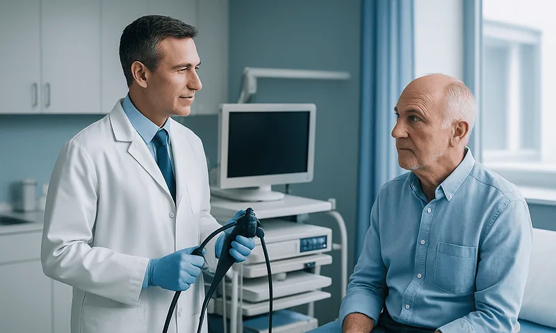
What Happens During Band Ligation for Esophageal Varices: The Procedure Explained
Band ligation for esophageal varices is an endoscopic procedure in which a doctor passes a thin flexible camera down the throat, suctions a swollen…

After Variceal Banding: Soft Foods, Chest Discomfort and the First Days of Recovery
Recovery after variceal banding is usually measured in days. Most people go home the same day, drink clear liquids first, then eat soft foods…