Gastrointestinal Bleeding Treatment
Gastrointestinal bleeding care focuses on finding the bleeding source, stabilizing the patient and stopping bleeding, often with urgent endoscopy and supportive hospital treatment.
Quick answer
Gastrointestinal bleeding is blood loss from any part of the digestive tract, oesophagus, stomach, small intestine, colon, rectum or anus. It is a sign of an underlying condition such as an ulcer, diverticular disease, varices, polyps or cancer. Care involves stabilising the patient, finding the source with endoscopy, colonoscopy or imaging, then stopping the bleeding and treating the cause.
What Is Gastrointestinal Bleeding?
Gastrointestinal bleeding is blood loss from any point along the digestive tract: the oesophagus, stomach, small intestine, colon, rectum or anus. It is a sign, not a disease in its own right: something along that pathway is damaged, inflamed or abnormal, and it is bleeding. Care for gastrointestinal bleeding therefore has three jobs, usually in this order: keep you stable, find the source, and stop the bleeding while treating whatever caused it.
The way gastrointestinal bleeding shows itself varies widely. Some people notice bright red blood in the toilet or black, tar-like stools. Others vomit blood, or material that looks like coffee grounds. Some feel faint, unusually weak or short of breath, and only learn later that a falling blood count was the reason. And some have no visible blood at all: the bleeding is discovered because a routine test shows iron deficiency anaemia with no obvious explanation.
The amount of blood you can see is a poor guide to how serious the situation is. A small, steady leak can cause significant anaemia over weeks or months without ever producing a dramatic symptom. A sudden bleed from an ulcer or an enlarged vein can become dangerous within hours. This is why the evaluation focuses on the source and the cause, not the appearance in the toilet bowl.
Is Gastrointestinal Bleeding a Form of Internal Bleeding?
Yes, internal bleeding means blood loss inside the body, and the digestive tract is one of the most common places it happens. What makes a gastrointestinal bleed different from, say, bleeding into the abdominal cavity or a muscle is that the blood eventually travels through the gut and appears in vomit or stool. That gives doctors visible clues about the location: blood exposed to stomach acid turns dark and tar-like, while blood from the lower bowel usually stays red or maroon. Blood that never becomes visible can still be detected, through stool testing or through its effect on the blood count.
Bleeding, Hemorrhaging and Haemorrhage: Do the Words Mean Different Things?
No, bleeding and hemorrhaging describe the same process: blood escaping from a damaged blood vessel. “Haemorrhage” is simply the formal medical term, and you may see phrases such as “upper GI haemorrhage” on hospital reports or discharge summaries. Doctors do distinguish between acute bleeding, which starts suddenly and can progress quickly, and chronic bleeding, which continues slowly over time. The two are investigated with the same tools but on very different timescales.
Upper and Lower Gastrointestinal Bleeding
Upper gastrointestinal bleeding comes from the oesophagus, the stomach or the duodenum: the first part of the small intestine. It may cause vomiting of fresh blood, vomit that resembles coffee grounds, or black, sticky stools known as melaena. The dark colour comes from blood being partly digested on its way through the gut. Upper sources include peptic ulcers, inflamed stomach or oesophageal lining, and enlarged veins related to liver disease.
Lower gastrointestinal bleeding comes from the colon, rectum or anus and usually appears as bright red or maroon blood in or around the stool. Common sources include diverticular disease, haemorrhoids, anal fissures, inflammatory bowel disease, polyps and, less often, colorectal cancer. Because the blood has a short distance to travel, it tends to keep its red colour.
Bleeding from the middle section of the small intestine is less common and harder to locate. The small bowel is several metres long and not fully reachable with a standard endoscope or colonoscope, so it is usually investigated only after the more accessible upper and lower sources have been ruled out.
The distinction matters because it determines which test comes first. A colour rule of thumb is useful but not absolute: a brisk upper bleed can move through the gut fast enough to appear as red blood in the stool. Clinical assessment, not colour alone, decides the pathway.
Symptoms and Warning Signs
Bleeding can announce itself loudly or hide for months. Both patterns deserve evaluation, because neither the visible amount of blood nor the absence of pain reliably indicates the cause.
What Are the Warning Signs of Gastrointestinal Bleeding?
The warning signs of gastrointestinal bleeding fall into two groups: signs of the blood itself, and signs of what blood loss does to the body. The first group includes:
- Black, tar-like or sticky stools
- Bright red or maroon blood in or on the stool, or on toilet paper
- Vomiting fresh blood or material resembling coffee grounds
The second group includes unexplained fatigue, pale skin, dizziness on standing, a racing heart, breathlessness on mild exertion and reduced exercise tolerance: the everyday footprint of anaemia. Emergency teams treat the combination of visible bleeding with faintness, confusion, chest discomfort or a rapid pulse as a medical emergency, because it suggests the circulation is struggling to compensate for the blood loss.
What Are 5 Signs of Internal Bleeding?
The signs of internal bleeding that clinicians watch for most closely are:
- Dizziness or fainting, especially on standing up, as blood volume falls
- A rapid heartbeat with low blood pressurethe body compensating for lost volume
- Pale, cool or clammy skin as circulation is redirected to vital organs
- Breathlessness and unusual weakness, because fewer red cells are carrying oxygen
- Visible blood where it should not bein vomit or stool, or as black, tarry stools
These signs overlap heavily with the signs of a significant GI bleed, because the digestive tract is one of the most frequent sites of internal blood loss. None of them proves where the bleeding is coming from; that is what the diagnostic work-up establishes.
How Long Can Someone Survive With a GI Bleed?
There is no single answer: how long someone can go on with a GI bleed depends entirely on how fast the bleeding is, what is causing it, and how much reserve the person has. A slow, chronic leak, from angiodysplasia or a small ulcer, for instance, can continue for months while the body partly keeps pace, producing gradually worsening anaemia rather than collapse. A brisk arterial bleed from a deep ulcer, or variceal bleeding in liver disease, can become life-threatening within hours. Age, heart disease, kidney disease and blood-thinning medication all narrow the margin. Because nobody can tell from home which pattern they have, medical assessment is what separates a nuisance from an emergency.
Occult Bleeding: When There Is No Visible Blood
Occult bleeding means blood loss too slow or too small to be seen. It is often discovered indirectly: a routine blood test shows low haemoglobin or depleted iron stores, or a stool test detects traces of blood. Occult bleeding deserves the same structured evaluation as visible bleeding, because it can be the first sign of a peptic ulcer, inflammatory bowel disease, a polyp or a colorectal cancer, conditions that are far easier to treat when found early.
What Causes Gastrointestinal Bleeding?
Bleeding can arise from dozens of conditions along the digestive tract, from a minor anal fissure to a major vascular malformation. The likely causes shift depending on where the bleeding starts, how old the patient is, what medication they take and what other diseases they have.
What Is the Most Common Cause of Gastrointestinal Bleeding?
Peptic ulcer disease is the most common cause of significant upper gastrointestinal bleeding. Ulcers in the stomach or duodenum are frequently linked to Helicobacter pylori infection or to regular use of aspirin and other nonsteroidal anti-inflammatory drugs, which weaken the protective lining of the gut. In the lower tract, diverticular disease is among the most frequent causes of major bleeding, while haemorrhoids are the most common explanation for small amounts of bright red blood after a bowel movement. “Most common” is not the same as “most likely in you”, which is why evaluation is individual rather than statistical.
Causes in the Oesophagus, Stomach and Duodenum
Beyond peptic ulcers, upper gastrointestinal bleeding may come from gastritis, oesophagitis, enlarged veins (varices) caused by liver disease, Mallory-Weiss tears at the junction of oesophagus and stomach after forceful vomiting, vascular malformations, tumours, or injury to the lining caused by medication. Variceal bleeding deserves special mention: it occurs in people with cirrhosis or other causes of raised pressure in the liver’s blood supply, it can be abrupt and heavy, and it requires a treatment pathway of its own that combines endoscopic banding, specific medication and liver-focused care.
Blood in Stool Causes: From Haemorrhoids to Colorectal Cancer
Blood in stool causes range from minor anorectal conditions to serious bowel disease. The list includes haemorrhoids and anal fissures, diverticular disease, inflammatory bowel disease such as ulcerative colitis and Crohn’s disease, ischaemic colitis (reduced blood flow to the bowel), infectious colitis, angiodysplasia (fragile abnormal blood vessels in the bowel wall), polyps, colorectal cancer and radiation-related changes after previous pelvic treatment. Two cautions apply. First, the presence of an obvious minor cause, such as haemorrhoids, does not exclude a second, more serious source higher up. Second, a brisk upper gastrointestinal bleed can appear as red blood in the stool, mimicking a lower source. Both are reasons why colour and quantity alone never settle the diagnosis.
Small-Bowel Bleeding: The Hidden Middle
Small-bowel bleeding is suspected when upper endoscopy and colonoscopy are both normal yet anaemia persists or bleeding recurs. Possible causes include vascular lesions such as angiodysplasia, ulcers, Crohn’s disease, tumours, medication-related injury to the bowel lining, and rarer inherited vascular conditions. Because the small intestine sits beyond the reach of standard instruments, dedicated tools, capsule endoscopy, device-assisted enteroscopy or specialised imaging, are used to examine it. Finding a small-bowel source can take patience, and a negative first round of tests does not mean the search is over.
Who Needs Structured Evaluation?
Visible blood in vomit or stool always warrants medical assessment, however small the amount and however quickly it seems to settle. Some causes are minor, but there is no reliable way to distinguish them from serious ones without examination and, usually, endoscopy. The same is true of unexplained iron deficiency anaemia found on a routine blood test: a common route by which slow bleeding first comes to light, often through an internal medicine assessment.
Some groups carry higher risk and need particularly careful planning. These include people taking anticoagulants or antiplatelet medication, patients with liver cirrhosis, individuals with kidney disease, older adults, patients with heart disease, and anyone who has bled from the gut before. In these patients, treatment must balance bleeding control against the risk of clots, stroke or heart strain: a balance that usually involves more than one specialty.
Patients who have already been evaluated elsewhere sometimes pursue a second review when bleeding recurs, the source was never found, anaemia continues despite treatment, or surgery has been proposed. The most useful reviews draw on previous endoscopy and colonoscopy reports with images, pathology results, CT or MRI studies, laboratory trends over time, medication lists and discharge summaries. With that material, specialists can often avoid repeating tests unnecessarily and identify precisely which step comes next.
How Gastrointestinal Bleeding Is Diagnosed
The pathway depends on how you present. Someone actively vomiting blood with low blood pressure is stabilised first and investigated urgently. Someone stable with chronic anaemia is investigated in a planned sequence. In both cases, the goal is the same: to move from uncertainty to a clear, medically sound plan.
Initial Assessment and Stabilisation
For an urgent presentation, the sequence typically runs in this order:
- Assessment of airway, breathing, circulation, mental state and vital signs
- Intravenous access, so fluids, medication or blood products can be given quickly
- Blood samples for haemoglobin, clotting, organ function and blood type
- Continuous monitoring, with transfusion or medication as the results dictate
- A decision on the timing and type of endoscopy or imaging
For patients on blood thinners, the team also assesses whether the medication should be paused, reversed or continued. This decision sits with the treating physicians, because stopping anticoagulation abruptly can create clotting risks that are just as serious as the bleeding itself.
Blood Tests and Monitoring
Laboratory work does much of the early detective work. Haemoglobin and its trend over time indicate how much blood has been lost and whether loss is continuing. Platelet counts and clotting studies show whether the blood can form clots normally. Kidney and liver function tests, inflammation markers and iron studies help point towards a cause. Repeat testing during the hospital stay is normal, because a single snapshot can lag behind an active bleed. If you are preparing for planned investigations, it helps to know how to prepare for blood tests, scans and pre-procedure checks in advance.
Endoscopy and Colonoscopy
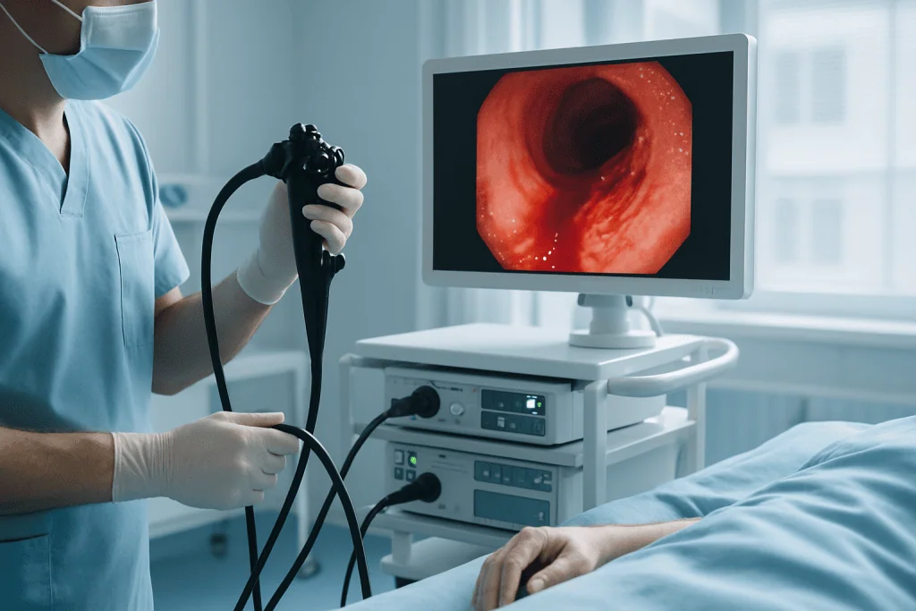
Upper endoscopy is usually the first test for suspected upper bleeding. A flexible camera is passed through the mouth to examine the oesophagus, stomach and duodenum, usually under sedation. Colonoscopy examines the colon and rectum from below and requires bowel preparation, a cleansing routine taken over several hours or overnight, so the physician can see the lining clearly. Both tests share a decisive advantage: they can treat many bleeding sources at the moment they find them, in the same session. Flexible sigmoidoscopy, a shorter examination of the rectum and lower colon, is used in selected cases.
CT, Angiography and Capsule Endoscopy
When bleeding is brisk, or endoscopy is not immediately possible, contrast-enhanced computed tomography can reveal active bleeding, vascular abnormalities, inflammation, masses or complications. Angiography goes a step further: an interventional radiologist threads a catheter through the blood vessels to locate the bleeding artery directly, and, in many cases, treat it during the same procedure. When the standard examinations find nothing and suspicion falls on the small bowel, capsule endoscopy is used: you swallow a camera the size of a large pill, which photographs the intestine as it passes through. Device-assisted enteroscopy can then reach deep into the small bowel to treat what the capsule finds.
How Is Gastrointestinal Bleeding Treated?
Treatment works on two levels at once: stopping the active bleeding, and addressing the condition behind it so it is less likely to return. Depending on the source, that may mean endoscopic therapy, medication, transfusion, an interventional radiology procedure or, in selected cases, surgery, often in combination.
Endoscopic Treatment
Bleeding gastrointestinal lesions, ulcers with visible vessels, oesophageal varices, angiodysplasia, Mallory-Weiss tears, bleeding polyps, are the main targets of endoscopic therapy, and for many patients endoscopy is both the diagnosis and the treatment. Depending on what is found, the gastroenterologist may place mechanical clips to close a bleeding point, apply heat to seal tissue, inject medication around the site, band enlarged veins, apply topical haemostatic agents or remove a bleeding polyp entirely. Sedation or anaesthesia support is arranged according to urgency, the procedure planned and your overall condition, with monitoring throughout. Endoscopic therapy controls many bleeds without any need for open surgery, though a repeat procedure is sometimes medically appropriate, if bleeding returns, if blood or stool limited the view during an emergency examination, or if the underlying condition needs staged treatment.
Medication and Supportive Care
Medication works alongside procedures rather than instead of them. Acid-suppressing therapy supports healing in ulcer-related bleeding. When variceal bleeding is suspected, antibiotics and specific vasoactive drugs form part of the standard pathway. If H. pylori infection is confirmed, eradication treatment reduces the risk of the ulcer, and the bleeding, coming back. After chronic blood loss, iron replacement rebuilds depleted stores, given by mouth or intravenously depending on severity and tolerance. A structured medication review is also central: aspirin, anti-inflammatory painkillers, anticoagulants, antiplatelet drugs, steroids and some supplements can all contribute to bleeding risk. Whether any of these is paused, reversed, restarted or replaced is a decision for the treating doctors, weighed against the reason each drug was prescribed, often with input from cardiology, neurology or haematology.
When a Blood Transfusion Is Needed
Transfusion is considered when anaemia is severe, symptoms are concerning or blood loss is ongoing. The decision is individual rather than automatic: age, heart disease, how stable the circulation is and the trend of the blood counts all feed into it, guided by clinical protocols. Some patients with significant bleeding never need transfusion; others need blood products urgently before any procedure can begin.
Interventional Radiology and Surgery
When endoscopy cannot control the bleeding, or cannot reach it, interventional radiology offers a minimally invasive alternative. Through a small puncture in a blood vessel, the radiologist guides a catheter to the bleeding artery and delivers material that blocks blood flow to that precise area: a technique called embolisation, particularly useful for certain lower gastrointestinal bleeds and bleeding after previous surgery. Surgery itself is needed less often than in the past, because endoscopic and radiological methods handle most cases. It remains important in specific situations: bleeding that cannot be controlled by other means, perforation, certain tumours, severe colitis, or recurrent bleeding from a source no other approach can manage. When surgery is planned, the team weighs your overall health, the exact location, the likelihood of recurrence and the least invasive approach that will actually work.
Can Gastrointestinal Bleeding Stop on Its Own?
Yes, frequently, and this is precisely why it is misleading. Diverticular bleeding, for example, often stops spontaneously, and minor anorectal bleeding may settle within a day. But bleeding that stops has not been treated: the ulcer, vessel, polyp or tumour that caused it is still there, and recurrence is common. A quiet interval is an opportunity to investigate in controlled conditions rather than a reason to consider the matter closed.
Hospital Stay and Recovery
How long care takes varies with the severity and the cause. An upper endoscopy itself is a short procedure, but preparation, sedation recovery, monitoring and treatment planning extend the total time in hospital. Colonoscopy adds the hours needed for bowel preparation. Patients with minor bleeding and stable blood counts may go home the same day or after brief observation; those with major bleeding, severe anaemia, liver disease, heart disease or recurrent bleeding may need several days as an inpatient, sometimes in a high-dependency or intensive care setting.
| Time Period | What Patients Can Expect |
|---|---|
| Day 1 | Assessment, blood tests, monitoring and stabilisation; endoscopy or imaging if bleeding is active or the risk is judged significant. |
| First week | Medication adjustments by the treating team, diet guidance, iron or acid-suppressing therapy where appropriate, and observation for any sign of rebleeding. |
| First month | Strength and blood counts gradually improve. Follow-up visits or repeat tests are scheduled according to the diagnosis. |
| Longer term | Ongoing management focuses on preventing recurrence, treating the underlying condition and keeping medication use safe. |
Recovery is about more than the bleeding stopping. It means rebuilding blood levels, restoring energy, and confirming that the cause has been dealt with. Before discharge, patients typically receive written guidance on diet, medication, follow-up tests and when normal activity can resume. It is worth keeping that written plan and sharing it with your regular doctors: the guide on warning signs after returning home from treatment sets out which symptoms after discharge should prompt a fresh medical review rather than watchful waiting.
Why Acting Early Matters
Gastrointestinal bleeding can change quickly. A patient who seems stable can become weak, hypotensive or severely anaemic if bleeding continues. Early assessment lets medical teams identify who is at higher risk and intervene before complications develop, and it distinguishes minor anorectal bleeding from causes that need urgent endoscopy or imaging.
Delay carries measurable physical costs: continued blood loss, fainting and falls, strain on the heart, kidney injury, and the need for emergency transfusion that planned care might have avoided. In people with coronary artery disease, severe anaemia reduces oxygen delivery to the heart muscle itself. In older adults and patients with multiple conditions, the physiological reserve is smaller, so the same blood loss does more damage.
Delay also postpones diagnosis. Recurrent or hidden bleeding can be the first sign of colorectal cancer, inflammatory bowel disease, a bleeding ulcer, liver-related varices or small-bowel disease. Earlier investigation means the underlying condition is found at a more manageable stage, and reduces the likelihood of repeated hospital admissions for the same unsolved problem.
What Timely Treatment Can Achieve
Set against those risks, structured care delivers practical benefits at each stage.
| Benefit | What It Means for You |
|---|---|
| Rapid stabilisation | Fluids, blood tests, monitoring and transfusion when needed reduce the immediate risks of significant blood loss. |
| Accurate identification of the source | Endoscopy, colonoscopy, imaging and small-bowel tests establish where the bleeding comes from and why. |
| Treatment during diagnosis | Many bleeding points can be treated during the same endoscopy that finds them, avoiding additional procedures. |
| Lower risk of recurrent bleeding | Treating the underlying cause, reviewing medication and planning follow-up reduce the chance of repeat episodes. |
| Detection of serious underlying disease | Evaluation may identify ulcers, inflammatory disease, polyps, cancer, liver-related complications or vascular lesions that need targeted care. |
| A personalised plan | Care adapts to your age, medical history, blood thinner use and overall risk profile. |
Factors That Influence Outcomes
Several factors shape how an episode of gastrointestinal bleeding turns out. The first is severity at presentation. Patients who arrive with very low blood pressure, a rapid heart rate, severe anaemia or ongoing loss face a harder course and need more intensive treatment; early stabilisation and timely intervention change that trajectory materially.
The cause matters just as much. A bleeding ulcer often responds well to endoscopic therapy and medication when managed promptly. Variceal bleeding in liver disease is more complex and needs coordinated liver-focused care. Diverticular bleeding may stop on its own but can recur. Bleeding from cancer or inflammatory bowel disease demands treatment of the disease itself, not only the bleeding episode.
Medication is a third major factor. Blood thinners and antiplatelet drugs are essential for many patients: those with heart rhythm disorders, coronary stents, previous strokes or clotting conditions. The clinical challenge is to control bleeding without creating avoidable clotting risk, which is why these decisions are made by the treating doctors, often across gastroenterology, cardiology, haematology, neurology and surgery.
Visibility during endoscopy affects both diagnosis and treatment. Blood, food or stool in the tract can obscure the view, especially in emergency examinations. Good bowel preparation before colonoscopy improves the odds of finding the source, and a repeat procedure is sometimes the honest, medically correct next step after an initial examination performed under difficult conditions.
Finally, patient factors count: age, heart, kidney and liver disease, diabetes, immune status, nutrition, previous surgery and overall resilience. In chronic bleeding, the depth and duration of iron deficiency determine how quickly energy returns. Attending follow-up, completing prescribed treatment and avoiding drugs the treating team has flagged as risky all reduce the chance of a repeat episode.
A good outcome is not just visible bleeding stopping on one day. It means the cause is understood, the risk of another episode has been actively reduced, broader health has been protected, and a clear follow-up plan exists that your own doctors can act on.
How Acibadem Organises Gastrointestinal Bleeding Care
Because gastrointestinal bleeding is a clinical sign with many possible causes, no single specialty owns it. At Acibadem, care is organised around rapid assessment and coordinated teams: gastroenterology, emergency medicine, surgery, interventional radiology, anaesthesiology and intensive care work together as the severity and cause require, with hepatology, haematology, oncology and pathology joining when the diagnosis points their way. Hospital infrastructure supports the full pathway in one place, endoscopy units with therapeutic capability, laboratory medicine, cross-sectional imaging, angiography and monitored inpatient care.
Multidisciplinary discussion matters most in the harder cases: bleeding linked to cancer or inflammatory bowel disease, variceal bleeding in liver disease, complex anticoagulation, or recurrent anaemia with no source found. In these situations the team reviews findings together and aligns decisions with evidence-based protocols, rather than treating the bleeding episode in isolation. A patient with a bleeding tumour needs gastroenterology, surgery, oncology, radiology and pathology working from the same picture; a patient with varices needs gastroenterology, hepatology, interventional radiology, intensive care and nutrition planning.
Care is individualised because the same symptom can mean very different things. A young patient with an anal fissure, an older patient with iron deficiency anaemia, a patient on long-term anticoagulation and a patient with cirrhosis who vomits blood follow four different pathways. Personalisation covers risk assessment, the timing of endoscopy, sedation planning, transfusion decisions and follow-up strategy. Where a patient has already been investigated elsewhere, recent endoscopy reports, images and pathology results are reviewed rather than repeated by default, and discharge documentation is prepared so that follow-up can continue smoothly with the patient’s own doctors.
Living Beyond a Bleeding Episode
Gastrointestinal bleeding should never be filed away as resolved just because the symptoms have faded. Some bleeding stops temporarily and returns; some causes are trivial, while others need urgent treatment or careful long-term management. The reliable way to know which applies is a professional assessment that weighs symptoms, history, medication and test results together.
Long-term, the priorities are straightforward: confirm the cause has been treated, complete any eradication or maintenance therapy the team has prescribed, rebuild iron stores where anaemia developed, keep medication use under regular review with the prescribing doctors, and attend the surveillance endoscopy or colonoscopy appointments the diagnosis calls for. That plan is written down before discharge, which tests to repeat, on what schedule, and what the results should be measured against, so that care continues seamlessly with your own doctors.
Can Gastrointestinal Bleeding Be Prevented?
Often, yes, many recurrences of gastrointestinal bleeding are preventable once the cause is known. Eradicating H. pylori substantially lowers the chance of an ulcer re-forming and bleeding again. If anti-inflammatory painkillers contributed, the prescribing doctor can consider alternatives or add protective acid suppression alongside them. In liver disease, treating the underlying condition and monitoring varices reduces the risk of a further variceal bleed. Removing polyps completely and attending surveillance colonoscopy stops some of them from ever progressing. Limiting alcohol, stopping smoking and making sure every doctor who prescribes for you knows about your bleeding history all add a layer of protection. What prevention never replaces is follow-up: the schedule set at discharge exists precisely because the conditions behind gastrointestinal bleeding are managed over time, not in a single admission.
Preparation
- Gastrointestinal bleeding may require urgent assessment, blood tests, vital sign monitoring and imaging or endoscopy planning. Patients are usually asked to fast before endoscopy, and blood thinners or other medications are reviewed. In severe cases, IV fluids, blood transfusion and intensive monitoring may be started before the procedure.
Aftercare
- After treatment, patients are monitored for recurrent bleeding, blood pressure changes and anemia. Diet is usually restarted gradually, and medications such as acid reducers may be prescribed depending on the cause. Follow-up endoscopy, colonoscopy or specialist review may be recommended to prevent recurrence.
Frequently Asked Questions
What affects the cost of gastrointestinal bleeding care?
Cost depends on urgency, hospital admission level, diagnostic tests, endoscopy type, anaesthesia, blood support, medications, specialist consultations and whether interventional radiology or surgery is required.
Can I receive a fixed quote before travelling?
A preliminary estimate may be possible after reviewing medical reports, endoscopy findings, blood tests and imaging. Because gastrointestinal bleeding can change quickly, the final plan and cost may change after in-person assessment.
Is gastrointestinal bleeding suitable for medical travel?
Active or severe bleeding should be treated as an emergency in the nearest appropriate hospital. International care is considered only when the patient is stable enough to travel safely or needs planned evaluation after an acute episode.
What is usually included in an international patient care plan?
Depending on the case, coordination may include specialist review, diagnostic planning, endoscopy scheduling, inpatient arrangements, interpreter support, medical report translation and follow-up guidance after discharge.
How can I get a personalised quote from Acibadem?
You can request a free consultation and share current medical reports, medication details, laboratory results, endoscopy reports and imaging if available. The team can then guide you on the appropriate pathway and provide a personalised estimate.
Medically reviewed by the Acıbadem International Medical Board August 31, 2026
See our medical review board →
Update history
- PublishedJune 8, 2026
- Medical review approvedAugust 31, 2026
- Last content updateSeptember 8, 2026
References1
- Gastrointestinal Bleeding medlineplus.gov
Trusted care for international patients
From the Acibadem Blog

Recovering After a Gastrointestinal Bleed: Anemia Checks, Diet Steps and Regaining Energy
Recovery after a GI bleed usually unfolds over weeks rather than days. The lining heals first, then the blood count and iron stores follow,…