Mastoid Surgery
Mastoid surgery removes infected or diseased mastoid air cells behind the ear, often for chronic ear infections or cholesteatoma. It aims to clear disease and protect hearing-related structures.
Quick answer
Mastoid surgery, or mastoidectomy, removes infected tissue or cholesteatoma from the mastoid bone behind the ear. It is performed under general anaesthesia through an incision behind the ear, using an operating microscope and fine drill. The goals are a safe, dry ear, protection of hearing and the facial nerve, and prevention of spreading infection. It is often combined with eardrum repair, and many patients go home within a day.
Mastoid Surgery for Cholesteatoma and Chronic Ear Disease
Mastoid surgery is an operation to remove infected, inflamed or abnormal tissue from the mastoid bone, the honeycomb-like section of the skull directly behind your ear. It is most often performed for cholesteatoma: a skin-lined growth in the middle ear and mastoid that slowly erodes the structures around it, or for chronic ear infection that no longer responds to medication. The aim is a safe, dry, stable ear, and the protection of hearing, balance and the facial nerve.
For most patients, the decision to consider mastoid surgery does not arrive suddenly. It comes after months or years of ear problems that never fully settle: repeated ear drainage, a feeling of blockage, hearing changes, dizziness, ear pressure, or infections that improve briefly with antibiotics and then return. Some patients are told they have a cholesteatoma after a routine examination. Others have chronic mastoiditis, a perforated eardrum that keeps becoming infected, or complications from long-standing middle ear disease. By the time surgery is discussed, many people have already tried several courses of drops and antibiotics without lasting benefit.
It is understandable to feel concerned. The ear is small, delicate, and closely connected to hearing, balance, facial movement and important structures inside the skull. Patients reasonably ask whether surgery will affect their hearing, whether the disease can come back, how long recovery takes, and how urgent the operation really is. These are not minor questions, and honest answers depend on the specific ear, which is why careful evaluation by an experienced ear surgeon matters more than any general reassurance.
When chronic ear disease is active or progressive, delay allows it to become more complex. Timely assessment does not always mean immediate surgery. It means finding out whether medication, monitoring, hearing rehabilitation or an operation is the safest path for your ear, before the choice narrows.
What is mastoid surgery?
Mastoid surgery is an operation that removes diseased air cells from within the mastoid bone, the part of the temporal bone located just behind the ear. These air cells connect directly with the middle ear, so infection and abnormal tissue can pass between the two spaces. When infections become chronic, or when a cholesteatoma develops, disease can spread into the mastoid and become impossible to clear with medication alone, because infected material sits trapped inside a closed bony honeycomb where drops and antibiotics cannot reach it effectively.
Mastoid surgery may be performed alone or combined with other ear procedures. It is frequently paired with tympanoplasty, which repairs the eardrum, or ossiculoplasty, which reconstructs the tiny hearing bones if they have been eroded by disease. In selected cases the surgeon recommends staged surgery: a first operation clears the disease, and a later procedure checks for recurrence or rebuilds the hearing mechanism once the ear is stable.
The main goals are consistent across all versions of the operation: create a safe, dry, stable ear; remove chronic infection or cholesteatoma completely; prevent complications; and preserve or improve hearing where that can be done safely. Hearing improvement is not the primary goal in every case. In advanced disease, the priority is to protect the ear and the structures around it from further harm, with hearing rehabilitation planned as a separate step. A responsible surgical plan is built on what can actually be achieved in that specific ear, not on what sounds appealing.
Cholesteatoma: The Most Common Reason for Mastoid Surgery
Cholesteatoma is an abnormal collection of skin cells and keratin debris that grows within the middle ear and mastoid. Despite the alarming name, it is not a cancer. It is, however, destructive in its own slow way: as it expands, it can erode the hearing bones, the bony canal of the facial nerve, the balance organ, and the thin plate of bone separating the ear from the brain. You may also see the condition written as cholesteotomaa common misspelling of the same diagnosis. Because the growth is lined with skin that continually sheds debris into a space with no exit, it tends to enlarge and become repeatedly infected over time.
What is the cause of cholesteatoma?
Most cholesteatomas develop from long-standing poor ventilation of the middle ear, usually because the Eustachian tube, the channel connecting the middle ear to the back of the nose, does not work well. Persistent negative pressure pulls a segment of the eardrum inward, forming a retraction pocket. Skin cells shed into that pocket, cannot escape, and gradually accumulate into a growing mass. Cholesteatoma can also form at the edges of a chronic eardrum perforation, after ear surgery or injury, or, rarely, be present from birth (congenital cholesteatoma) behind an intact eardrum. In children, poor middle ear ventilation is sometimes linked to enlarged adenoids, which is one reason adenoid surgery is occasionally considered as part of managing recurrent ear disease.
Does a cholesteatoma go away?
No. A cholesteatoma does not disappear on its own, and it does not resolve with antibiotics or ear drops. Medication can quieten the infection around it for a while, which is why symptoms often improve temporarily, but the skin-lined sac itself remains and continues to enlarge. This is the central reason surgery is regarded as the standard treatment: the abnormal tissue must be physically removed. In a small number of frail patients who cannot undergo an operation, regular cleaning and observation may be used to slow the disease, but this manages the condition rather than removing it.
What does a cholesteatoma feel like?
A cholesteatoma ear typically feels blocked or full, with gradually worsening hearing on that side and recurrent discharge that often carries an unpleasant odour. Discomfort is frequently mild or absent, which is part of the danger: the growth can enlarge quietly for years while causing little more than a sense of pressure. Some patients notice tinnitus, episodes of dizziness, or an ear that becomes infected after every swim or cold. Because the symptoms overlap with ordinary chronic ear infection, cholesteatoma is often found only when a specialist examines the eardrum under magnification and sees the retraction pocket or debris behind it.
How do you treat a cholesteatoma in the ear?
The treatment for a cholesteatoma in the ear is surgical removal, usually through a mastoidectomy combined with tympanoplasty. The surgeon clears the skin-lined sac and all its debris from the middle ear and mastoid, then repairs the eardrum and, where appropriate, reconstructs damaged hearing bones. Ear drops and antibiotics still have a role: they settle active infection before and after the operation and make surgery safer, but they are supporting measures, not the treatment itself. Because residual disease can regrow from microscopic remnants, treatment also includes structured follow-up after surgery, sometimes with repeat imaging or a planned second-look procedure.
The Mastoid Bone: Where the Operation Takes Place
The mastoid bone is the rounded, bony prominence you can feel behind your earlobe, and it forms part of the temporal bone at the side of the skull. Unlike most bone, the mastoid is not solid: it contains a network of interconnected air cells, rather like a honeycomb, which communicate with the middle ear through a narrow passage. In a healthy ear these air cells are lined with thin membrane and filled with air. In chronic ear disease they fill with inflamed tissue, fluid, infected debris or cholesteatoma, material that medication cannot reliably reach.
The mastoid also sits in demanding company. The facial nerve, which moves the muscles of your face, runs through a bony canal directly through the surgical field. The balance canals of the inner ear lie along its inner wall. A large vein draining the brain (the sigmoid sinus) passes behind it, and only a thin plate of bone, the tegmen, separates the mastoid air cells from the lining of the brain above. This anatomy explains both why untreated disease here is taken seriously and why mastoid surgery is performed slowly, under high magnification, by surgeons trained specifically in temporal bone work.
Who May Need Mastoid Surgery?
Mastoid surgery may be recommended for patients with chronic or complicated ear disease that has not responded adequately to medical treatment. It is commonly considered when infections keep returning, when abnormal tissue is damaging the middle ear or mastoid, or when imaging shows disease hidden behind the eardrum or deep within the mastoid air cells.
Typical symptoms include ongoing or recurrent ear discharge, hearing loss, ear fullness, an unpleasant smell from the ear, ear pain, pressure behind the ear, tinnitus, dizziness, or infections that keep recurring despite appropriate treatment. Importantly, some patients have very few symptoms even when disease is significant. Cholesteatoma in particular can grow slowly and quietly, which is why persistent ear findings deserve specialist examination rather than another repeat prescription.
Diagnosis usually begins with a detailed ear, nose and throat examination. The surgeon inspects the ear canal and eardrum under magnification with an otoscope or microscope, often cleaning debris to see the drum properly. Audiology testing measures your hearing levels, distinguishes conductive from sensorineural hearing loss, and records a baseline before any operation. Imaging is often essential: a high-resolution CT scan of the temporal bone maps the mastoid air cells, middle ear, ossicles, ear canal, facial nerve pathway and surrounding bone. MRI, including specialised diffusion-weighted sequences, is used in selected patients, particularly when cholesteatoma is suspected, has recurred, or is difficult to distinguish from scar tissue or fluid on CT.
Patients reach surgery by different routes: years of intermittent infections, a newly diagnosed cholesteatoma, a complication such as a mastoid abscess, or recurrent disease after a previous ear operation. Both children and adults can require mastoid surgery, though the indications, anatomy and follow-up needs differ between them. Seeking a second opinion is common and reasonable, especially when a previous operation has not resolved the problem or when the recommended surgery has not been fully explained.
Conditions Treated With a Mastoidectomy
A mastoidectomy treats diseases that involve the mastoid air cells, the middle ear spaces, or both. The most frequent indication is chronic otitis mediaa long-standing middle ear infection often associated with a perforated eardrum, persistent drainage and conductive hearing loss. When inflammation extends into the mastoid, medication cannot fully clear it, because infected tissue remains trapped within the air cell system where drops and tablets cannot penetrate effectively.
The second major indication is cholesteatoma, described above. Because the growth erodes bone and does not resolve with medication, surgical removal through the mastoid is generally the standard of care, whether the disease is acquired or congenital.
Mastoid surgery is also performed for acute mastoiditis that fails to respond to intravenous antibiotics or that forms an abscess; for complications of ear infection threatening nearby structures; for recurrent drainage after previous ear surgery; and as part of managing certain skull base and ear canal conditions. In some operations the mastoid is opened not because it is diseased, but because it provides the safest corridor to reach a specific area of the middle ear or temporal bone for reconstruction or disease removal.
The operation is not identical for every diagnosis. A patient with limited chronic infection needs a different plan from a patient with extensive cholesteatoma wrapped around the facial nerve or eroding towards the inner ear. Detailed imaging, hearing tests and unhurried counselling exist precisely so the operation can be tailored to the disease, rather than a single technique being applied to every ear.
How Mastoid Surgery Is Performed
Preparing for the operation
Preparation starts well before the operating theatre. You will usually have an ear examination under magnification, hearing tests and imaging. The surgeon reviews your current medications, previous ear operations, allergies, recent infections, and any history of dizziness, facial weakness or significant hearing loss. If the ear is actively draining, topical or oral treatment may be arranged beforehand to reduce inflammation and give the surgeon a cleaner operating field. Blood tests and an anaesthesia assessment are organised according to your age, medical history and the planned extent of surgery.
For patients travelling for treatment, preparation also covers practicalities: how long to remain near the hospital after the operation, when it is safe to fly, what follow-up is needed, and which tests are worth completing before arrival. You will be asked not to eat or drink for a set period before general anaesthesia. Before you consent, the team explains the planned incision, the dressing, the recovery restrictions and the signs of a problem that should prompt review, so that nothing about the days after surgery comes as a surprise.
During surgery: step by step
Mastoid surgery is typically performed under general anaesthesia, so you are asleep throughout. Although details vary with the disease, most operations follow a recognisable sequence:
- Step 1: Access. An incision is made behind the ear, and the soft tissue is lifted to expose the surface of the mastoid bone. In some limited cases, access through the ear canal is possible instead.
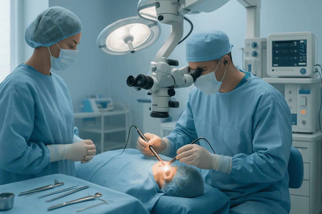
- Step 2: Opening the mastoid. Using an operating microscope and a fine high-speed drill with continuous irrigation, the surgeon removes the outer bone and opens the diseased air cells one layer at a time.
- Step 3: Removing disease. Infected tissue, cholesteatoma, granulation tissue and trapped debris are cleared with micro-instruments, following the disease into the middle ear where necessary.
- Step 4: Checking hidden spaces. An endoscope may be used to inspect narrow recesses of the middle ear that the microscope cannot see in a straight line, places where cholesteatoma remnants classically hide.
- Step 5: Reconstruction. If needed, the eardrum is repaired with a graft and the hearing bones are reconstructed, either now or at a planned later stage.
- Step 6: Closure. The ear canal is packed with dressing material, the incision is closed, and a protective bandage is applied.
Throughout the procedure the surgeon works close to the facial nerve, the balance canals, the hearing bones, the eardrum and the bony plate beneath the brain. Preoperative imaging maps these structures in advance, and intraoperative facial nerve monitoring is used in many cases, particularly in revision surgery or when disease lies against the nerve, to support safety during drilling and dissection.
Types of mastoidectomy
In a canal wall up mastoidectomy, the bony back wall of the ear canal is preserved. This keeps the ear closer to its natural anatomy and avoids an open cavity, but hidden recurrence is harder to detect afterwards, so it demands close surveillance, sometimes including a planned second-look operation or follow-up MRI.
In a canal wall down mastoidectomy, part of the canal wall is removed so that the mastoid and ear canal become one open cavity. The cavity can be inspected and cleaned directly in clinic, which makes recurrence easier to spot, but it usually requires periodic professional cleaning and lifelong water precautions. This approach is often preferred when disease is extensive, recurrent or otherwise unsafe to leave behind a preserved wall.
A modified radical mastoidectomy sits between these poles: it is used when disease is widespread, but the surgeon aims to preserve or reconstruct as much of the hearing mechanism as remains feasible. Neither wall-up nor wall-down surgery is universally better. The right choice depends on how far the disease extends, your anatomy, your hearing goals, and, honestly, how reliably you will be able to attend follow-up over the coming years.
How long does mastoid surgery take?
Most mastoid operations take a few hours. A limited mastoidectomy for contained infection sits at the shorter end, while extensive cholesteatoma surgery, revision procedures, or operations combined with eardrum repair and ossicular reconstruction take longer. Duration is not a quality measure in either direction: careful drilling near the facial nerve and inner ear is deliberately slow, and a longer operation often simply reflects more disease to remove. Your surgeon can give you a realistic estimate for your specific case once the imaging has been reviewed.
Repairing the eardrum and hearing bones
If the eardrum is perforated or has been consumed by the disease, the surgeon usually repairs it during the same operation using a graft taken from your own tissue, most often fascia from above the ear or cartilage from the ear itself. If the ossicles have been eroded, reconstruction may be performed at the same sitting or deferred to a second stage. That decision hinges on infection control, the stability of the ear, and the likelihood of a durable hearing result: in some ears it is genuinely safer to clear disease first, confirm the ear stays healthy, and rebuild hearing later than to attempt everything at once.
Risks and Safety of Mastoid Surgery
Is mastoid surgery dangerous?
Mastoid surgery is a routine operation for trained ear surgeons, and serious complications are uncommon, but the surgery does take place next to delicate structures, so the risks deserve a plain description rather than a wave of the hand. It is also worth holding the comparison honestly: the operation exists because progressive mastoid disease left untreated carries risks of its own, some of them more serious than those of a planned procedure.
Risks your surgeon should discuss with you include:
- Hearing changestemporary reduction is expected while packing is in place; in a small number of ears, hearing can be permanently worse, particularly when disease already involved the inner ear or hearing bones.
- Dizziness or imbalanceoften short-lived after surgery, occasionally more persistent if the balance organ was irritated by disease or the operation.
- Tinnitusringing may appear, improve or persist after surgery.
- Taste disturbancea nerve carrying taste from the tongue (the chorda tympani) crosses the middle ear and can be stretched or divided, causing altered taste that is usually temporary.
- Facial weaknessrare, because the facial nerve runs through the surgical field; monitoring and meticulous technique are used to protect it, the same nerve surgeons safeguard during parotidectomy surgery.
- Cerebrospinal fluid leakrare, related to the thin bone between the mastoid and the brain lining; repairable, usually at the time it is identified.
- Wound infection, bleeding, or anaesthetic complicationsas with any operation under general anaesthesia.
- Residual or recurrent diseasecholesteatoma can regrow from microscopic remnants, which is why follow-up is part of the treatment, not an optional extra.
The honest summary: no ear operation is without risk, and no responsible surgeon will tell you otherwise. What changes the balance is experience with temporal bone anatomy, good preoperative imaging, appropriate use of monitoring, and a follow-up plan that catches problems early.
Recovery After Mastoid Surgery
Recovery depends on the type of mastoidectomy, whether reconstruction was performed, the presence of active infection, and your general health, but most patients follow a staged healing pattern over weeks to months rather than days.
| Time period | What to expect |
|---|---|
| Day 1 | You wake with packing in the ear and often a bandage around the head. Ear fullness, mild dizziness and discomfort are common. Some patients go home the same day; others stay overnight, particularly after extensive surgery or in children. |
| First week | Rest matters. You keep the ear dry, avoid heavy activity, and use the drops or medication your surgeon prescribed. Hearing on the operated side is reduced: this reflects swelling and packing, not the final result. |
| First month | The surgeon checks healing and removes or adjusts packing. Light daily activities usually resume, but swimming, strenuous exercise and flying may remain restricted until you are cleared. |
| Two to three months | Swelling settles and hearing can be reassessed accurately. If the hearing bones were reconstructed, the result becomes clearer as the ear heals. |
| Longer term | Patients with cholesteatoma or an open mastoid cavity need ongoing follow-up, periodic cleaning, imaging or staged surgery depending on the original disease and the surgical approach. |
Caring for your ear at home
Early aftercare follows a consistent set of principles, adjusted by your surgeon for your case:
- Keep the ear strictly dry, protected showering only, no swimming until cleared.
- Avoid blowing your nose forcefully, and sneeze with your mouth open, so pressure does not push against the healing eardrum.
- Avoid heavy lifting and straining in the early weeks.
- Do not fly until your surgeon confirms the ear can tolerate pressure changes.
- Use prescribed drops and medication exactly as directed by your treating doctor.
- Attend every follow-up appointment, even if the ear feels fine: a comfortable ear is not necessarily a fully healed one.
Follow-up itself is part of the treatment. The surgeon removes or changes packing, checks the incision, monitors healing, and reviews the pathology report if tissue was sent for analysis. Hearing testing is repeated once the ear has healed, because early postoperative hearing is distorted by swelling and dressing material. For cholesteatoma, surveillance continues long term: even after meticulous surgery, residual or recurrent disease can occur, and scheduled examinations, sometimes with diffusion-weighted MRI, detect it before it causes new damage.
Why Early Treatment Matters
Chronic ear disease is deceptively slow. A patient can live with intermittent drainage or reduced hearing for years and simply grow used to it. Meanwhile, persistent infection and cholesteatoma quietly erode bone and creep towards structures that are far harder to repair once damaged. Early specialist assessment does not automatically mean surgery, many ears can be monitored, but it separates the ears that can safely wait from the ears that cannot.
Delaying mastoid surgery when it is clearly indicated raises the risk of worsening hearing loss, persistent drainage, balance problems, facial nerve irritation, abscess formation, or spread of infection beyond the ear. Rarely, untreated disease contributes to serious complications involving the inner ear, the bone around the brain, or the intracranial space, situations that may require joint management with neurosurgery. Such complications are uncommon, but they are the reason otologists treat chronic ear disease as more than a nuisance.
Timing also shapes the operation itself. Limited, well-defined disease can often be cleared while preserving anatomy. Recurrent inflammation, scarring from previous infections, or extensive cholesteatoma makes surgery more demanding and the reconstruction options narrower. Acting early gives the surgical team the chance to plan a controlled operation, rather than respond to an emergency with fewer choices on the table.
Benefits of Mastoid Surgery
The benefits depend on your diagnosis and the extent of disease, but they cluster around three things: infection control, protection of the ear’s structures, and long-term stability.
| Benefit | What it means for you |
|---|---|
| Removal of chronic infection or cholesteatoma | The operation clears diseased tissue that medication cannot fully reach, creating a safer, more stable ear. |
| Protection of hearing-related structures | Removing disease near the eardrum, ossicles and inner ear can help prevent further damage and preserve the hearing that remains. |
| Reduction in persistent ear drainage | Recurrent discharge and odour drive many patients to surgery. A dry ear is a central goal of treatment and a real quality-of-life change. |
| Lower risk of serious complications | Treating progressive mastoid or middle ear disease reduces the chance of infection spreading to nearby nerves, balance organs, bone or intracranial spaces. |
| Opportunity for reconstruction | Where appropriate, eardrum repair or hearing bone reconstruction can be performed during the same operation or planned once the ear is stable. |
| Clearer long-term monitoring | After disease removal, examinations and imaging can track healing and detect any recurrence earlier, while it is still small. |
What Influences the Outcome of Mastoid Surgery?
The first factor is the extent and type of disease. Limited chronic infection is a different problem from extensive cholesteatoma that has eroded bone or reached the inner ear, the facial nerve canal, or a previous surgical site. The more advanced the disease, the more complex the plan, and the more the goal shifts from restoring the ear to protecting it.
Hearing before surgery matters as much. If the hearing bones are intact and the inner ear is healthy, preserving or improving hearing is more achievable. If the ossicles are severely damaged or long-standing infection has affected the inner ear, reconstruction has real limits. You should expect a clear conversation about this: a safe, dry ear may be the primary endpoint of the operation, with hearing rehabilitation pursued separately through ossicular reconstruction, hearing aids, or implantable hearing options in selected cases. It also helps to understand what mastoid surgery is not: it does not address fixation of the stirrup bone, for example, which is treated with a different operation, stapedectomy.
The condition of the eardrum and the middle ear lining influences healing, as do Eustachian tube function, smoking, allergies, chronic sinus disease and repeated infections. Children carry different recurrence dynamics because their Eustachian tube function and immune patterns are still developing, which is why paediatric follow-up schedules are often closer than adult ones.
The surgical approach shapes long-term care. A canal wall up procedure preserves anatomy but requires vigilant surveillance for hidden recurrence. A canal wall down procedure reduces hidden spaces but needs periodic cavity cleaning and water precautions. Neither is universally better; the right choice balances safety, disease access, anatomy, hearing goals and your realistic ability to attend follow-up over years.
Finally, experience counts. Mastoid surgery demands familiarity with temporal bone anatomy, disease patterns, microscopic and endoscopic visualisation, and the management of unexpected findings mid-operation. And it does not end at the operating table: chronic ear disease is managed through the combination of surgery, healing care, hearing assessment and long-term observation. An excellent operation with poor follow-up is an incomplete treatment.
How Mastoid Surgery Is Organised at Acibadem
At Acibadem, mastoid surgery is approached as a planned ear and skull base procedure, not as simple drainage of an infection. Evaluation typically includes detailed ENT examination under magnification, audiology testing, high-resolution temporal bone CT, and MRI when clinically appropriate. Complex cases, extensive cholesteatoma, disease near the facial nerve, paediatric patients, or revision surgery after operations elsewhere, are discussed across specialties, drawing on otology, radiology, audiology, anaesthesiology, infectious disease and, when needed, neurosurgical or paediatric input. The purpose is practical: understand the disease fully before the operation, so the removal and any reconstruction follow a considered plan rather than improvisation.
Treatment recommendations follow evidence-based protocols and are individualised rather than standardised: mastoidectomy alone, mastoidectomy with tympanoplasty, ossicular chain reconstruction, staged surgery, or postoperative hearing rehabilitation, depending on the ear in question. Surgeons explain the goals, the limits, the risks and the follow-up requirements in plain terms, including, where relevant, why a safe dry ear is the realistic first objective and hearing work comes second.
Technology supports each stage. High-resolution imaging defines anatomy and disease extent before surgery. Operating microscopes and endoscopes let the surgeon see into the narrow recesses of the middle ear and mastoid. Fine drilling systems and micro-instruments allow controlled removal of diseased bone, and facial nerve monitoring is used in appropriate cases, especially revision surgery or disease lying against the nerve. Audiology services measure hearing before and after treatment and guide rehabilitation options.
For patients travelling from abroad, Acibadem International coordinates care in more than 20 languages, medical record review, appointment scheduling, interpreter support, hospital admission, discharge instructions and communication with the treating team. This coordination matters particularly for mastoid surgery, because wound care, ear precautions, medication use, the timing of flying and the follow-up schedule all need to be clearly understood before you return home.
Weighing the Decision
Mastoid surgery is a significant operation, and it is entirely reasonable to take time over the decision, including seeking a second opinion, which is common practice in ear surgery, particularly when the plan involves hearing reconstruction, revision of a previous operation, or a choice between canal wall up and canal wall down techniques. A good second opinion reviews your examination findings, hearing tests, imaging and previous treatments, and either confirms the original plan or explains, specifically, why a different one fits your ear better.
Whoever operates, certain questions are worth having answered before you consent: what exactly the disease involves, which structures it threatens, what the primary goal of the operation is, what hearing outcome is realistic, which approach is planned and why, whether staged surgery is likely, and what follow-up will be required, and for how long, after the ear has healed.
For the right patient, mastoid surgery is an important step: it controls chronic disease that medication cannot clear, protects delicate structures from progressive damage, and puts the ear on a footing where it can finally be monitored rather than repeatedly rescued. The strongest position to make that decision from is a clear understanding of what is happening in your ear, what risks it carries, and what each option can honestly deliver.
Preparation
- An ENT specialist evaluates symptoms, hearing tests, ear examination, and imaging such as CT when needed. Blood tests and anesthesia assessment may be required before surgery. Patients are usually asked to stop certain blood-thinning medicines and avoid eating or drinking for several hours before anesthesia.
Aftercare
- The ear is usually covered with a dressing, and prescribed antibiotics or ear drops may be used as directed. Keep the ear dry, avoid heavy lifting, and attend follow-up visits for wound and ear canal checks. Temporary dizziness, fullness, or taste changes can occur, but worsening pain, fever, or drainage should be reported promptly.
Frequently Asked Questions
What affects the cost of mastoid surgery?
The main factors are the diagnosis, extent of mastoid and middle ear disease, type of mastoidectomy, need for eardrum or ossicle repair, imaging, anaesthesia, hospital stay, surgeon expertise, and follow-up requirements.
How can I get a personalised quote from Acibadem?
You can request a free consultation by sharing your diagnosis, ear examination notes, hearing test results, imaging reports, previous surgery details if any, and current symptoms. The clinical team can then advise which treatment plan and package may be appropriate.
Is mastoid surgery usually offered as a package for international patients?
Packages may include the specialist assessment, standard pre-operative tests, surgery, anaesthesia, hospital stay, routine medications, and early follow-up. The exact inclusions should always be confirmed before travel.
Will the quote change after I arrive?
It can change if the specialist finds more extensive disease, additional tests are required, or the planned surgery needs to include hearing reconstruction or revision procedures. A final plan is usually confirmed after in-person assessment.
Does insurance cover mastoid surgery abroad?
Coverage depends on your insurer, policy terms, pre-authorisation rules, and whether treatment abroad is included. Patients should confirm coverage directly with their insurer before making travel arrangements.
Medically reviewed by the Acıbadem International Medical Board September 8, 2026
See our medical review board →
Update history
- PublishedJune 8, 2026
- Medical review approvedSeptember 8, 2026
- Last content updateSeptember 8, 2026
References1
- Mastoiditis medlineplus.gov
Trusted care for international patients
From the Acibadem Blog

Mastoid Surgery Recovery: Dressings, Ear Drops and When Dizziness and Fullness Settle
Mastoidectomy recovery usually means a padded head dressing for the first day or so, dissolvable or removable packing in the ear canal for several…

After Mastoid Surgery: Which Symptoms Are Expected and Which Mean It Is Time to Call?
After mastoid surgery, a dull ache, a blocked or muffled ear, mild unsteadiness, numbness behind the ear and a little blood-tinged discharge are usually…