Pituitary Tumor Treatment
Pituitary tumor treatment manages benign or rarely malignant growths near the pituitary gland using monitoring, medication, endoscopic surgery, or radiosurgery depending on size, hormones, and vision impact.
Quick answer
A pituitary tumor is an abnormal growth in the pituitary gland, the small hormone-controlling gland at the base of the brain. Most are benign adenomas. Treatment depends on size and hormone activity and may involve monitoring with scans, hormone-lowering medication, endoscopic surgery through the nose, or focused radiation, followed by long-term hormone and imaging follow-up.
Pituitary Tumor: What It Is and Why It Matters
A pituitary tumor is an abnormal growth of cells in or around the pituitary gland, the pea-sized gland that sits in a bony pocket at the base of the brain and directs hormone production throughout the body. The great majority of these growths are benign adenomas, not cancers. Treatment is designed for people whose tumour presses on nearby structures, disturbs hormone levels, or grows on repeat scans, and, just as importantly, it recognises the many people whose tumour needs nothing more than careful monitoring.
Learning that you have a pituitary tumor can feel unsettling, precisely because the pituitary gland sits deep inside the head and controls hormones that affect so many parts of the body. Some people reach the diagnosis after months of headaches, changes in vision, fatigue, menstrual changes, low libido, unexplained weight change, or difficulty conceiving. Others are told about a pituitary lesion after an MRI performed for an entirely unrelated reason. In either situation, the first questions are usually urgent and personal. Is it cancer? Will it affect my vision? Do I need brain surgery? Can my hormones return to normal? This page answers those questions as directly as the evidence allows.
Benign does not always mean harmless. A pituitary tumor may press on the optic nerves, affect nearby brain structures, or produce excess hormones that disturb metabolism, blood pressure, growth, reproduction, stress response, and overall well-being. Rarely, pituitary tumours behave aggressively or are malignant, and those cases need specialised evaluation and long-term management. The point of treatment is therefore fourfold: protect vision, control hormone imbalance, reduce or remove pressure from the tumour, and preserve as much normal pituitary function as possible.
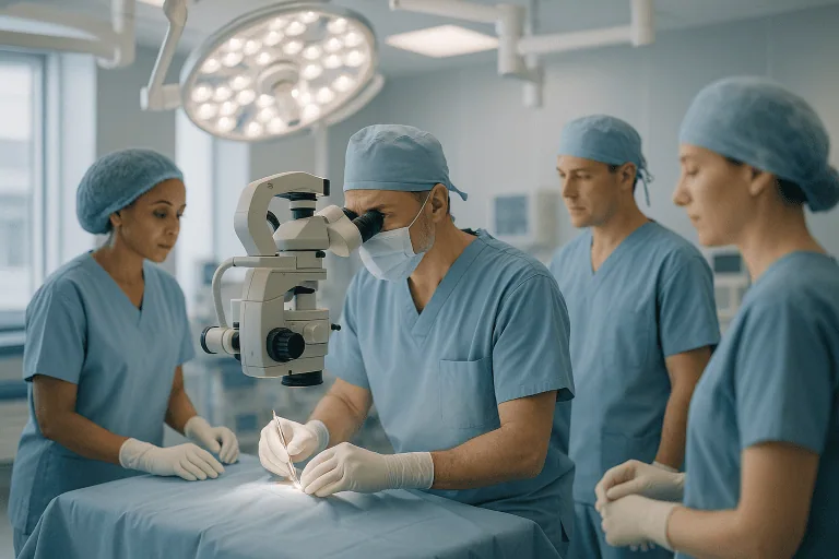
Because the pituitary gland is small but highly influential, decisions about it are best made by a team experienced in pituitary disorders rather than any single specialist working alone. Neurosurgeons, endocrinologists, neuroradiologists, radiation oncologists, ophthalmologists, pathologists, and specialised nursing teams each see a different part of the picture. For patients travelling from abroad, the quality of the diagnosis, the clarity of the explanation, and the coordination of follow-up matter as much as the procedure itself.
Is a pituitary tumor a brain tumor?
A pituitary tumor is usually counted among brain tumors in medical classifications because of where it sits, but it does not arise from brain tissue itself. It grows from the hormone-producing cells of the pituitary gland, which hangs beneath the brain in a bony hollow called the sella turcica. This distinction matters in practice. Because the tumour starts in gland tissue rather than brain tissue, most pituitary tumours are benign, grow slowly, do not spread to distant organs, and can often be reached surgically through the nose rather than through an opening in the skull. The tumour becomes a neurological problem mainly when it grows large enough to press upward on the optic nerves or outward into neighbouring structures.
What is the main function of the pituitary gland?
The main function of the pituitary gland is to act as the body’s hormonal control centre: it is often called the master gland. The pituitary gland and the hormones it releases regulate other glands, including the thyroid, the adrenal glands, the ovaries, and the testes. It also governs growth, milk production, water balance, and elements of the stress response. Because so many systems answer to it, a small disturbance in the pituitary can produce symptoms in places that seem unrelated: skin, bones, mood, weight, blood pressure, blood sugar, fertility, and sleep. This is why pituitary problems are so often mistaken, for a time, for stress, ageing, or menopause.
Types of Pituitary Adenoma
A pituitary adenoma is a benign tumour arising from the hormone-producing cells of the gland, and it is by far the most common type of pituitary tumor. In older medical records and some European reports you may see the term adenoma of hypophysisthe hypophysis is simply the anatomical name for the pituitary gland, so the phrase means exactly the same thing. Adenomas are classified in two ways that shape every treatment decision: by size and by hormone activity.
By size. A microadenoma measures smaller than 10 millimetres; a macroadenoma measures 10 millimetres or larger. The threshold is not arbitrary. Larger tumours are more likely to reach the optic chiasm, the point above the gland where the optic nerves cross, and compression there causes a characteristic loss of side vision. Larger tumours are also more likely to squeeze the normal gland tissue around them and reduce its hormone output.
By hormone activity. A functioning adenoma produces too much of a specific hormone and causes a recognisable clinical syndrome, such as acromegaly or Cushing disease. A nonfunctioning adenoma produces no active hormone excess but can still cause real harm through pressure alone. The two behave very differently: a tiny functioning tumour can make a person seriously unwell, while a larger nonfunctioning tumour may cause no symptoms at all until it presses on the optic nerves.
What size pituitary tumor should be removed?
There is no single size at which a pituitary tumor must be removed; the decision rests on what the tumour is doing, not only on how large it is. Broadly, surgery is considered when a tumour of any size compresses the optic nerves or chiasm, when it produces hormones that cannot be controlled with medication, when it is clearly growing on serial MRI scans, when it has damaged normal pituitary function, or when the diagnosis is uncertain and tissue is needed. Many macroadenomas that touch or lift the optic chiasm are recommended for surgery to protect vision. Conversely, a small nonfunctioning microadenoma found by chance, causing no symptoms and no hormone disturbance, is very often simply monitored with periodic scans and blood tests. And a prolactin-producing tumour, even a large one, may be treated first with medication rather than surgery, because that tumour type frequently shrinks with tablets. Size opens the conversation; behaviour decides it.
Symptoms and Diagnosis of a Pituitary Tumor
Symptoms of a pituitary tumor come from two sources: pressure on nearby structures, and disturbance of the hormones the pituitary gland controls. Some patients have a clear hormone syndrome. Many others have nonspecific complaints that develop slowly over months or years and are easy to attribute to stress, ageing, menopause, weight change, or other conditions, which is why the diagnosis is so often delayed or made incidentally.
How do you tell if you have a problem with your pituitary gland?
A pituitary problem usually announces itself through some combination of visual change, headache, and hormone-related symptoms, and it is confirmed with blood tests and an MRI: it cannot be reliably self-diagnosed from symptoms alone. Complaints that lead doctors to test the pituitary include persistent headaches, blurred or double vision, loss of side vision, menstrual irregularity, infertility, erectile dysfunction, reduced libido, unexpected milk production from the breasts, fatigue, dizziness, unexplained weight gain or loss, easy bruising, muscle weakness, changes in facial appearance, enlarged hands or feet, excessive sweating, high blood pressure, diabetes, and increased thirst and urination. No single item on that list is specific to the pituitary; it is the pattern, and the laboratory results behind it, that point to the gland.
How do people find out they have a pituitary tumor?
In practice, people discover a pituitary tumor by one of three routes. The first is a hormone syndrome: a woman investigated for absent periods and milky nipple discharge is found to have high prolactin; a man with gradually enlarging hands and jaw is tested for growth hormone excess. The second is a vision problem: an optician or ophthalmologist detects a loss of peripheral vision and orders imaging. The third, and it is common, is pure chance: an MRI done for headaches, sinus trouble, a head injury, or dizziness happens to show a lesion in the sella. Patients who share their stories online often describe the same arc: vague symptoms dismissed for months, then a single scan or blood test that suddenly explains everything. If that is your story, it is a familiar one to any pituitary team.
What testing confirms the diagnosis?
Diagnosis is confirmed with a combination of imaging, hormone blood tests, and vision testing, usually in that order of prominence. A high-resolution MRI of the pituitary region defines the tumour’s size, its location, its relationship to the optic nerves, and any extension into neighbouring structures such as the cavernous sinus. Blood tests measure the pituitary hormones themselves and the hormones of the glands the pituitary controls, depending on the suspected tumour type, this may include prolactin, growth hormone-related markers, cortisol pathway testing, thyroid function, sex hormones, and adrenal function. In selected cases, dynamic endocrine testing, measuring how hormone levels respond to a stimulus or suppression, is needed to characterise production precisely. These hormone panels are different in purpose from the tumor marker blood tests used in some other cancers; for the pituitary, the hormones themselves are the markers. Finally, formal visual field testing can detect subtle loss of peripheral vision before you would notice it in daily life, and an ophthalmologic assessment evaluates visual acuity, eye movement, and optic nerve health. Together these results tell the team whether the tumour is active, whether it is threatening vision, and whether normal gland function is already reduced.
Conditions Pituitary Tumor Treatment Addresses
Pituitary tumor treatment covers a spectrum of adenomas and related lesions of the sellar region, and the pathway differs meaningfully from one condition to the next.
Nonfunctioning pituitary adenomas cause no hormone-excess syndrome, so they tend to be diagnosed only when large enough to cause headaches, visual field loss, or pituitary hormone deficiency, often at macroadenoma size. Surgery is commonly considered when they compress the optic apparatus or continue to grow; small, stable ones are frequently observed.
Prolactinomas produce excess prolactin. They can cause irregular or absent menstrual periods, infertility, breast milk production unrelated to pregnancy, low testosterone, erectile dysfunction, reduced libido, and bone loss over time. Prolactinomas occupy a special position: they are usually treated first with medication rather than surgery, because dopamine agonist drugs can lower prolactin and shrink the tumour. Surgery becomes relevant when medication is not tolerated, does not work adequately, or when urgent decompression is needed.
Growth hormone-producing adenomas cause acromegaly in adults: gradual enlargement of the hands, feet, jaw, and facial features, along with joint pain, sleep apnoea, sweating, diabetes, hypertension, and increased cardiovascular strain. Surgery is often the cornerstone of treatment, supported by medication or radiotherapy when hormone levels remain elevated.
ACTH-producing adenomas cause Cushing disease, driven by excess cortisol. Features may include central weight gain, a rounded facial appearance, thin skin, easy bruising, muscle weakness, high blood pressure, diabetes, mood changes, osteoporosis, and increased susceptibility to infection. Accurate diagnosis is essential and sometimes genuinely difficult, because cortisol disorders can mimic other conditions and often require repeated or specialised endocrine testing before surgery is planned.
TSH-producing adenomas are uncommon tumours that overstimulate the thyroid, producing hyperthyroid symptoms such as rapid heartbeat, weight loss, tremor, heat intolerance, and anxiety. Management requires close coordination between endocrinology and neurosurgery, since thyroid hormone excess must be addressed around the time of any operation.
Recurrent or residual pituitary tumoursthose that regrow or remain after previous surgery, medication, or radiation, form their own category. These cases benefit from a careful review of prior imaging, operative reports, pathology, hormone trends, and any earlier radiation exposure before a new plan is made.
Finally, some lesions near the pituitary gland are not adenomas at all but can mimic them on imaging: Rathke cleft cysts, craniopharyngiomas, meningiomas, inflammatory lesions, and, rarely, metastases from cancers elsewhere. Distinguishing among these matters, because both the treatment and the follow-up schedule differ significantly.
What are four diseases due to pituitary disorder?
Four well-recognised diseases caused by pituitary disorders are acromegaly (growth hormone excess), Cushing disease (cortisol excess driven by an ACTH-producing adenoma), hyperprolactinaemia from a prolactinoma (with its effects on periods, fertility, and libido), and hypopituitarism (deficiency of one or more pituitary hormones). A fifth condition worth knowing is diabetes insipidus, a disturbance of water balance that can follow damage to the gland or its stalk; it causes marked thirst and large urine volumes and is managed medically. Each of these conditions can be produced by a tumour, by its treatment, or occasionally by unrelated pituitary disease, which is why endocrine follow-up continues long after any operation.
Treatment Options for a Pituitary Tumor
Treatment for a pituitary tumor is a personalised strategy built from five components, used alone or in combination depending on the tumour’s size, hormone activity, location, growth pattern, your symptoms, and your general health.
- Monitoring: regular MRI scans, hormone testing, and visual evaluations. This is a genuine treatment choice, not an absence of one, and it is often the safest option for small, nonfunctioning tumours that cause no symptoms and show no growth.
- Medication: some hormone-producing tumours, prolactinomas above all, often respond to medication that lowers hormone production and may shrink the tumour. Drug therapy also has a role in acromegaly and Cushing disease, before surgery, after surgery, or when surgery is not the best first step.
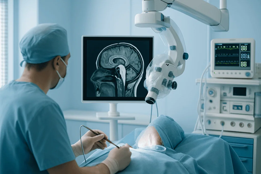
- Endoscopic transnasal transsphenoidal surgery: removal of the tumour through the nose using an endoscope, avoiding an external incision and minimising handling of normal brain tissue. This is the standard operation for most pituitary tumours that need surgery.
- Radiotherapy or stereotactic radiosurgery: focused radiation used when a tumour remains after surgery, recurs, cannot be removed safely, or keeps producing hormones despite other treatments.
- Hormone replacement or hormone-lowering therapy: medication to replace hormones the gland can no longer make, or to control hormone excess before or after tumour treatment. These prescriptions are tailored and adjusted by the treating endocrinologist over time.
Modern pituitary care is not simply about removing a mass. It means understanding the tumour’s biology, measuring hormone effects accurately, assessing vision, protecting the normal gland, and planning follow-up that may run for years. A good plan usually pairs immediate symptom control with deliberate long-term surveillance.
Can you shrink a pituitary tumor naturally?
No diet, supplement, herbal product, or lifestyle change has been shown to shrink a pituitary tumor, and this question deserves a straight answer because it is searched so often. The confusion arises partly because one tumour type, the prolactinoma, genuinely does shrink without surgery, but it does so in response to prescribed dopamine agonist medication, not to natural remedies. Healthy habits are worth keeping for their own sake: good sleep, regular activity, and sensible nutrition support recovery and general endocrine health. But they are companions to treatment, not substitutes for it. Any change to prescribed medication is a decision for the treating doctor; the practical risk of pursuing unproven remedies is not that they cause harm directly, but that they delay evaluation while a tumour continues to press on the optic nerves or drive hormone excess.
How Pituitary Tumor Treatment Is Performed
Preparation and treatment planning
Preparation begins with confirming the diagnosis and understanding what the tumour is doing to you specifically. The team reviews the MRI, hormone levels, visual test results, symptoms, current medications, previous surgeries, and relevant medical conditions. If you have been treated elsewhere, prior MRI images, laboratory results, pathology reports, medication lists, and a short written timeline of symptoms make the review far more accurate.
Endocrine optimisation may be needed before any procedure. Cortisol deficiency, for instance, must be recognised and managed because it affects surgical safety; excess cortisol, growth hormone, or thyroid hormone may need to be brought under control medically before an operation to reduce risk. Blood thinners, diabetes, high blood pressure, sleep apnoea, heart disease, and similar conditions are assessed and addressed in advance by the anaesthesia and medical teams.
When surgery is recommended, the surgeon explains the planned approach, the expected hospital stay, the possibility of needing hormone replacement afterwards, and the specific risks in your anatomy. Planning imaging maps the nasal corridor, the sphenoid sinus, the tumour’s boundaries, the optic nerves, the carotid arteries, and the cavernous sinus. This usually means high-resolution MRI, a CT scan when bony detail is needed, and image-guidance systems that act as a navigational reference during the operation itself.
Endoscopic transnasal transsphenoidal surgery, step by step
The most common operation for a pituitary tumor is endoscopic transnasal transsphenoidal surgery, in which the surgeon reaches the gland through the nostril rather than through the skull. Under general anaesthesia, the operation typically proceeds as follows:
- A slender endoscope, a camera on a thin tube, is passed through the nostril, giving the team a magnified view of the nasal passages on a screen.
- The surgeon works along the natural nasal corridor to the sphenoid sinus, the air-filled space directly in front of the sella turcica.
- The thin bony floor of the sella is opened, exposing the covering of the pituitary region.
- Tumour tissue is removed piece by piece with specialised instruments, while the surgeon works to distinguish and preserve the normal pituitary gland and the structures around it. Image guidance assists with orientation, particularly in complex anatomy or repeat surgery.
- The opening is reconstructed, in selected cases with specialised techniques, sometimes using the patient’s own tissue, to reduce the risk of cerebrospinal fluid leakage afterwards.
Many endoscopic procedures are completed within a few hours, though duration varies with tumour size, anatomy, prior treatment, and complexity. Some tumours extend into places that are difficult to clear completely, above all the cavernous sinus, which houses critical nerves and the carotid artery. In those situations the safest strategy may be deliberate partial removal: relieve the pressure, confirm the diagnosis, and treat what remains with medication or focused radiation. A small number of tumours that extend far above or beside the pituitary region need a different surgical route through the skull; this is less common than the nasal approach, and the broader practicalities of that kind of operation are described in our brain tumour surgery treatment guide.
Medication-based treatment
Medication is central for certain tumour types. Prolactinomas frequently respond to dopamine agonists, which lower prolactin levels and can reduce tumour size; patients taking them are monitored for effectiveness, side effects, and recovery of normal hormone rhythms. Drug therapy is also used in acromegaly and Cushing disease, sometimes before surgery to reduce risk, sometimes afterwards to control residual hormone excess, and sometimes as the main treatment when surgery is not the right first option.
Hormone replacement becomes relevant when the tumour or its treatment has reduced normal pituitary output. Depending on which hormones are affected, replacement may include thyroid hormone, hydrocortisone or other adrenal support, sex hormone replacement, growth hormone in selected adults, or medication for water balance if diabetes insipidus develops. All of this is prescribed and adjusted by endocrinologists on the basis of symptoms and laboratory results, and it changes over time, replacement needs in the first weeks after surgery often differ from needs a year later.
Radiotherapy and stereotactic radiosurgery
Radiation is considered for residual, recurrent, invasive, or medically resistant pituitary tumours. Stereotactic radiosurgery delivers highly focused radiation to a defined target, often in a single session; fractionated radiotherapy divides the dose across multiple smaller sessions. The choice between them depends on the size and location of the remaining tumour, its distance from the optic nerves, any prior radiation, hormone activity, and the overall goal of treatment.
Radiation can control tumour growth and gradually reduce hormone overproduction in selected patients, but honesty requires saying that its effects take time, often months to years for hormone levels to fall. Long-term endocrine follow-up is essential afterwards, because pituitary hormone deficiencies can develop months or years after treatment. The radiation oncology team plans jointly with neurosurgery and endocrinology to balance tumour control against protection of vision and remaining gland function.
Hospital stay and the first days after surgery
After endoscopic surgery, patients spend time in a monitored recovery area and then stay in hospital for observation while the team checks the things that matter most in the first days: sodium levels, urine output, cortisol status, vision, headache, and any sign of cerebrospinal fluid leak. Nasal congestion, fatigue, mild headache, and a temporarily altered sense of smell are common early experiences and usually settle as the nose heals. Length of stay varies from patient to patient; discharge follows once you are stable, eating, walking, and safe to leave. You will be advised to avoid heavy lifting, straining, and forceful nose blowing during the initial healing period, and follow-up MRI and hormone testing are scheduled according to the tumour type and the plan agreed before surgery.
What Happens If a Pituitary Tumor Goes Untreated?
An untreated pituitary tumor that is genuinely inactive, small, nonfunctioning, and stable, may cause no harm at all, which is why monitoring is a legitimate plan. The concern is the tumour that is growing or hormonally active. Left unevaluated, a growing tumour can press progressively on the optic nerves, and vision lost to prolonged compression does not always return even after the pressure is relieved. Hormone-producing tumours damage the body more quietly: sustained excess of growth hormone, cortisol, or thyroid stimulation raises the long-term burden of cardiovascular disease, diabetes, bone loss, sleep apnoea, and mood disturbance.
There is a second, less obvious cost. A tumour that compresses the normal gland can gradually switch off essential hormone production, including the adrenal axis, and unrecognised adrenal insufficiency is medically dangerous, particularly during illness or an operation for something else. Rarely, sudden bleeding or loss of blood supply inside a pituitary tumour, pituitary apoplexy, causes severe headache, visual loss, eye movement problems, low blood pressure, or altered consciousness; apoplexy is treated as a medical emergency in every health system.
None of this means every diagnosis leads to surgery. Timely specialist review means something more modest and more useful: a clear risk assessment, an honest explanation of the choices, and the right plan started before complications progress. For patients whose prior recommendations conflict, or where the timing of treatment is genuinely uncertain, a structured second opinion from a pituitary-experienced team is a reasonable and common step.
What happens when your pituitary gland fails?
When the pituitary gland fails, a condition called hypopituitarism, the glands it controls stop receiving their instructions, and their hormones fall in turn. Depending on which pituitary hormones are lost, the result can include profound fatigue, low blood pressure, intolerance of illness and stress (from adrenal insufficiency), weight change and cold intolerance (from thyroid deficiency), loss of periods, libido, and fertility (from sex hormone deficiency), and disturbed water balance with marked thirst and urination. Failure can arrive gradually, as a tumour compresses the gland, or abruptly after apoplexy. The condition is manageable: each missing hormone can be replaced with medication supervised by an endocrinologist, and some deficiencies improve after the compressing tumour is removed. But it is a lifelong consideration for many patients, which is why hormone testing continues at intervals long after the tumour itself has been dealt with.
Benefits of Pituitary Tumor Treatment
The realistic benefits of treatment depend on the tumour type, its size, its hormone activity, and your condition before treatment began. The table below summarises what each benefit means in practice, noting that no outcome can be promised in advance for any individual patient.
| Benefit | What It Means for You |
|---|---|
| Protection or improvement of vision | Removing or reducing pressure on the optic nerves may help preserve sight and, in many patients, improve visual field problems caused by compression, particularly when compression has been recent rather than long-standing. |
| Control of excess hormones | Treatment can reduce abnormal hormone production that affects metabolism, fertility, blood pressure, blood sugar, body composition, and long-term cardiovascular and bone health. |
| Relief from pressure-related symptoms | Patients with large tumours may experience improvement in headaches, pressure symptoms, or nerve-related complaints once the mass effect is reduced. |
| Preservation of normal pituitary function | Careful surgical and radiation planning aims to protect healthy gland tissue, and structured follow-up identifies any hormone deficiency early so it can be treated appropriately. |
| Clear diagnosis and a long-term plan | Pathology, imaging, and endocrine follow-up define whether monitoring, medication, radiation, or further care is needed after the initial treatment, replacing uncertainty with a schedule. |
Recovery Timeline After Pituitary Tumor Treatment
Recovery depends on whether your treatment involves observation, medication, surgery, radiation, or a combination, and no two patients follow an identical course. The timeline below reflects common expectations after endoscopic pituitary surgery; your own team’s instructions take precedence over any general schedule.
| Time Period | What Patients Can Expect |
|---|---|
| Day 1 | Monitoring focuses on vision, neurologic status, urine output, sodium level, cortisol function, pain control, and nasal symptoms. Many patients are encouraged to walk with assistance the same day or the next. |
| First week | Fatigue, nasal congestion, mild headache, and reduced smell may be present. Activity restrictions apply, no heavy lifting, straining, or forceful nose blowing, and the team explains warning signs, medications, and follow-up testing before discharge. |
| First month | Energy gradually improves. Endocrine testing may be repeated, nasal healing is assessed, and the team reviews whether hormone replacement or additional therapy is needed. |
| Longer term | MRI surveillance and hormone monitoring continue on a planned schedule. Some patients need medication adjustments, radiation planning, fertility guidance, or ongoing endocrine follow-up depending on tumour type. |
Two points about this timeline deserve emphasis. First, hormonal recovery and physical recovery run on different clocks: you may feel physically well while your endocrine system is still settling, which is why blood tests continue even when you feel fine. Second, follow-up imaging is not a sign that anything is wrong: it is how a slow-growing disease is responsibly supervised, often for years, sometimes indefinitely for certain tumour types.
Factors That Influence Outcomes
Several medical and technical factors shape what treatment can achieve, and understanding them helps you interpret any recommendation you receive.
Size, location, and behaviour. Tumour size matters, but location and behaviour matter as much. A small hormone-producing tumour can cause major systemic illness, while a larger nonfunctioning tumour may cause only pressure symptoms. Tumours confined to the sella are generally more accessible surgically than tumours invading the cavernous sinus or wrapping around critical blood vessels, and for invasive tumours, a planned partial removal followed by medication or radiosurgery may honestly be the best available strategy rather than a compromise.
Hormone type. The hormone a tumour produces strongly shapes the plan. Prolactinomas often respond well to medication; many growth hormone-producing and ACTH-producing tumours are treated with surgery as a central component; nonfunctioning macroadenomas usually reach surgery to protect vision and relieve pressure. Residual tumour after an operation does not automatically mean treatment has failed, in some cases a deliberate subtotal removal with surveillance or radiosurgery afterwards is the strategy that best protects the patient.
Vision and endocrine status before treatment. Patients whose optic nerves have been compressed for a shorter time generally have a better chance of visual improvement than those with long-standing severe loss. Similarly, if normal pituitary function was already reduced before treatment, some deficiencies may improve after decompression while others require ongoing replacement. Baseline testing before treatment is what makes this distinction measurable afterwards.
Surgical experience and team coordination. Pituitary surgery demands familiarity with endoscopic skull base anatomy, tumour consistency, vascular relationships, cerebrospinal fluid leak prevention, and postoperative endocrine management. A good result depends not only on the hours in the operating theatre but on everything around them: preparation, hormone assessment, visual testing, pathology review, and disciplined long-term follow-up.
Patient factors. Age, general health, cardiovascular risk, diabetes, sleep apnoea, bone health, pregnancy plans, medication tolerance, and prior treatments all influence the plan. For patients treated far from home, the ability to continue follow-up after returning is a genuine planning consideration, not an afterthought: a well-designed plan includes clear documentation, medication instructions, imaging recommendations, and communication with your local physicians where appropriate.
Pituitary Tumor Care at Acibadem
Patients who travel for pituitary tumor treatment usually need more than a surgical appointment: an accurate diagnosis, a carefully reasoned recommendation, clear communication in their own language, and a hospital environment prepared for complex endocrine and neurosurgical care. At Acibadem, pituitary care is organised around multidisciplinary evaluation, cases are reviewed by the relevant specialist teams, which may include neurosurgery, endocrinology, neuroradiology, radiation oncology, ophthalmology, pathology, anaesthesiology, and intensive care when needed. This collaborative structure matters most for exactly the tumours described on this page: those that touch hormones, vision, fertility, metabolism, and the skull base all at once.
Diagnostic pathways use modern imaging and laboratory assessment to define the tumour before any treatment decision. High-resolution pituitary MRI evaluates tumour boundaries, optic nerve compression, cavernous sinus involvement, and, in recurrent cases, the altered anatomy left by previous surgery. Endocrine results are interpreted in context, because pituitary hormone disorders can be subtle and sometimes require repeated or dynamic testing before they are certain. Visual field analysis establishes whether the optic pathway is affected and provides the baseline against which any later recovery is measured.
When surgery is needed, endoscopic transnasal techniques allow surgeons to reach the pituitary region through the natural nasal corridor in many patients, with magnified visualisation, image guidance, microsurgical instruments, and careful reconstruction supporting precision and limiting avoidable trauma to surrounding structures. For tumours not best managed by surgery alone, focused radiation techniques and medical therapies are weighed within the same multidisciplinary plan rather than as separate afterthoughts.
For patients coming from abroad, Acibadem International coordinates the practical side of care, review of medical records before travel, appointment scheduling, language assistance in more than 20 languages, and guidance on logistics, so that the clinical pathway is not complicated by the journey around it. Planning is individual by design: a young patient prioritising fertility, a patient with acromegaly and cardiac risk, a patient with Cushing disease and fragile health, and a patient with a nonfunctioning tumour threatening vision each need different priorities. In every case the aim is the same: the least invasive effective approach that preserves long-term health, vision, and endocrine function as far as possible.
Living Beyond a Pituitary Tumor Diagnosis
A pituitary tumor diagnosis raises hard questions, but it also replaces months of unexplained symptoms with something that can be measured, explained, and managed. Many pituitary tumours are treatable; some can be safely watched without any intervention at all; others need timely medication, surgery, radiation, or combined care to protect vision and hormone health. What separates a good outcome from a frustrating one is rarely a single dramatic decision: it is the quality of the evaluation at the start and the discipline of the follow-up afterwards.
Whatever pathway applies to you, the long-term shape of pituitary care is consistent: periodic MRI, periodic hormone testing, honest reassessment when anything changes, and adjustment of replacement therapy as your needs evolve. Patients who understand why each scan and blood test is scheduled tend to keep to the schedule, and with this disease, keeping to the schedule is most of the battle. Where recommendations from different doctors conflict, or the right timing of treatment is genuinely unclear, an independent review by a team experienced in pituitary disease is a normal part of careful decision-making, not a sign of distrust.
Preparation
- Preparation usually includes pituitary MRI, hormone blood tests, visual field testing, and assessment by neurosurgery and endocrinology teams. Patients may need to adjust hormone medications, stop certain blood thinners, and fast before surgery if an operation is planned.
Aftercare
- Aftercare focuses on monitoring vision, hormone levels, fluid balance, and possible nasal or sinus symptoms after transsphenoidal surgery. Follow-up MRI and endocrinology visits help detect recurrence and guide hormone replacement, medication, or radiosurgery if needed.
Frequently Asked Questions
What affects the cost of pituitary tumor treatment?
Cost is influenced by tumor size and location, hormone activity, vision involvement, the need for surgery or radiosurgery, imaging and laboratory tests, hospital stay, medications, and follow-up with neurosurgery, endocrinology, and ophthalmology.
How can I get a personalised quote?
You can request a free consultation and share your MRI reports, hormone test results, visual field tests, current medications, and previous treatment history. A specialist team can then review your case and prepare a personalised treatment plan and quote.
Is surgery always needed for a pituitary tumor?
No. Some pituitary tumors can be monitored, and some hormone-producing tumors may respond to medication. Surgery or radiosurgery may be considered when there is growth, hormone excess, pressure on vision pathways, or other medical indications. Suitability is decided by a specialist.
What is usually included in an international patient package?
Depending on the hospital and treatment plan, a package may include specialist consultations, preoperative tests, hospital services, surgery-related care, translation support, care coordination, and assistance with travel logistics. Inclusions should always be confirmed before treatment.
Will I need follow-up after treatment?
Yes. Pituitary tumor care usually requires follow-up to monitor hormone levels, vision, MRI findings, and medication needs. Follow-up may be arranged locally or coordinated with the treating hospital, depending on your travel plans and medical needs.
Medically reviewed by the Acıbadem International Medical Board August 31, 2026
See our medical review board →
Update history
- PublishedJune 8, 2026
- Medical review approvedAugust 31, 2026
- Last content updateSeptember 8, 2026
References3
- Pituitary Tumors medlineplus.gov
- Pituitary Adenoma my.clevelandclinic.org
- Pituitary Tumors Treatment (PDQ) - Patient Version cancer.gov
Trusted care for international patients
From the Acibadem Blog

Which Symptoms After Pituitary Surgery Need Reporting? Thirst, Headache and Nasal Signs
After pituitary surgery, report new or worsening thirst with large volumes of pale urine, a severe or sudden headache, fever or a stiff neck,…

Recovery After Pituitary Tumor Surgery: Nasal Care, Fluid Balance and Vision Checks
Pituitary surgery recovery usually involves a short hospital stay followed by several weeks of gradual healing at home. The three things care teams watch…