Vulvar Cancer Treatment
Vulvar cancer treatment may include surgery, radiation therapy, chemotherapy or combined care, depending on tumor stage, location and overall health. Early diagnosis supports more effective, organ-sparing treatment options.

Quick answer
Vulvar cancer is a malignant tumour of the external female genitals, most often a squamous cell carcinoma of the vulvar skin. Treatment usually begins with surgery to remove the tumour and assess the groin lymph nodes; radiation therapy and chemotherapy may be added, or used instead of surgery, depending on the stage, the tumour type and the patient's overall health.
Vulvar Cancer: Understanding the Diagnosis and the Decision Ahead
Vulvar cancer is a malignant tumour that develops on the vulva: the external part of the female genitals, which includes the labia majora, labia minora, clitoris, the openings of the vagina and urethra, and the perineum. Most vulvar cancers are squamous cell carcinomas, arising in the thin, flat cells on the surface of the skin. Treatment ranges from removing a small tumour with a margin of healthy tissue to combined surgery, radiation therapy and chemotherapy for more advanced disease, and it is planned within specialist gynaecological oncology care.
A diagnosis of vulvar cancer can feel deeply personal and emotionally difficult. The vulva is an intimate part of the body, and many patients worry not only about the cancer itself, but about pain, appearance, sexual health, urinary function, lymph node surgery, recovery and whether life will feel normal again. These are legitimate concerns, and a good treatment plan takes them seriously rather than treating them as afterthoughts. If you are reading this page after a biopsy, or because a symptom has persisted despite creams and other treatments, the honest starting point is this: vulvar cancer is treatable, treatment decisions are genuinely individual, and understanding the logic behind those decisions makes the path ahead easier to walk.
Vulvar cancer is uncommon compared with other gynaecological cancers such as cervical cancer, endometrial cancer or ovarian cancer, but it requires careful evaluation by specialists who understand both cancer control and preservation of function. What is recommended for you depends on the size and location of the tumour, whether lymph nodes are involved, the exact cancer type, previous treatments, other medical conditions and your personal priorities. When the disease is found early, treatment can often be planned with the goal of removing or controlling the cancer while preserving as much normal anatomy and function as safely possible.
Timing matters. Vulvar cancers may begin as a persistent sore, lump, thickened patch, itching, burning, bleeding or pain that does not improve. Because these symptoms can resemble infections, skin conditions or simple irritation, diagnosis is sometimes delayed, occasionally for months. Once the diagnosis is confirmed, an organised treatment plan reduces uncertainty and gives you a clearer path forward.
What is vulvar cancer?
Vulvar cancer is a cancer that starts in the tissues of the vulva, most often in the skin of the labia. Vulva cancer and vulvar cancer are two names for the same disease; British sources often write it as vulval cancer as well. The most common type by far is squamous cell carcinoma, which develops in the surface layer of the vulvar skin. Less common types include vulvar melanoma, adenocarcinoma, basal cell carcinoma, Bartholin gland carcinoma, Paget disease of the vulva and rare soft-tissue sarcomas.
The distinction between types is not academic. A squamous cell carcinoma, a melanoma and a Paget disease with invasion behave differently, spread differently and are treated differently. That is why the pathology report from your biopsy, not the appearance of the lesion, and not anyone’s first impression, is the foundation of every decision that follows.
What Vulvar Cancer Treatment Involves
Vulvar cancer treatment is the medical and surgical care used to remove, destroy or control cancer of the vulvar tissues. For some patients, surgery alone is enough. For others, radiation therapy, chemotherapy or a combination is recommended, to shrink the tumour before an operation, to treat lymph nodes, to lower the risk of recurrence, or to manage cancer that has spread beyond the vulva. The central aim throughout is to treat the cancer effectively while preserving urinary, sexual and bowel function whenever possible.
Surgical treatment may range from a local excision of the tumour with a rim of healthy tissue to a partial vulvectomy or, in selected cases, more extensive vulvar surgery. Lymph node assessment is often a core part of the plan, because vulvar cancer tends to spread first to the lymph nodes in the groin. In selected early-stage cases, sentinel lymph node biopsy is used to identify the first node or nodes most likely to receive cancer cells from the tumour area, which can spare appropriate patients a more extensive groin dissection and its long-term consequences.
Radiation therapy uses carefully planned high-energy beams to target cancer cells while limiting exposure to surrounding tissue. Chemotherapy may be given together with radiation, an approach called chemoradiation, because certain drugs make cancer cells more sensitive to radiation. Chemotherapy may also be used on its own for advanced or recurrent disease. Which combination is right for you is guided by pathology results, imaging findings, tumour stage, your general health and your own preferences after a detailed discussion of expected benefits and possible side effects.
It is worth stating plainly what treatment cannot do: no plan removes all uncertainty. What a well-constructed plan does is match the intensity of treatment to the actual extent of disease, neither undertreating a cancer that needs more, nor overtreating one that needs less.
Vulvar Cancer Symptoms: What to Watch For
Vulvar cancer symptoms, sometimes listed in clinic letters and search results as carcinoma of the vulva symptoms, usually begin quietly and persist. The pattern that matters most is not any single sign but the failure of a symptom to resolve despite creams, antibiotics, antifungal treatments or time. Common warning signs include:
- Ongoing vulvar itching, burning or tenderness that does not settle
- A lump, thickened area or wart-like growth
- A sore or ulcer that does not heal
- Bleeding not related to menstruation
- Changes in the colour or texture of vulvar skin, paler, darker, redder or thicker patches
- Pain during sexual activity
- Discomfort or stinging when urinating
- A visible or palpable swelling in the groin
Some people have no obvious symptoms in the early stages. Others live for years with a chronic vulvar skin condition, such as lichen sclerosus, and notice only a subtle change within skin that has long felt abnormal. Both situations deserve examination and, where a lesion persists, a biopsy.
What are usually the first symptoms of vulvar cancer?
The most commonly reported first symptom is persistent itching, often present for a long time before diagnosis. Many patients describe having treated themselves, or having been treated, for presumed thrush or eczema repeatedly before the true cause was found. Other frequent first symptoms are a sore that will not heal, a new lump, soreness or burning, and bleeding or blood-stained discharge. The uncomfortable truth is that early vulvar cancer often feels like an ordinary skin problem. That is exactly why persistence is the signal: a vulvar symptom that has not resolved after appropriate treatment warrants direct examination and usually a tissue sample.
Spots on vag lips: when visible changes need proper assessment
Many people search for spots on vag lips when they first notice a change, and most of what they find has nothing to do with cancer. Spots on the labia are far more often cysts, blocked glands, ingrown hairs, folliculitis, genital warts or benign skin tags than malignancy. The features that shift a spot from “probably harmless” to “needs assessment” are persistence beyond a few weeks, steady growth, ulceration, bleeding, hardness, irregular colour or an area that repeatedly breaks down and re-forms. A single new spot that appears and resolves is rarely significant. A spot that stays, changes or bleeds should be examined rather than watched indefinitely.
What does vulvar cancer look like?
Vulvar cancer has no single appearance, which is precisely what makes visual self-diagnosis unreliable. A squamous cell carcinoma may look like a raised lump, a flat thickened plaque, a wart-like growth, an open ulcer or a rough red, white or dark patch. Vulvar melanoma typically appears as a dark or irregularly pigmented lesion, though some melanomas carry little pigment. Paget disease often looks like a red, scaly, eczema-like patch. Lesions most commonly arise on the labia majora or minora, but any part of the vulva can be involved. Because benign conditions can mimic every one of these appearances, and cancer can mimic benign disease, a biopsy, not a look, makes the diagnosis.
What does stage 1 vulvar cancer look like?
Stage 1 vulvar cancer is, by definition, a tumour confined to the vulva or perineum with no spread to lymph nodes, and it often looks unremarkable. It may be a small firm nodule, a shallow ulcer, a slightly raised rough patch or a subtle area of thickened, discoloured skin, frequently no larger than a fingertip. There is nothing about its appearance that reliably announces “cancer”, and nothing that reliably distinguishes it from a benign lesion. What defines stage 1 is not how the lesion looks but what the pathologist finds under the microscope and what examination and imaging show about the lymph nodes.
Cancer vulva images: why photographs can mislead
Searching for cancer vulva images is understandable, and it is usually unhelpful. Published photographs skew towards advanced, dramatic disease, because those images photograph clearly, so comparing your own subtle change against them is falsely reassuring. The reverse error happens too: benign conditions such as lichen sclerosus, warts and cysts can look alarming in photographs and prompt unnecessary panic. Images cannot show depth of invasion, cannot show lymph node status and cannot substitute for histology. Use images, if at all, only as a prompt to seek examination, never as a tool to rule cancer in or out.
Can a gynecologist see vulvar cancer?
Yes: a gynaecologist can see a suspicious vulvar lesion during an ordinary external examination, and the vulva is one of the few gynaecological cancer sites that is directly visible without instruments or imaging. This is also why attending routine gynaecological examinations matters even after the age when cervical screening ends. What a gynaecologist cannot do by looking is confirm that a lesion is cancer or exclude it: visual examination identifies what needs a biopsy, and the biopsy provides the answer. If a lesion is found, examination usually extends to the whole vulva, the vagina, the cervix and the groin, because related abnormalities can coexist.
What Causes Vulvar Cancer?
Vulvar cancer does not have one single cause. Research points to two broad pathways. The first is linked to infection with human papillomavirus (HPV), the same virus family involved in cervical cancer, and tends to affect younger women, often through a precancerous stage called vulvar intraepithelial neoplasia. The second pathway is independent of HPV and is associated with chronic inflammatory skin conditions of the vulva, particularly lichen sclerosus, and with a precursor called differentiated VIN; this pathway is more typical in older women.
What is the most common cause of vulvar cancer?
No single factor causes most cases outright, but HPV infection and chronic vulvar skin disease are the two most important known drivers, and each dominates in a different age group. HPV-related disease is the leading contributor in younger patients; long-standing lichen sclerosus and related skin changes are the more common background in older patients. Additional factors that raise risk include smoking, a weakened immune system, a previous history of cervical or vaginal precancer, and increasing age. Having a risk factor does not mean cancer will develop, and some patients have no identifiable risk factor at all, which is why persistent symptoms, not risk profiles, should drive the decision to be examined.
Two practical points follow from this. Chronic vulvar skin conditions deserve ongoing care and periodic review, not just a prescription and discharge, because they mark skin at higher long-term risk. And HPV-related disease elsewhere in the lower genital tract, cervix or vagina, is a reason to take new vulvar symptoms seriously rather than assume irritation.
Who May Need Vulvar Cancer Treatment
Treatment may be needed when a biopsy confirms invasive cancer, or when a precancerous vulvar condition carries a high enough risk of progression that removal or ablation is safer than surveillance. Most patients arrive at diagnosis because a symptom persisted despite treatment; some are diagnosed when a chronic skin condition changes character; a few are diagnosed with no symptoms at all, during examination for something else.
Treatment is typically considered in several distinct situations:
- Newly diagnosed early-stage vulvar cancerusually managed surgically, with lymph node assessment tailored to tumour size and depth
- Locally advanced diseasetumours involving or approaching the urethra, vagina, anus or deeper tissues, where combined treatment is often planned
- Cancer involving the groin lymph nodeswhich changes both the surgical plan and the case for radiation or chemoradiation
- Recurrent vulvar cancerafter previous surgery or radiation, where earlier treatment shapes what remains possible
- Rare tumour typesmelanoma, Paget disease, sarcoma and gland-derived cancers, each needing type-specific planning
- High-risk precancer (VIN)particularly differentiated VIN, which carries a meaningful risk of progression if left untreated
Diagnosis begins with a careful examination of the vulva, groin, vagina, cervix and surrounding tissues. A biopsy is essential: visual examination alone cannot reliably distinguish cancer from benign skin disease. The pathologist identifies the cancer type, the depth of invasion and other features that shape treatment. If cancer is confirmed, further evaluation may include pelvic examination, imaging such as ultrasound, MRI, CT or PET-based studies when appropriate, and laboratory tests to assess overall health before treatment. A second opinion can be genuinely valuable when the proposed surgery is extensive, when sexual function or body image concerns are significant, or when the pathology involves a rare subtype.
Conditions and Indications Vulvar Cancer Treatment Addresses
The most common indication is invasive squamous cell carcinoma of the vulva. Treatment is also planned for vulvar melanoma, which shares biology with melanoma of the skin elsewhere and may involve different systemic therapies, as well as extramammary Paget disease with invasion, Bartholin gland carcinoma, adenocarcinoma, selected basal cell carcinomas, rare vulvar sarcomas, and recurrent tumours after previous surgery or radiation.
Care planning also covers precancerous conditions. Vulvar intraepithelial neoplasia (VIN) is not invasive cancer, but some forms can progress if untreated. Differentiated VIN, usually arising in skin affected by lichen sclerosus, carries a higher progression risk than HPV-related low-grade change and is generally treated rather than watched. Options for precancer include local excision, laser treatment, topical therapy or structured surveillance, depending on the diagnosis and extent.
For invasive cancer, staging is central to treatment selection. Early-stage tumours confined to the vulva are usually treated surgically, with deliberate attention to preserving normal tissue and minimising the risk of long-term leg swelling from lymph node surgery. Larger tumours, tumours close to the urethra, clitoris, anus or vagina, and cancers with lymph node involvement often require combined treatment. In some cases radiation and chemotherapy are given first to shrink the tumour so that a less extensive operation becomes possible; in others, surgery comes first and radiation or chemoradiation follows if pathology reveals higher-risk features such as close margins or positive nodes.
Treatment also matters for symptom control. Advanced vulvar cancer can cause pain, bleeding, infection, difficulty sitting or walking, urinary discomfort and real emotional distress. Even when disease cannot be fully removed, carefully selected treatments can control symptoms, improve comfort and support quality of life: a goal that deserves the same rigour as any other.
How Vulvar Cancer Treatment Is Performed
Evaluation and treatment planning
Care begins with confirming the diagnosis and mapping the full extent of disease. The team reviews the biopsy and pathology detail, examination findings, imaging, medical history, current medicines and any prior gynaecological or cancer treatment. The pathology report matters most: it records the cancer type, depth of invasion, grade, margin status where prior surgery was done, and features such as lymphovascular invasion.
Imaging is chosen to answer specific questions. MRI helps show how deeply a tumour extends and whether nearby structures are involved. CT or PET-based imaging is considered when spread to lymph nodes or beyond is suspected. Ultrasound is often used to evaluate groin nodes, sometimes with a needle biopsy if a node looks abnormal. A typical diagnostic pathway runs as follows:
- Step 1 specialist examination of the vulva, vagina, cervix and groin
- Step 2 biopsy of the suspicious area, with pathology review
- Step 3 imaging matched to the clinical question (MRI, ultrasound, CT or PET)
- Step 4 needle sampling of any suspicious groin node
- Step 5 staging, multidisciplinary review and a written treatment plan
Complex cases are discussed in multidisciplinary tumour boards, where gynaecological oncology, radiation oncology, medical oncology, radiology, pathology, nuclear medicine, reconstructive surgery, anaesthesia and supportive care align around a single plan rather than a sequence of separate opinions.
Preparation before treatment
Preparation depends on what is planned. Before surgery, patients typically undergo blood tests, anaesthesia assessment and evaluation of chronic conditions such as diabetes, heart disease, clotting risk or immune suppression. Medicines that affect bleeding or healing are reviewed by the treating team, which decides whether any change is needed: this is never something to adjust on your own. Stopping smoking is strongly encouraged, because smoking impairs wound healing in an area where healing is already demanding. If extensive surgery or reconstruction is expected, a plastic and reconstructive surgeon joins the planning early, not as a rescue measure later.
Before radiation therapy, a planning session maps the treatment area precisely, usually with CT-based simulation and positioning devices that reproduce the same body position at every session. The plan targets the tumour bed and at-risk lymph node regions while limiting dose to the bladder, rectum, bowel and healthy skin. If chemotherapy is included, the medical oncology team checks kidney function, blood counts and overall fitness beforehand.
Surgery for vulvar cancer
Surgery remains the main treatment for most vulvar cancers, and its extent is matched to the tumour, no more, no less. A wide local excision removes the tumour with a surrounding margin of normal tissue. A partial vulvectomy removes a larger portion of the vulva when the tumour’s size or position requires it. More extensive procedures are reserved for cancers involving a broader area or adjacent structures. The surgeon’s aim is complete removal with adequate margins while preserving as much function and appearance as is medically defensible, and a good surgeon will explain exactly where that line falls in your case and why.
Wound care is a genuine part of the operation’s success, not a footnote. The vulvar area is sensitive, constantly mobile and difficult to keep dry; healing is influenced by moisture, movement, diabetes, smoking, prior radiation and infection risk. Some patients need reconstruction with local tissue flaps to close the wound securely and protect function. Pain control, urinary catheter care, clot prevention and early mobilisation are standard parts of the postoperative pathway.
Sentinel lymph node biopsy and groin surgery
Because vulvar cancer spreads first to the groin nodes, node assessment shapes everything that follows. For selected patients with early-stage tumours and no suspicious nodes on examination or imaging, sentinel lymph node biopsy uses tracer techniques to identify the first draining node or nodes from the tumour site. If those nodes are clear, many patients can avoid full groin dissection: an operation that carries a higher long-term risk of leg swelling (lymphoedema), wound problems and numbness. If nodes are enlarged, suspicious or confirmed to contain cancer, fuller node surgery or radiation-based treatment of the groin is planned instead. The choice between these approaches is one of the most consequential decisions in vulvar cancer care, and it deserves an explicit discussion.
Radiation therapy and chemotherapy
Radiation therapy is used after surgery when pathology shows close or positive margins, lymph node involvement or other higher-risk features; as a primary treatment for tumours not best treated surgically; or combined with chemotherapy for locally advanced disease. Treatment is usually delivered over several weeks in short outpatient sessions, with image guidance confirming positioning each time.
Chemoradiation, chemotherapy given alongside radiation to sensitise cancer cells, is a standard approach in selected patients, particularly when the goal is to shrink a large tumour before surgery or to treat disease that surgery alone cannot address. Chemotherapy on its own is considered for advanced, metastatic or recurrent disease. Drug choice depends on the cancer type, previous treatments, kidney function, blood counts and treatment goals; rare types such as vulvar melanoma follow different systemic strategies based on tumour biology. Throughout, technology serves precision rather than spectacle: high-resolution imaging defines the target, planning systems shape the dose around it, and surgical mapping refines the operation, all in service of decisions, not instead of them.
How long does treatment take, and what does recovery involve?
A surgical procedure may take one to several hours, depending on the extent of tumour removal, node assessment and any reconstruction. Hospital stay ranges from a short admission to several days, particularly after groin or reconstructive surgery. Recovery after limited surgery may take a few weeks; more extensive operations need a longer healing period, sometimes with ongoing wound care at home.
Radiation therapy runs as a planned course over multiple weeks, with chemotherapy visits coordinated around it when both are used. Side effects can include skin irritation in the treated area, fatigue, urinary or bowel discomfort, temporary blood count changes, nausea and appetite changes. Many settle after treatment ends, though some patients need longer-term help with swelling, scarring, sexual discomfort or skin sensitivity. Follow-up after any treatment includes examination of the vulva and groin, symptom review and imaging when indicated, plus practical support with skin care, pelvic floor function, lymphoedema prevention, sexual health and emotional recovery.
Why Acting Early Matters
When vulvar cancer is diagnosed at an earlier stage, treatment is usually less extensive and more likely to preserve normal anatomy and function. Smaller tumours may be managed with local excision and selective node assessment; larger or neglected tumours more often require complex surgery, radiation, chemotherapy or reconstruction. Delay allows the cancer to grow into nearby structures, urethra, vagina, anus, deeper tissue, and increases the chance of spread to the groin nodes, which is one of the most important factors driving how intensive treatment must become.
Acting early does not mean rushing into treatment before you understand your options. It means moving through the right steps efficiently: expert examination, biopsy confirmation, appropriate imaging, pathology review, staging and a written plan. Days spent getting the diagnosis right are well spent; months spent treating a persistent symptom as thrush are not.
Can vulvar cancer kill you?
Yes, vulvar cancer can be fatal, particularly when it is advanced at diagnosis, has spread to lymph nodes or beyond, or goes untreated. That is the honest answer, and it is also incomplete on its own. Vulvar cancer found while still confined to the vulva is generally treatable with surgery, and many patients are treated with the intention of removing the disease completely. Outlook depends on stage, node status, tumour type and depth of invasion, and on the response to treatment, which is why the same diagnosis can mean very different journeys for different patients, and why early assessment of persistent symptoms carries real weight.
Benefits of Vulvar Cancer Treatment
What treatment can realistically offer depends on the stage and biology of the disease, but the goals are consistent: control the cancer, preserve function where possible, and protect quality of life.
| Benefit | What It Means for You |
|---|---|
| Cancer control | Treatment is designed to remove or destroy cancer cells in the vulva and, when needed, treat lymph nodes or other areas at risk. |
| More tailored surgery in early disease | When cancer is found early, some patients are candidates for less extensive tissue removal and selective lymph node assessment. |
| Symptom relief | Treatment can reduce pain, bleeding, discharge, ulceration, infection, and discomfort with walking, sitting, urination or sexual activity. |
| Preservation of function | Careful planning aims to protect urinary, bowel, sexual and mobility function as much as safely possible. |
| Coordinated multidisciplinary care | Complex decisions are reviewed by the relevant specialists so that surgery, radiation therapy, chemotherapy, reconstruction and follow-up align. |
| Ongoing surveillance | Regular follow-up helps detect recurrence or new vulvar changes early and manage the long-term effects of treatment. |
Recovery Timeline After Vulvar Cancer Treatment
Recovery varies with the type and extent of treatment, but knowing the general milestones helps with planning work, travel and daily life. Treat this as orientation, not a schedule: your team will adjust it to your operation and your healing.
| Time Period | What Patients Can Expect |
|---|---|
| Day 1 | After surgery, monitoring covers pain control, bleeding, urinary function, wound care and safe movement. After radiation or chemotherapy sessions, most patients return to their accommodation the same day unless additional care is needed. |
| First week | Surgical swelling, tenderness, bruising and fatigue are common. Instructions typically cover gentle hygiene, wound protection, activity limits and medication management by the treating team. Radiation-related skin changes may begin gradually during the treatment course. |
| First month | Steady healing is usual after limited surgery; more extensive operations may still need wound care and activity adjustments. If radiation is ongoing, fatigue and skin irritation may become more noticeable. |
| Six to twelve weeks | Healing is usually well advanced, and return to exercise, work, travel and sexual activity is discussed. Some patients need continued care for wound healing, skin sensitivity or groin swelling. |
| Longer term | Follow-up focuses on surveillance for recurrence, management of scarring or lymphoedema, sexual health, vulvar skin care and emotional recovery. Some effects improve gradually over months. |
Factors That Influence Outcomes and a Good Result
Outcomes in vulvar cancer are shaped by several medical and personal factors, and it helps to know which ones carry the most weight. Stage at diagnosis matters most: tumours that are small, superficial and confined to the vulva generally need less extensive treatment than cancers involving lymph nodes or nearby organs. Lymph node status is particularly significant, because cancer in the groin nodes usually means additional treatment and closer follow-up.
The exact pathology matters too. Squamous cell carcinoma is treated differently from melanoma, invasive Paget disease, adenocarcinoma or sarcoma. Tumour depth, grade, margin status after surgery, lymphovascular invasion and response to radiation or chemotherapy all shape both prognosis and future care. Careful pathology review is not a formality; it is the foundation of every good plan.
A good result is not defined only by removing the cancer. It also includes safe healing, preserved urinary and bowel function, acceptable sexual comfort where possible, mobility, body image and emotional recovery. The best surgical plan balances adequate margins with thoughtful reconstruction; the best radiation plan targets what needs treating while sparing what does not; the best systemic plan weighs tumour biology against your overall health. If a proposed plan cannot explain how it protects these things, ask why.
Patient factors play a role as well. Diabetes, smoking, obesity, immune suppression, vascular disease, prior radiation and nutritional status all influence healing. Before treatment, your team may recommend concrete preparation: optimising blood sugar, stopping tobacco, treating infection, improving nutrition, reviewing medicines that affect healing, or arranging physiotherapy support. None of this is decorative: it measurably changes how surgery and radiation are tolerated.
Finally, follow-up is not optional. Vulvar cancer can recur locally or in the lymph nodes, and treated patients can also develop new precancerous or cancerous changes elsewhere in the vulvar skin. Regular examinations catch these early. New itching, pain, ulcers, lumps, bleeding, groin swelling or skin changes should be reported to your treating team promptly rather than saved for the next scheduled visit.
How Vulvar Cancer Care Is Organised at Acibadem
Vulvar cancer sits at the intersection of several specialties, and Acibadem organises care accordingly. Treatment planning may involve gynaecological oncologists, radiation oncologists, medical oncologists, radiologists, pathologists, nuclear medicine specialists, reconstructive surgeons, anaesthesiologists, oncology nurses, wound care professionals, pain specialists and rehabilitation teams. This structure matters most when decisions are genuinely nuanced: whether sentinel node biopsy is appropriate, whether radiation should come before or after surgery, whether reconstruction is needed, or how to approach recurrence after previous treatment.
Diagnostic pathways support the decision rather than the other way round: expert examination, biopsy and pathology review, cross-sectional imaging, lymph node evaluation and pre-anaesthesia assessment feed into multidisciplinary discussion before treatment begins. When rare tumour types are involved, additional pathology and oncology review helps refine the plan.
Privacy and dignity are central in vulvar cancer care. Examinations and discussions involve intimate symptoms, sexual health, body image and fears many patients find hard to voice. A respectful clinical environment makes room for the direct questions that matter: How much tissue will be removed? Will I need lymph node surgery? Will I urinate normally? When can I sit, walk, travel or resume sexual activity? What side effects should I expect? What happens if the cancer returns? Those questions deserve clear, medically honest answers, and a plan that has already considered them.
Moving Forward with Clarity
Vulvar cancer is a serious diagnosis, but it is one where careful planning genuinely changes the experience of treatment. Early-stage disease can often be managed with focused surgery; more advanced cancer is usually approached with coordinated surgery, radiation therapy, chemotherapy, reconstruction and supportive care. The right plan starts with an accurate diagnosis, expert staging and a team that weighs cancer control alongside the person living through it. Understanding the reasoning behind each recommendation, why this operation, why these nodes, why radiation now rather than later, is not a luxury. It is what turns a frightening diagnosis into a sequence of decisions you can actually follow, question and own.
Preparation
- Evaluation usually includes a pelvic examination, biopsy review, imaging and blood tests to define the cancer stage. Your care team reviews medications, anesthesia risks and whether lymph node assessment is needed. Smoking cessation and infection prevention may be recommended before surgery or radiation.
Aftercare
- Follow-up visits monitor wound healing, lymph node areas and possible side effects of radiation or chemotherapy. Patients may need pain control, wound care, hygiene guidance and support for sexual and urinary symptoms. Long-term surveillance is important to detect recurrence early.
Frequently Asked Questions
What affects the cost of vulvar cancer treatment?
The main factors are the disease stage, tumor location, type of surgery, lymph node assessment, pathology review, imaging, hospital stay and whether radiation therapy or chemotherapy is needed. Support services such as interpreters, transfers and follow-up coordination may also affect the final estimate.
How can I get a personalised quote from Acibadem?
You can request a free consultation by sharing your medical reports, biopsy results, imaging and previous treatment details. A specialist team can review your case and provide a personalised treatment plan and cost estimate based on your clinical needs.
Is surgery always required for vulvar cancer?
Surgery is commonly used, especially for early disease, but it is not the only option. Some patients may need radiation therapy, chemotherapy or combined care, and the decision is made by a gynecologic oncology specialist after full assessment.
Can early diagnosis reduce treatment complexity and cost?
Early diagnosis may allow more limited, organ-sparing treatment in suitable cases, which can reduce the need for extensive surgery or combined therapy. However, the final plan depends on pathology, imaging and specialist evaluation.
What is usually included in an international patient treatment package?
Packages may include specialist consultation, diagnostic review, surgery or oncology treatment, hospital stay, nursing care, interpreter support and appointment coordination. Inclusions vary by treatment plan, so patients should confirm what is covered before travel.
Is this information a medical or financial recommendation?
No. This is general educational information and not a substitute for medical or financial advice. A personalised recommendation and quote should be based on specialist review of your medical records and current health status.
Medically reviewed by the Acıbadem International Medical Board August 30, 2026
See our medical review board →
Update history
- PublishedJune 5, 2026
- Medical review approvedAugust 30, 2026
- Last content updateSeptember 8, 2026
References3
- Vulvar Cancer Treatment (PDQ) - Patient Version cancer.gov
- Vulvar Cancer medlineplus.gov
- Vulval cancer nhs.uk
Trusted care for international patients
From the Acibadem Blog
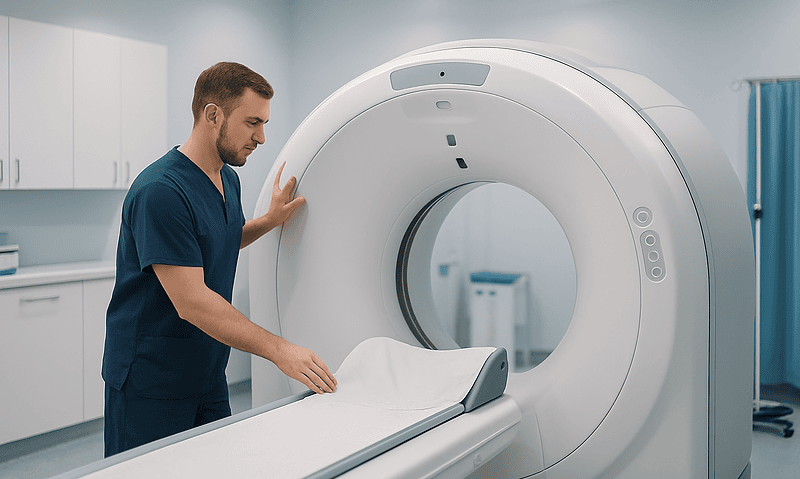
When Is Radiation Chosen Over Surgery for Vulvar Cancer, and When Are Both Used?
Surgery is usually the first treatment for vulvar cancer that can be removed with a clear margin of healthy tissue while preserving bladder, bowel…
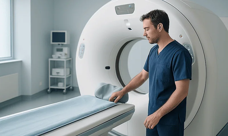
What Is Normal Healing After Vulvar Cancer Surgery and What Needs a Prompt Call
After vulvar cancer surgery, mild swelling, bruising, thin pinkish drainage, numbness and soreness that eases week by week are usually part of normal healing.…
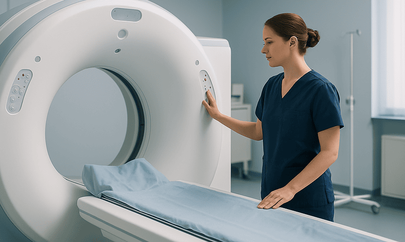
How Vulvar Cancer Treatment Is Planned by Stage and Tumor Location
Vulvar cancer treatment is planned by stage, which describes how deep the tumor has grown and whether it has reached lymph nodes or other…