Sarcoma Cancer
Sarcoma cancer care focuses on diagnosing and treating rare bone and soft-tissue tumors with surgery, chemotherapy, radiotherapy or targeted therapy. Treatment is personalized by tumor type, stage and location.
Quick answer
Sarcoma cancer is a rare group of malignant tumours that begin in bone or in soft tissues such as muscle, fat, nerves and blood vessels. Treatment is planned around the exact subtype, grade and stage, and usually combines surgery to remove the tumour with radiotherapy, chemotherapy or targeted therapy, followed by rehabilitation and long-term imaging surveillance.
Sarcoma Cancer: What It Is and Why Expert Diagnosis Matters
Sarcoma cancer is a group of rare malignant tumours that begin in the body’s connective tissues — muscle, fat, nerves, blood vessels, tendons and the deep layers beneath the skin — or in bone. It behaves differently from the common organ cancers and it is treated differently too: management usually combines surgery with radiotherapy, chemotherapy or targeted therapy, chosen according to the exact subtype, its grade and whether it has spread. Sarcoma can occur at any age and in almost any part of the body, which is part of what makes it hard to recognise.
A sarcoma diagnosis often feels particularly unsettling because most people have never heard of this cancer until it affects them or someone close to them. A sarcoma may first appear as a painless lump, a persistent swelling, unexplained pain, a bone that breaks after minor trauma, or symptoms caused by pressure on nearby organs. Because these signs overlap with many harmless conditions, sarcomas are sometimes mistaken for cysts, lipomas or old injuries. An accurate diagnosis — proper imaging, a carefully planned biopsy and expert pathology review — must come before any treatment decision is made.
If this diagnosis has just entered your life, your questions are probably layered. You may want to know whether surgery is necessary, whether chemotherapy or radiotherapy will be recommended, whether a limb can be preserved, and how long treatment takes. You may be weighing a second opinion after being told that a tumour is difficult to remove or that options are limited. These are reasonable questions, and they deserve careful, specific answers rather than reassurance. This page sets out what sarcoma is, how it is diagnosed and treated, and what recovery genuinely involves — including the limits of what any medical team can honestly promise.
One point matters more than any other: sarcoma care works best when it is planned from the start by a team experienced in rare bone and soft-tissue tumours. The tumour’s location, size, grade, subtype, molecular features and spread all shape the plan. In some cases surgery is the main treatment; in others chemotherapy, radiotherapy, targeted therapy — or a staged combination — is recommended. The aim is not only to treat the cancer but to preserve function, limit complications and protect quality of life. At Acibadem, sarcoma care sits within a broader oncology and cancer treatment programme built around multidisciplinary evaluation, modern imaging and pathology review, and individualised planning. That structure matters most in rare cancers, where the first treatment decision strongly influences long-term results.
What is sarcoma, and how does it differ from other cancers?
Sarcoma is a cancer of connective tissue and bone, whereas most common cancers — the carcinomas — begin in the lining cells of organs. That is the practical answer to the question of what is sarcoma: it is not a cancer of one organ but of the structural tissues found throughout the body. The distinction has real clinical consequences. Sarcomas are classified by the tissue they resemble under the microscope and by their molecular profile, not by the organ where they happen to sit. They also tend to spread through the bloodstream rather than the lymphatic system, which is why the lungs are a common site of distant spread for many subtypes. Diagnosis, surgery and drug treatment for sarcoma therefore follow their own pathways, distinct from those used for cancers such as breast cancer or colon cancer, and the evidence base comes from sarcoma-specific research rather than from the larger, more familiar tumour types.
What kind of cancer is sarcoma?
Sarcoma is a malignant tumour of mesenchymal tissue — the biological term for the cells that build muscle, fat, cartilage, bone, blood vessels and fibrous tissue. There are two broad families. Soft-tissue sarcomas arise in muscle, fat, nerves, vessels and connective tissue and can develop in the limbs, trunk, head and neck, abdomen or the retroperitoneum, the space behind the abdominal cavity. Bone sarcomas, such as osteosarcoma, Ewing sarcoma and chondrosarcoma, arise within the skeleton itself. Within these families sit dozens of distinct subtypes, each with its own typical age range, behaviour and treatment approach. This is why the word “sarcoma” on its own is never enough information to plan treatment; the subtype, grade and stage define what should happen next.
What Sarcoma Cancer Treatment Involves
Sarcoma cancer treatment is a personalised medical and surgical plan designed to control or remove a malignant tumour arising from bone or soft tissue. Unlike more common cancers that are categorised by organ, sarcomas are categorised by the tissue they resemble under the microscope and by their molecular characteristics. There are many subtypes, including liposarcoma, leiomyosarcoma, synovial sarcoma, undifferentiated pleomorphic sarcoma, angiosarcoma, rhabdomyosarcoma, osteosarcoma, chondrosarcoma and Ewing sarcoma, among others — and the subtype drives the plan.
Because sarcomas differ so widely in behaviour, there is no single treatment that fits every patient. A small, low-grade soft-tissue sarcoma in the forearm is managed differently from a high-grade sarcoma in the pelvis, a bone sarcoma in a teenager or a retroperitoneal sarcoma pressing against major organs and blood vessels. Decisions depend on the tumour’s location, size, grade, subtype and resectability, on whether it has reached lymph nodes or distant organs, and on your overall health and priorities.
The main methods are surgery, radiotherapy, chemotherapy, targeted therapy and, in selected subtypes or advanced disease, immunotherapy or clinical-trial-based approaches. Surgery aims to remove the tumour with an appropriate margin of healthy tissue whenever this can be done safely. Radiotherapy may be given before or after surgery to reduce the risk of the tumour returning at the same site, particularly for certain soft-tissue sarcomas. Chemotherapy is a standard part of treatment for some bone sarcomas and is considered for selected high-risk soft-tissue sarcomas. Targeted therapies come into play when a tumour carries specific biological features or when standard options have been exhausted.
Modern sarcoma care also includes everything that surrounds the tumour-directed treatment: pain control, wound care, nutrition, physiotherapy, prosthetic planning when reconstruction is needed, psychological support and structured surveillance afterwards. For some patients the treatment journey is short and focused. For others it involves several months of staged therapy and years of follow-up. Both are normal; neither means something has gone wrong.
Soft Tissue Sarcoma and Bone Sarcoma: The Two Main Groups
Soft tissue sarcoma is cancer that begins in the body’s supporting tissues — muscle, fat, nerves, blood vessels, tendons and the fibrous tissue that wraps and connects them. These tumours can develop almost anywhere, but the arms and legs, the trunk wall, the head and neck region, the abdomen and the retroperitoneum are the usual sites. Bone sarcoma, the second group, begins within the skeleton and often behaves quite differently, which is why the two families are assessed and treated along separate pathways even though both carry the name sarcoma.
Common soft-tissue subtypes
Liposarcoma arises from fat cells and ranges from slow-growing, well-differentiated forms to aggressive variants. Leiomyosarcoma arises from smooth muscle and can occur in the limbs, the uterus, the abdomen or the walls of blood vessels. Synovial sarcoma, despite its name, does not come from joint lining and often affects younger adults near — but not inside — large joints. Undifferentiated pleomorphic sarcoma is a high-grade tumour usually found in the limbs of older adults. Angiosarcoma arises from the cells lining blood or lymph vessels. Rhabdomyosarcoma, a tumour of skeletal-muscle-type cells, occurs mainly in children. Gastrointestinal stromal tumours (GISTs) are a distinct sarcoma of the digestive tract with their own, largely targeted, treatment pathway. Each of these diagnoses carries different implications, which is why expert pathology is not a formality but the foundation of the plan.
Bone sarcomas
The main bone sarcomas are osteosarcoma, Ewing sarcoma and chondrosarcoma. Osteosarcoma and Ewing sarcoma occur most often in children, adolescents and young adults, although they can appear at other ages; both usually require systemic chemotherapy in addition to surgery, because microscopic spread may already be present even when scans look localised. When these tumours occur in younger patients, care is coordinated with specialists in pediatric cancers, since growth, schooling and long-term function all enter the planning. Chondrosarcoma, a cartilage-forming tumour of adults, is usually treated primarily with surgery, with the approach depending on grade, location and subtype. Bone sarcomas frequently involve reconstructive decisions — endoprostheses, grafts or joint reconstruction — that are best made before the first operation, not after it.
Is Kaposi sarcoma skin cancer?
No — Kaposi sarcoma is not a skin cancer in the usual sense, even though it typically shows itself on the skin. It arises from the cells lining blood and lymphatic vessels and appears as purple, red or brown patches, plaques or nodules; it can also involve the mouth, lymph nodes and internal organs. It is linked to human herpesvirus 8 and occurs more often when the immune system is suppressed. This makes it biologically different from true skin cancers such as melanoma skin cancer, which develop from the skin’s own cells, and its treatment pathway — often involving the immune context as much as the tumour itself — is different too.
Symptoms: When a Lump or Bone Pain Needs Specialist Assessment
What are the first signs of sarcoma?
The most common first sign of a soft-tissue sarcoma is a lump or swelling — often painless at first — that grows over weeks or months. Features that warrant careful assessment include a mass that keeps enlarging, sits deep to the muscle layer, is larger than a golf ball, becomes painful, or comes back after removal. Bone sarcomas usually announce themselves through persistent localised pain, pain at night, swelling over a bone, reduced movement in a nearby joint, limping, or a fracture after minimal injury. It is worth being clear about the other side of this: most lumps are benign, and a swelling that appeared straight after trauma and is steadily settling usually reflects one of the ordinary soft tissue injuries rather than a tumour. The concern is the mass or pain that persists and progresses without a good explanation. In deep locations — the pelvis, abdomen or retroperitoneum — sarcomas can grow to a considerable size before causing any symptoms at all, and are sometimes found incidentally on scans done for another reason.
What causes sarcoma cancer?
In most people who develop sarcoma cancer, no cause is ever identified — the tumour arises from genetic changes in tissue cells without an external trigger that can be named. A minority of cases are linked to known risk factors. Previous radiotherapy to a body region can, years later, be associated with a sarcoma in the treated area. Certain inherited syndromes raise the risk, including Li-Fraumeni syndrome, neurofibromatosis type 1 and hereditary retinoblastoma. Long-standing lymphoedema is associated with angiosarcoma, and some chemical exposures, such as vinyl chloride, have been linked to specific liver sarcomas. Kaposi sarcoma is associated with human herpesvirus 8, particularly in the context of immune suppression. Importantly, an injury does not cause sarcoma; a knock or strain often simply draws attention to a lump that was already there.
How is sarcoma diagnosed?
Diagnosis begins with a detailed history and physical examination, followed by imaging chosen for the tumour’s location. Magnetic resonance imaging is usually preferred for tumours of the limbs, trunk wall and pelvis, because it maps the mass against muscles, nerves, vessels and joints. Computed tomography is used for the chest, abdomen and retroperitoneum, and for checking the lungs, the most frequent destination when sarcomas spread. Positron emission tomography can help in selected cases to assess metabolic activity and stage the disease, and bone scans or specialised imaging may be added for certain bone tumours.
The biopsy is the step where sarcoma care most often goes right or wrong. The track of the biopsy needle or incision can compromise a later operation if it is placed without surgical planning, so the biopsy should ideally be performed at, or in coordination with, the centre that will carry out the surgery. Pathology review then goes beyond the microscope: immunohistochemistry, molecular testing and genetic profiling are used when needed to pin down the subtype, because two tumours that look similar on a slide can require entirely different treatments. Patients who benefit from this level of assessment include those with a new suspicious mass, a confirmed diagnosis, a tumour close to vital structures, a tumour that has returned after treatment, or disease that has spread and needs an updated strategy.
Conditions and Indications Addressed by Sarcoma Treatment
Sarcoma treatment addresses a broad group of rare cancers arising from connective tissue or bone. Whatever the subtype, the shared aims are to control the disease at its original site, reduce the risk of spread and treat metastatic disease when present. In localised disease, treatment is planned with the intention of removing the cancer completely; in advanced disease, the emphasis shifts to controlling growth; and in some situations, the priority is relief of symptoms and protection of quality of life. Being honest about which of these applies to your situation is part of good care.
Retroperitoneal sarcomas deserve a specific mention because they demand particular expertise. They grow in the space behind the abdominal cavity, close to the kidneys, pancreas, bowel and major blood vessels and nerves, and they are often large by the time they are found. Surgical planning may involve several specialties at once, weighing what can be removed against the functional consequences of removing it. The first operation is usually the best opportunity for complete removal, which is one more argument for having the plan made by an experienced team.
Treatment is also indicated for recurrent sarcoma, metastatic sarcoma, sarcoma arising in a previously irradiated area, sarcoma linked to inherited cancer syndromes, and gastrointestinal stromal tumours, which are often managed with targeted medicines alongside surgery when appropriate. In every one of these situations, the plan is shaped by the specific diagnosis rather than by the word “sarcoma” alone.
Because the disease is rare, a second pathology review can change everything. A tumour initially reported as a different cancer, a benign mass or an unclassified soft-tissue lesion may be reclassified after review by pathologists who see these tumours regularly — and reclassification can substantially change the recommended treatment. For rare cancers, this review is not a luxury; it is often the single most valuable step before therapy begins.
How Sarcoma Treatment Is Performed, Step by Step
Although every plan is individual, most sarcoma pathways follow a recognisable sequence:
- Step 1 — Confirmation: review of existing scans, biopsy material and reports; new imaging or repeat biopsy if anything is incomplete or unclear.
- Step 2 — Staging: imaging of the tumour site and of the chest, since the lungs are the usual site of distant spread for many subtypes.
- Step 3 — Multidisciplinary planning: the case is discussed by a tumour board and a sequenced plan is agreed.
- Step 4 — Treatment: surgery, radiotherapy and systemic therapy delivered in the order the board has recommended.
- Step 5 — Recovery and surveillance: rehabilitation, review of final pathology and scheduled follow-up imaging over the following years.
Preparation and diagnostic planning
Treatment begins with confirming the diagnosis and staging the disease precisely. The team reviews earlier scans, pathology reports, operative notes if surgery has already been performed, biopsy material and laboratory results. If earlier imaging is incomplete or out of date, new scans are arranged; the point is to understand the tumour fully before deciding anything. Gathering earlier records in advance — imaging in its original digital format rather than printed summaries, complete pathology reports and any operative notes — makes this stage faster and helps avoid repeating tests that have already been done well.
The diagnostic work-up may include MRI for local mapping, CT for staging, chest imaging, PET-based imaging in selected cases, ultrasound- or CT-guided biopsy, blood tests, cardiac assessment before certain chemotherapy regimens, and molecular or genetic testing where the subtype demands it. The case is then discussed in a multidisciplinary tumour board that may include surgical oncologists, orthopaedic oncologists, medical oncologists, radiation oncologists, radiologists, pathologists, nuclear medicine specialists, plastic and reconstructive surgeons, paediatric oncologists when needed, and rehabilitation physicians. The board exists so that no recommendation rests on a single viewpoint: the diagnosis, the imaging and the realistic options are examined together before anything irreversible is done.
Surgery for sarcoma
Surgery is the central treatment for most localised sarcomas that can be removed safely. The goal is removal of the tumour together with a margin of normal tissue around it, because that margin reduces the chance of microscopic cancer cells remaining at the site. In limb sarcoma, modern planning usually allows limb-sparing surgery rather than amputation, though the safest approach always depends on the tumour’s extent, involvement of nerves and vessels, any previous surgery and the response to preoperative therapy — and an honest surgical team will say so plainly rather than promise a particular operation before the imaging has been studied.
For soft-tissue sarcomas of the arms or legs, the operation removes the tumour and surrounding tissue, with reconstruction where needed; plastic surgical techniques close complex wounds, protect exposed structures and support healing after radiotherapy. When sarcoma involves bone, part of the bone may be removed and reconstructed with an endoprosthesis, a graft or another orthopaedic solution, sometimes including joint reconstruction. Retroperitoneal and pelvic sarcomas are more complex still: these tumours can sit against major vessels, the kidneys, bowel, pancreas, bladder or reproductive organs, and surgery may involve several specialties working from a carefully mapped preoperative plan. The team weighs not only whether the tumour can come out, but what the functional consequences will be and how complications can be reduced. Operating time varies accordingly — a small superficial tumour needs a short procedure, while complex limb, pelvic, abdominal or reconstructive surgery can take several hours — and length of hospital stay varies with the extent of surgery, wound management, pain control and rehabilitation needs.
Radiotherapy for sarcoma
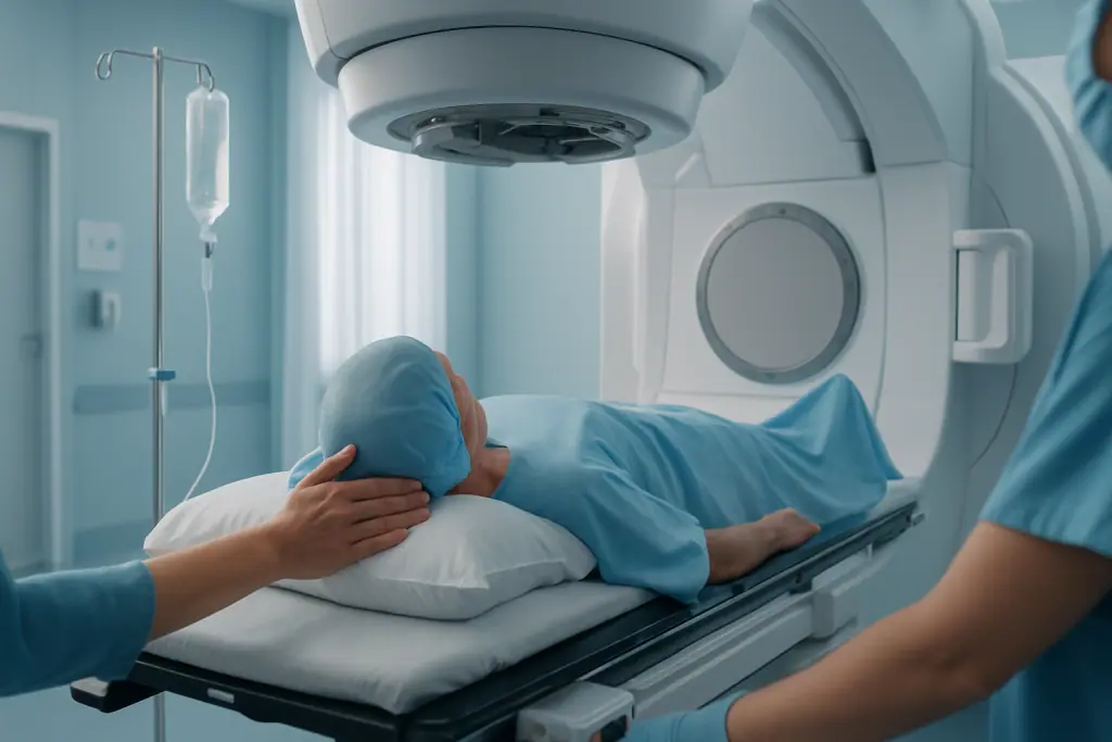
Radiotherapy uses precisely planned radiation beams to damage cancer cells and reduce the risk of the tumour returning at its original site. It may be given before surgery, to shrink the tumour or sterilise its margins, or after surgery when the final pathology shows higher-risk features; the timing depends on location, size, grade, wound-healing considerations and the surgical plan. Modern planning systems use detailed imaging to shape the radiation dose around the tumour while sparing nearby healthy tissue as far as possible — which matters most when the sarcoma lies near nerves, bowel, bladder, spinal cord, skin or the growth plates of younger patients. Treatment is delivered over multiple sessions, usually on weekdays, with the schedule tailored to the clinical situation rather than to a fixed template.
Chemotherapy, targeted therapy and immunotherapy
Systemic therapy treats cancer cells wherever they are in the body. Chemotherapy is a standard component of treatment for osteosarcoma and Ewing sarcoma and is considered for selected high-grade soft-tissue sarcomas. It can be given before surgery, to treat microscopic disease and gauge how the tumour responds; after surgery, to reduce the risk of recurrence; or in metastatic disease, to control growth. Targeted therapy is appropriate for certain subtypes with specific molecular features — gastrointestinal stromal tumours are the clearest example, where targeted medicines play a major role — and other targeted agents may be considered in advanced disease depending on subtype, previous treatment and molecular test results. Immunotherapy currently has a more limited but evolving role in sarcoma and is considered for selected patients on the basis of tumour biology, clinical context or trial availability; it is not a routine option for most subtypes, and it would be misleading to present it as one. Planning for systemic therapy includes assessment of organ function and blood counts, heart function checks before certain medicines, fertility considerations for younger patients, and supportive medication to manage side effects, with monitoring between treatment cycles.
Recovery and follow-up after treatment
Recovery depends on what was done. After surgery, you may need pain management, wound care, mobility support, physiotherapy and a gradual return to daily activity. After limb surgery, rehabilitation concentrates on strength, range of motion, gait, balance and adaptation to any reconstruction; when an endoprosthesis or graft has been used, the physiotherapy programme is built around protecting the reconstruction while steadily restoring load and confidence. After abdominal or pelvic surgery, recovery includes monitoring of bowel function, nutritional support and careful management of drains and incisions. During radiotherapy, fatigue, skin irritation, swelling or stiffness in the treated area are common and are actively managed. Chemotherapy can cause fatigue, nausea, lowered blood counts with infection risk, hair loss or nerve symptoms depending on the medicines used; these effects are monitored and treated rather than simply endured. Follow-up then continues for years: physical examinations and imaging at defined intervals check for recurrence or spread — especially to the lungs in many subtypes, which is why chest imaging is a fixture of sarcoma surveillance — with the schedule set by tumour type, grade, stage and time since treatment. Visits are typically closer together in the first years, when recurrence risk is highest, and gradually spaced out. Rehabilitation and survivorship care carry on for as long as they are useful.
Why Acting Early Matters
Early evaluation of a suspicious mass or persistent bone pain matters because sarcomas grow locally and can spread through the bloodstream, most often to the lungs. A tumour that is small and localised is generally easier to remove while preserving function. As it enlarges, it may involve nerves, blood vessels, joints, organs or skin, making surgery more complex and raising the risk of complications. None of this means panic; it means a growing lump or unexplained persistent pain deserves imaging rather than watchful hoping.
Delay can also compromise treatment quality in a specific, avoidable way. An unplanned excision — sometimes called a “whoops” surgery — happens when a sarcoma is removed without proper imaging, biopsy planning or a margin strategy because it was presumed benign. Microscopic disease may be left behind, and the later, definitive operation often has to be more extensive than the first would have been. This is the strongest practical argument for assessing a deep or growing soft-tissue mass before anyone removes it.
In bone sarcoma, delayed evaluation increases the risk of fracture, worsening pain and more complex reconstruction, and in aggressive subtypes, postponing systemic therapy gives microscopic disease time to progress. Acting early does not mean rushing into treatment without information. It means obtaining the correct diagnosis and an expert plan promptly — and then proceeding deliberately.
Benefits of Specialised Sarcoma Treatment
What specialised treatment can realistically offer depends on the subtype, stage, location and your overall health, but expert care is structured around several concrete goals.
| Benefit | What It Means for You |
|---|---|
| Accurate diagnosis | Expert imaging, biopsy planning and pathology review define the exact sarcoma subtype, which is the essential first step in choosing the right treatment. |
| Local tumour control | Surgery and radiotherapy remove or control the tumour at its original site and reduce the risk of local recurrence. |
| Function preservation | Where possible, treatment is planned to preserve limb function, organ function, mobility and independence — not just to remove tissue. |
| Personalised systemic therapy | Chemotherapy, targeted therapy or other medicines are selected according to tumour biology, stage and the risk of spread. |
| Coordinated complex care | Multidisciplinary planning aligns surgery, radiotherapy, medical treatment, reconstruction, rehabilitation and follow-up into one sequence. |
| Long-term surveillance | Structured follow-up aims to detect recurrence or treatment effects early, when further management can do the most good. |
Recovery Timeline After Sarcoma Treatment
Recovery varies from patient to patient and depends heavily on the type of surgery and whether radiotherapy or chemotherapy follows. The timeline below is a general orientation, not a schedule to hold yourself to.
| Time Period | What Patients Can Expect |
|---|---|
| Day 1 | Monitoring after surgery or treatment, pain control, early mobilisation when safe, wound assessment and a review of immediate recovery needs. |
| First week | Gradual increase in activity, physiotherapy if needed, management of drains or dressings, discharge planning and review of pathology results where available. |
| First month | Continued wound healing, rehabilitation, adjustment of mobility, and discussion of any additional treatment such as radiotherapy or chemotherapy. |
| During additional therapy | Regular monitoring for side effects, blood tests or imaging as needed, supportive care and coordination between the oncology, radiation and rehabilitation teams. |
| Longer term | Surveillance imaging, ongoing functional recovery, return to work or school when appropriate, management of any late effects and continuing survivorship support. |
What Influences Outcomes in Sarcoma Care
Outcomes in sarcoma depend on many factors, and every case has to be read individually. The subtype is among the most important: some tumours grow slowly and stay localised, others are inherently aggressive. Grade matters — high-grade tumours are more likely to recur or spread than low-grade ones — and so does stage at diagnosis. Localised disease generally offers more options than metastatic disease, although selected patients with limited metastases can still benefit from combined local and systemic treatment. Tumour size and depth influence surgical planning and recurrence risk, tumours near major nerves, vessels, organs or joints demand more complex decisions, and prior surgery or radiotherapy narrows what can safely be done.
The quality of the first treatment plan is itself an outcome factor. Correct biopsy technique, complete staging, expert pathology review and appropriate surgical margins all contribute to better local control, and for selected sarcomas the timing and choice of chemotherapy or radiotherapy shape the result as well. Response to preoperative therapy provides useful information about the tumour’s sensitivity and helps guide what comes next. Patient-related factors also count: age, general health, nutrition, heart and kidney function, mobility, smoking status, diabetes, immune status and the ability to take part in rehabilitation all affect how treatment is tolerated and how recovery goes.
Is sarcoma a serious cancer?
Yes — sarcoma is a serious diagnosis and should always be treated as one, but seriousness is not the same as hopelessness. The spectrum is wide: some low-grade sarcomas, removed completely with clear margins, may never trouble the patient again, while high-grade or metastatic disease requires intensive, prolonged treatment. What makes sarcoma dangerous in practice is often delay and mismanagement — a tumour dismissed as a harmless lump, or removed casually without a margin strategy — rather than the diagnosis alone. Serious disease handled by an experienced team from the outset is a very different situation from the same disease handled late or piecemeal.
Is sarcoma cancer deadly?
Sarcoma cancer can be life-threatening, particularly when it is high grade, large, deep or has already spread — and no honest clinician will tell you otherwise. But it is not uniformly so. Outcome depends on the subtype, the grade, the stage at diagnosis, the completeness of the first surgery and the tumour’s response to any systemic treatment. Two patients who both carry the word “sarcoma” in their reports can face entirely different situations. The meaningful question is never whether sarcoma in general is deadly, but what your specific subtype, grade and stage mean — and that answer comes from your own pathology and imaging, discussed with your treating team.
What is life expectancy with sarcoma?
There is no single life expectancy figure for sarcoma, and any page that offers one is oversimplifying. Prognosis varies enormously with the subtype, grade, size, depth and location of the tumour, whether it has spread, whether it can be removed completely, and how it responds to treatment. Published averages pool together very different tumours and very different patients, so they say little about any individual. What a specialist team can do is interpret your specific findings — pathology, imaging, staging — and explain what they mean for you, including the genuine uncertainty. That conversation, grounded in your own results, is worth more than any general number.
A good result is also not defined only by scan findings. For many patients it means walking well after limb surgery, returning to daily activities, controlling pain, preserving independence, maintaining body image, managing long-term side effects and feeling informed at every stage. Sarcoma care should aim at both cancer control and functional recovery, and a treatment plan that ignores the second is incomplete.
How Sarcoma Care Is Organised at Acibadem
Rare cancers demand two things above all: genuine expertise and coordination across the many specialties involved, and at Acibadem sarcoma care is structured around both. Cases are evaluated with input from the specialties the tumour actually requires — depending on type and location, that may include orthopaedic oncology, surgical oncology, medical oncology, radiation oncology, radiology, pathology, nuclear medicine, plastic and reconstructive surgery, paediatric oncology, vascular surgery, urology, thoracic surgery, gastroenterology, physiotherapy and pain management. Multidisciplinary tumour boards review the diagnosis, imaging and goals together, so that recommendations reflect a shared assessment rather than a single opinion.
Diagnostic and treatment technology supports that planning: high-resolution cross-sectional imaging maps the tumour against nearby structures, image-guided biopsy supports accurate sampling, pathology methods including immunohistochemical and molecular testing clarify the subtype where needed, and radiotherapy planning systems shape treatment fields around the tumour while limiting exposure of healthy tissue. In surgery, detailed preoperative planning, reconstructive techniques and intraoperative decision-making support safe removal and, where possible, preservation of function.
Treatment itself remains individual: a newly diagnosed extremity sarcoma may need biopsy confirmation, preoperative radiotherapy and limb-sparing surgery with rehabilitation; osteosarcoma may need chemotherapy before and after surgery; a retroperitoneal sarcoma may need complex surgery involving abdominal and vascular expertise; metastatic disease may need systemic therapy, symptom control and selective local treatment. These are different journeys, and the plan should reflect the difference — which is exactly why the same diagnosis can lead to quite different treatment calendars for two different patients, and why neither calendar is “wrong”.
Preparing for a Specialist Sarcoma Opinion
A specialist review — whether a first assessment or a second opinion — is only as good as the material it rests on. The documents that make the biggest difference are prior imaging in its original digital format rather than printed summaries, the biopsy and pathology reports, pathology slides or tissue blocks where they can be obtained, operative notes from any previous surgery, and a summary of treatments already given. With these in hand, a sarcoma team can confirm or refine the diagnosis, judge whether additional molecular testing would change the plan, assess whether surgery is feasible and what it would involve, and clarify the role of chemotherapy or radiotherapy in your particular case.
It also helps to arrive at that review with your own questions written down: whether the biopsy result is definitive or provisional, what grade and stage have been assigned and on what evidence, what the surgical options are and what each would mean for function, whether treatment order could change depending on how the tumour responds, and what the follow-up schedule will look like. A good team welcomes these questions, because a patient who understands the reasoning behind a plan is better placed to follow it through months of treatment and years of surveillance.
In a rare cancer such as sarcoma, this kind of structured review before major treatment is not a sign of distrust in anyone; it is standard good practice. The first surgical decision has lasting consequences, pathology reclassification is a recognised reality in soft-tissue tumours, and treatment protocols differ meaningfully between subtypes. Taking the time to establish exactly what the tumour is — and to have the plan made by people who treat these tumours regularly — is the single most useful thing a patient facing sarcoma can do.
Preparation
- Preparation usually includes imaging, biopsy review, pathology confirmation and staging tests to define the sarcoma type and spread. Your oncology team reviews medications, general health and previous treatments before creating a personalized plan. If surgery is planned, fasting and anesthesia assessment may be required.
Aftercare
- Follow-up includes wound care if surgery was performed, management of chemotherapy or radiotherapy side effects, and scheduled imaging to monitor response. Rehabilitation or physiotherapy may be recommended to restore movement and function. Patients should report fever, increasing pain, swelling or new symptoms promptly.
Turkey vs UK, Germany & USA
Sarcoma cancer treatment is highly individualized because these tumors can arise in bone or soft tissue and may behave differently depending on subtype, grade and location. Comparing destinations can help patients understand how hospital expertise, diagnostics, treatment planning and travel logistics may influence the overall care pathway.
International sarcoma care costs and patient experience can vary by the complexity of diagnosis, the need for a specialist tumor board, the treatment plan and the level of support offered to international patients.
| Factor | Turkey | UK | Germany | USA |
|---|---|---|---|---|
| Price drivers | Often shaped by private hospital packages, advanced imaging, pathology review, surgery complexity and systemic therapy needs. | Private care costs may be influenced by consultant fees, hospital fees, diagnostics and access to specialist oncology services. | Costs typically reflect specialist center fees, detailed diagnostics, surgery, radiotherapy planning and medication protocols. | Costs may vary widely due to hospital billing models, specialist fees, imaging, surgery, medications and insurance arrangements. |
| Hospital and surgeon factors | International hospitals may offer multidisciplinary sarcoma evaluation with orthopedic oncology, surgical oncology, medical oncology and radiation oncology teams. | Specialist sarcoma units and private oncology pathways may be available, with consultant-led treatment planning. | Care may involve university or specialist oncology centers with structured diagnostic and treatment pathways. | Major cancer centers may offer subspecialty sarcoma programs and access to advanced treatment modalities. |
| Accreditation and quality | Some hospitals serving international patients are JCI-accredited and use multidisciplinary tumor board review. | Quality oversight depends on the provider and hospital group, with established clinical governance systems. | Hospitals often follow national and European oncology standards, with specialist certification varying by center. | Accreditation and quality programs vary by hospital, with many major centers using comprehensive cancer care standards. |
| Waiting times | Private international pathways may allow coordinated scheduling for imaging, biopsy review and consultation after records are assessed. | Waiting time depends on public or private pathway availability and consultant scheduling. | Scheduling depends on center capacity, referral process and complexity of diagnostic workup. | Access can be prompt in some private systems, but timing depends on insurance approval, center capacity and referral review. |
| Travel and language logistics | International patient departments commonly assist with appointments, airport transfers, accommodation guidance and interpreter support. | Less travel may be needed for local patients; international patients may need to arrange visas, accommodation and language support if required. | International offices may be available in larger centers; language support varies by hospital. | Travel distance, visa requirements, accommodation and insurance coordination can significantly affect the patient experience. |
| Typical package scope | Packages may include consultation, imaging coordination, pathology review, treatment planning and hospital admission items, depending on the case. | Private quotes may separate consultant, diagnostic, hospital and treatment fees. | Quotes may be itemized by diagnostics, specialist consultations, procedures and inpatient care. | Billing may be highly itemized, with separate provider, facility, imaging, medication and anesthesia charges. |
- What affects your final cost:
- Sarcoma type, grade, stage and whether it is located in bone, limb, trunk, abdomen or another area.
- Need for advanced imaging, biopsy, molecular testing or expert pathology review.
- Type of surgery, reconstruction requirements and length of hospital stay.
- Use of chemotherapy, radiotherapy, targeted therapy, immunotherapy or a combined approach.
- Need for rehabilitation, wound care, prosthetic planning or follow-up imaging.
- Travel arrangements, interpreter support, accommodation and companion needs.
Compare your options
Sarcoma treatment may involve one modality or a combination of approaches. Suitability is decided by a specialist after imaging, pathology review and multidisciplinary evaluation.
| Option | What it is | Typical use | Key considerations |
|---|---|---|---|
| Specialist diagnosis and biopsy | Imaging, tissue sampling and pathology review to confirm sarcoma subtype and grade. | Used before treatment planning to distinguish sarcoma from other tumors and define the safest approach. | Biopsy planning is important because the biopsy route can affect later surgery. Expert pathology review may be recommended. |
| Surgery | Removal of the tumor with an aim to achieve clear margins when possible. | Commonly used for localized soft-tissue and bone sarcomas when the tumor can be safely removed. | Complexity depends on tumor location, nearby nerves and vessels, reconstruction needs and functional preservation. |
| Limb-sparing surgery and reconstruction | Surgical removal of the tumor while aiming to preserve limb function, sometimes with implants, grafts or soft-tissue reconstruction. | Considered for selected limb sarcomas where safe tumor removal and functional preservation are feasible. | Requires experienced surgical planning and may involve rehabilitation, physiotherapy and long-term follow-up. |
| Radiotherapy | Focused radiation treatment used to control cancer cells in a defined area. | May be used before or after surgery, or for tumors that are difficult to remove completely. | Planning depends on tumor size, location, surgical timing and protection of nearby organs and tissues. |
| Chemotherapy | Systemic medication that circulates through the body to treat sensitive sarcoma types. | Used for certain bone and soft-tissue sarcomas, metastatic disease or when recommended as part of combined treatment. | Benefit varies by sarcoma subtype. Side effects, general health and monitoring needs are reviewed by the oncology team. |
| Targeted therapy or immunotherapy | Medicines selected according to tumor biology, molecular findings or specific disease behavior. | May be considered for selected sarcoma subtypes or advanced disease when appropriate. | Eligibility depends on pathology, molecular testing, previous treatment and specialist assessment. |
| Supportive and palliative care | Care focused on symptom control, mobility, nutrition, pain management and quality of life. | Used alongside active treatment or when disease control and comfort are the main goals. | Can include rehabilitation, psychosocial support, pain services and coordinated follow-up. |
General information only — not medical or financial advice. Final costs depend on the factors above and your individual case; request a free, personalised quote.
Frequently Asked Questions
What affects the cost of sarcoma cancer treatment?
The main factors are sarcoma subtype, tumor location, stage, required imaging, biopsy and pathology review, surgery complexity, hospital stay, rehabilitation needs and whether chemotherapy, radiotherapy or targeted treatment is recommended.
How can I get a personalised quote for sarcoma care in Turkey?
You can request a free consultation by sharing your medical reports, imaging, pathology results and previous treatment history. A specialist team can review the case and prepare a personalised treatment plan and cost estimate.
Does a sarcoma treatment package include all care steps?
Package content varies by the treatment plan. It may include consultations, diagnostic coordination, surgery or oncology treatment components, hospital stay and international patient support, but medications, extra tests or extended care may be quoted separately.
Why is expert pathology review important for sarcoma?
Sarcoma is a rare and diverse group of cancers, and treatment depends strongly on the exact subtype and grade. Expert review can help confirm the diagnosis and guide surgery, radiotherapy or systemic therapy decisions.
Can international patients receive coordinated care for surgery, chemotherapy and radiotherapy?
Yes, many international hospitals coordinate multidisciplinary care across surgical oncology, orthopedic oncology, medical oncology, radiation oncology, radiology, pathology and rehabilitation. The exact pathway depends on specialist assessment.
Is the information provided here medical or financial advice?
No. This is general educational information. A personalised recommendation and quote can only be provided after a specialist reviews your diagnosis, medical condition and treatment goals.
Medically reviewed by the Acıbadem International Medical Board — August 30, 2026
See our medical review board →
Update history
- PublishedJune 8, 2026
- Medical review approvedAugust 30, 2026
- Last content updateAugust 30, 2026
References3
- Soft Tissue Sarcoma Treatment (PDQ) – Patient Version — cancer.gov
- Soft Tissue Sarcoma — medlineplus.gov
- Sarcoma — my.clevelandclinic.org
Trusted care for international patients
Doctors Performing This Treatment

Prof. Dr. Handan Onur Topuzlu
Medical Oncology
Prof. Dr. İsmet Aydoğdu
Hematology
Prof. Dr. Ahmet Öztürk
Hematology
Prof. Dr. Ayşen Timurağaoğlu
Hematology
Prof. Dr. Aziz Yazar
Medical Oncology
Prof. Dr. Ali Arıcan
Medical Oncology
Prof. Dr. Gülsan Sucak
Hematology
Prof. Dr. Siret Ratip
Hematology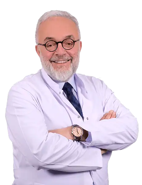
Prof. Dr. Mustafa Çetiner
Hematology
Prof. Dr. Gökhan Demir
Medical Oncology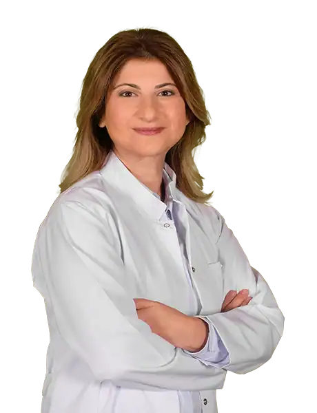
Prof. Dr. Yeşim Eralp
Medical Oncology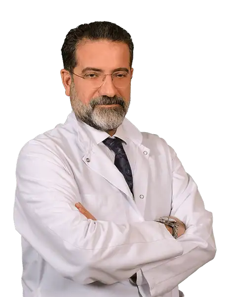
Prof. Dr. S. Sami Kartı
Hematology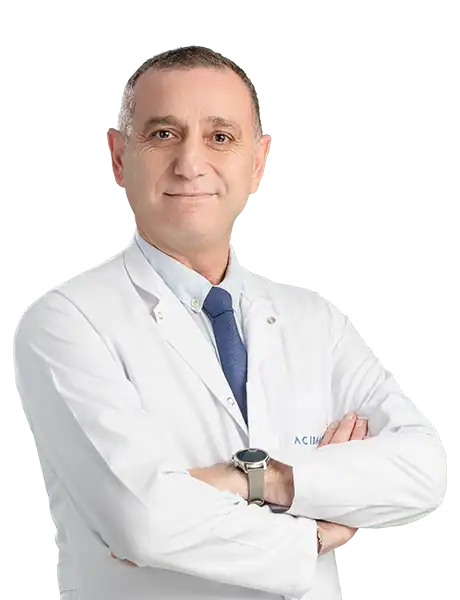
Prof. Dr. Bülent Karabulut
Medical Oncology
Prof. Dr. Gül Başaran
Medical Oncology
Prof. Dr. Hüseyin Engin
Medical Oncology
Prof. Dr. Özlem Er
Medical Oncology
Prof. Dr. Başak Oyan Uluç
Medical Oncology
Prof. Dr. Faysal Dane
Medical Oncology
Prof. Dr. Taner Korkmaz
Medical Oncology
Prof. Dr. Ömer Fatih Ölmez
Medical Oncology
Prof. Dr. İbrahim Yıldız
Medical Oncology
Prof. Dr. Türkan Öztürk Topcu
Medical Oncology
Prof. Dr. Özge Gümüşay
Medical Oncology
Prof. Dr. Meliha Nalçacı
HematologyMedical Units
Available at These Hospitals