

Moles and Skin Checks: What Is Normal and What Needs Evaluation

Normal moles are usually evenly colored, round or oval, and stable over time. The ABCDE rule helps people notice asymmetry, border changes, color variation, diameter, and evolution.
Key Takeaways
- Normal moles are usually evenly colored, round or oval, and stable over time.
- The ABCDE rule helps people notice asymmetry, border changes, color variation, diameter, and evolution.
- A new, changing, bleeding, itchy, painful, or unusual-looking spot should be assessed by a dermatologist.
- People with many moles, a personal or family history of skin cancer, or significant ultraviolet exposure may need more regular professional skin checks.
- Sun protection and avoiding tanning beds help reduce the risk of new sun-related skin damage.
Most moles are harmless and remain stable for years, but changes in size, shape, color, or symptoms deserve attention. Regular skin self-checks and dermatologist examinations help identify concerning lesions early and guide appropriate care.
Overview
Moles, medically called melanocytic nevi, are common skin growths made up of pigment-producing cells. They may be flat or raised, light brown to dark brown, pink, or close to a person’s natural skin tone. Many people develop moles during childhood, adolescence, and early adulthood, and some moles slowly fade with age.
Most moles are benign and do not need treatment. A normal mole often has a regular shape, an even color, and a stable appearance over months and years. However, because some skin cancers can resemble moles, it is useful to know what is typical and what should be evaluated by a qualified dermatologist.
Skin checks include both self-examination at home and professional examination in a clinic. The goal is not to worry about every mark, but to recognize meaningful changes. When a suspicious lesion is found, a dermatologist can examine it with appropriate tools and decide whether monitoring, dermoscopy, biopsy, or removal is needed.
What Normal Moles Often Look Like
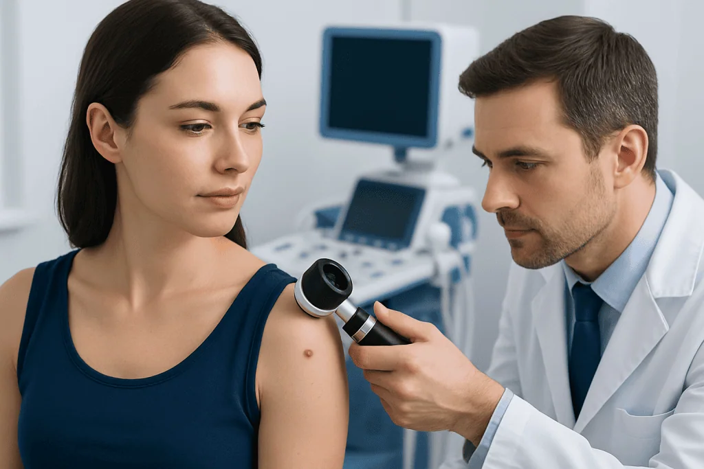
A typical mole is usually round or oval, with a smooth and fairly even border. It is often one color throughout, such as tan, brown, dark brown, pink, or skin-colored. It may be flat like a freckle or gently raised, and some moles may have hairs growing from them, which is not automatically a sign of danger.
Normal moles are commonly smaller than a pencil eraser, although size alone does not determine whether a mole is benign or concerning. The most reassuring feature is stability. A mole that has looked the same for a long time, especially when it resembles a person’s other moles, is less likely to be worrisome than one that is rapidly changing.
People may also have atypical moles, sometimes called dysplastic nevi. These can be larger, more irregular, or more varied in color than ordinary moles, yet many are still benign. Because atypical moles can be harder to assess by sight alone, people with several of them often benefit from regular dermatology follow-up and photographs for comparison.
Warning Signs: The ABCDE Mole Check
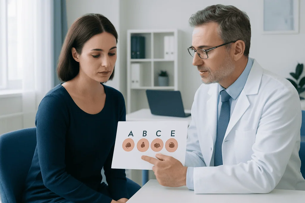
The ABCDE rule is a simple way to remember features that may need medical evaluation. It is not a diagnosis, and not every mole with one ABCDE feature is cancer. Still, it is a helpful screening tool for deciding when to make a dermatology appointment.
- A for Asymmetry: one half of the mole does not match the other half.
- B for Border: the edges are uneven, blurred, notched, or poorly defined.
- C for Color: the mole contains several colors or has uneven shades of brown, black, red, white, blue, or gray.
- D for Diameter: the spot is larger than about 6 millimeters, or it is growing, even if smaller.
- E for Evolution: the mole changes in size, shape, color, surface, or symptoms over time.
Evolution is often the most important warning sign. A spot that begins to bleed without injury, forms a persistent crust, becomes tender, itches repeatedly, or looks noticeably different from a person’s other moles should be checked. Dermatologists also use the concept of the unusual mole, meaning one lesion that stands out from the rest of a person’s skin pattern.
Some skin cancers do not follow classic rules, especially on darker skin tones, under nails, on the palms or soles, or in less sun-exposed areas. Any new or changing spot that seems unusual to the patient is a valid reason to seek professional assessment.
Causes and Risk Factors
Moles develop when melanocytes, the skin cells that produce pigment, grow in clusters. Genetics strongly influences how many moles a person has and what they look like. It is common for mole patterns to run in families, and some people naturally develop more moles than others.
Ultraviolet radiation from the sun and tanning beds contributes to skin damage and can increase the risk of certain skin cancers. Intermittent intense sun exposure and sunburns, especially earlier in life, are important risk factors. Tanning beds are not a safe alternative to sunlight because they also emit ultraviolet radiation that can damage skin cells.
Risk is also higher for people with a personal history of melanoma or other skin cancers, a strong family history of melanoma, many moles, atypical moles, fair skin that burns easily, or a weakened immune system. People with darker skin have a lower overall risk of melanoma than people with lighter skin, but melanoma can still occur and may be diagnosed later if unusual areas are not examined.
Hormonal changes may make moles appear darker or more noticeable, such as during pregnancy or adolescence. Mild, gradual changes can be normal, but rapid growth, irregular change, bleeding, or persistent symptoms should not be assumed to be hormonal and should be evaluated.
How to Do a Skin Self-Check
A monthly self-check can help a person become familiar with their own skin. The best time is often after a shower, in a well-lit room, using a full-length mirror and a hand mirror. A partner or family member can help check areas that are difficult to see, such as the scalp, back, and backs of the legs.
A complete self-check includes the face, ears, scalp, neck, chest, abdomen, back, arms, hands, fingernails, legs, feet, soles, toenails, and the areas between the toes. It is also important to look at skin folds and less visible areas. Skin cancer can develop in places that receive little sun, so a thorough approach is useful for everyone.
Taking clear photographs of moles or using a body map can make it easier to notice change over time. The same lighting, distance, and angle help make comparisons more reliable. People should avoid trying to diagnose a lesion from photos alone, as professional examination is still needed for suspicious or changing spots.
Diagnosis and Dermatology Evaluation
During a dermatology visit, the doctor will ask about the mole’s history, including when it appeared, whether it has changed, and whether there are symptoms such as bleeding, itching, crusting, or pain. The dermatologist will also review personal and family history, sun exposure, and previous skin conditions or biopsies.
The skin examination may include dermoscopy, a noninvasive technique that uses magnification and special light to see structures within the skin that are not visible to the naked eye. Dermoscopy helps trained clinicians distinguish many benign moles from lesions that need biopsy. Some clinics may also use digital mole mapping or serial photography for patients with many moles or high risk.
If a lesion looks suspicious, the dermatologist may recommend a biopsy, which means removing all or part of the spot for laboratory examination by a pathologist. Biopsy is the only way to confirm whether a mole is benign, atypical, or cancerous. The type of biopsy depends on the size, location, and clinical appearance of the lesion.
Not every mole needs removal. Many can be safely observed if they are clinically reassuring. When removal is advised, it may be for diagnosis, symptoms, repeated irritation, or cosmetic reasons after medical evaluation.
Treatment Options and Follow-Up
Benign moles usually do not require treatment. If a mole is removed, methods may include surgical excision or shave removal, depending on the reason for removal and the dermatologist’s assessment. Patients should follow wound care instructions carefully and attend follow-up appointments if pathology results require review.
If a biopsy shows an atypical mole, the next step depends on the degree of atypia, whether the lesion was completely removed, and the dermatologist’s judgment. Some atypical moles only need observation, while others may require complete excision. If skin cancer is diagnosed, treatment is planned according to the exact type, depth, location, and overall health of the patient.
Follow-up schedules are individualized. A person with one stable mole may not need frequent visits, while someone with many atypical moles, previous melanoma, or strong family history may need regular full-body skin examinations. Patients should ask their dermatologist how often they should be checked and what changes should prompt an earlier visit.
Prevention, Self-Care, and When to See a Doctor
Sun protection supports long-term skin health. Recommended habits include seeking shade during peak sun hours, wearing protective clothing, using broad-spectrum sunscreen on exposed skin, and reapplying sunscreen as directed, especially after swimming or sweating. Avoiding tanning beds is also an important preventive step.
A dermatologist should evaluate any mole or skin spot that is new and unusual, changing, asymmetrical, irregularly bordered, uneven in color, bleeding, crusting, painful, or persistently itchy. Evaluation is also appropriate for a sore that does not heal, a dark streak under a nail, or a spot on the palm, sole, or mucous membrane that changes or stands out.
People at higher risk should discuss a personalized screening plan with a qualified doctor. This includes those with many moles, atypical moles, a personal or family history of melanoma, significant sunburn history, or immune suppression. At Acibadem International, multidisciplinary specialists in JCI-accredited hospitals diagnose and treat dermatologic conditions, including suspicious moles and skin cancers, for international patients.
Frequently asked questions
Are all moles dangerous?
No. Most moles are benign and remain stable for many years. A mole becomes more concerning when it changes, looks very different from the others, or develops symptoms such as bleeding, crusting, pain, or persistent itching.
How often should someone check their skin?
Many people benefit from looking over their skin about once a month so they can recognize their usual pattern. The recommended frequency of professional skin checks depends on personal risk factors, such as many moles, atypical moles, or a history of skin cancer.
Can a mole with hair be cancerous?
Hair growing from a mole is usually a reassuring sign and is common in benign moles. However, hair does not completely rule out a problem. If the mole changes in shape, color, size, or symptoms, it should be assessed by a dermatologist.
Should a mole be removed just because it is raised?
A raised mole is not automatically abnormal. Many benign moles are raised, soft, or dome-shaped. Removal may be considered if it is irritated, cosmetically bothersome, or medically suspicious, but the decision should follow a professional skin examination.
What is dermoscopy?
Dermoscopy is a noninvasive examination method that uses magnification and special lighting to view patterns within a skin lesion. It helps dermatologists decide whether a mole looks benign, should be monitored, or needs biopsy.
Can melanoma occur on skin that is not exposed to the sun?
Yes. Although ultraviolet exposure is an important risk factor, melanoma can develop on areas with little sun exposure, including the soles, palms, under nails, and mucous membranes. Any changing or unusual spot in these areas should be checked.
Is it safe to monitor a changing mole at home?
Photographs can help track changes, but a changing mole should not be managed only at home. A dermatologist can examine it properly and decide whether monitoring or biopsy is needed. Early evaluation is often simpler and provides clearer guidance.
References
- American Academy of Dermatology
- Skin Cancer Foundation
- National Cancer Institute
- World Health Organization
- British Association of Dermatologists
This article is for general information only and is not a substitute for professional medical advice. Please consult a qualified doctor about your individual situation.
Explore treatments in Turkey — costs, top hospitals & a free quote
JCI-accredited · board-certified surgeons · reply within 24h
Add us as a Preferred Source to see more of our trusted health content across Google Search, AI Overviews and Discover.

Related Treatments
Related Conditions
More from the Health Library



Related Specialists

Prof. Dr. Sedef Şahin
Dermatology
Prof. Dr. Şükrü Yazar
Aesthetic Plastic & Reconstructive Surgery
Prof. Dr. Emel Öztürk Durmaz
Dermatology
Prof. Dr. Özlem Dicle
Dermatology