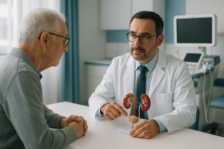
Cystoscopy
Cystoscopy is a minimally invasive test that lets a urologist view the bladder and urethra with a thin scope. It helps diagnose bleeding, infections, stones, tumors, and urinary symptoms.

Quick answer
A cystoscopy is an examination of the inside of the bladder and urethra using a thin telescope called a cystoscope, passed through the urethra. Urologists use it to investigate blood in the urine, recurrent infections, pain or poor urine flow, and sometimes to take biopsies or treat small problems such as stones. A diagnostic flexible cystoscopy usually takes only a few minutes and is done with local anaesthetic gel.
What Is a Cystoscopy?
A cystoscopy is an examination of the inside of the urethra and bladder using a thin, lighted telescope called a cystoscope. The instrument is passed gently through the urethra, the tube that carries urine out of the body, and gives your urologist a direct, real-time view of the bladder lining, the bladder outlet and the urethra itself. Urologists use cystoscopy to find the cause of urinary symptoms such as blood in the urine, recurrent infections, pain on urination or a weak stream, and, in selected cases, to carry out a minor treatment during the same session.
That direct view is what sets the test apart. Ultrasound, CT and MRI show the urinary tract from the outside; cystoscopy shows the inner surface itself. Inflammation, stones, strictures, tumours, bleeding points and structural changes that imaging leaves uncertain are often plainly visible through the cystoscope. The images may be viewed directly through the lens or on a monitor, depending on the equipment used and the type of examination being performed. Cystoscopy is rarely interpreted in isolation. It usually sits alongside urine analysis, urine culture, urine cytology, ultrasound, CT urography, MRI, urodynamic testing or prostate assessment: the exact combination depends on your symptoms and on what your urologist is trying to establish.
If the test has been recommended for you, there is a good chance you have been living with symptoms that are hard to ignore. Blood in the urine, repeated urinary tract infections, pain when you urinate or the need to pass urine far more often than usual are unsettling. Some people worry immediately about serious disease; others are simply frustrated after weeks or months without a clear explanation. Cystoscopy exists to replace that uncertainty with direct evidence. It answers questions such as where bleeding is coming from, whether recurrent infections have a structural cause, whether a bladder tumour is visible, or whether a blockage is contributing to your symptoms.
It is also worth being clear about what the test is not. Cystoscopy is not bladder surgery, although some limited procedures can be performed through the cystoscope. For most people it is primarily a diagnostic examination, brief, performed on an outpatient basis, and followed by a conversation about what was seen and what, if anything, needs to happen next.
Cystourethroscopy, cytoscopy and other names you may see
Cystourethroscopy is the full clinical name for the examination, because the urethra is inspected as well as the bladder. You will also come across the misspellings cytoscopy and cystocopy online; both refer to the same test. Whatever spelling appears in your referral letter, your report or your search results, the procedure being described is identical: a telescopic inspection of the lower urinary tract from the inside.
Flexible and rigid cystoscopy: two instruments, two purposes
There are two main types of cystoscope, and the choice between them shapes your whole experience of the test.
A flexible cystoscope is soft and bendable. It is the usual choice for diagnostic examinations in an outpatient setting, because it follows the natural curves of the urethra and generally causes less discomfort. Flexible cystoscopy can usually be performed with a local anaesthetic gel alone, no injections, no general anaesthetic, and often no fasting beforehand.
A rigid cystoscope is straight and slightly wider. It is chosen when a more detailed examination is needed, or when the urologist expects to do something as well as look: take tissue samples, remove a small stone, treat certain lesions or address a urethral narrowing. Because working instruments are passed through the scope, rigid cystoscopy is usually performed under sedation, spinal anaesthesia or general anaesthesia, depending on the case.
Neither type is better in any absolute sense; they answer different questions. Matching the instrument and the anaesthetic plan to the clinical problem is one of the decisions your urologist makes before the day of the procedure, and it is worth asking which type is planned for you and why.
Who May Need a Cystoscopy?
Your urologist may recommend cystoscopy when symptoms, test results or risk factors suggest that the bladder or urethra needs direct evaluation. The test matters most when symptoms persist, recur or cannot be fully explained by basic investigations.
Why would a urologist do a cystoscopy?
A urologist performs a cystoscopy when looking at the bladder and urethra directly is likely to change the diagnosis or the treatment plan. The most common triggers are blood in the urine, recurrent infections, persistent lower urinary tract symptoms, abnormal imaging findings and scheduled surveillance after bladder cancer treatment.
Blood in the urine, haematuria, is the single most important indication. Blood may be visible, turning the urine pink, red, tea-coloured or cola-coloured, or it may be microscopic, detected only on a urine test. Haematuria has many benign causes: infection, stones, vigorous exercise, prostate enlargement, medication effects. But it can also be associated with bladder tumours and other significant urinary tract conditions. That is why cystoscopy is routinely recommended for adults with persistent or unexplained blood in the urine, direct inspection of the bladder lining is the most reliable way to see whether a visible lesion is responsible.
Recurrent urinary tract infections are the second common trigger, particularly when infections are frequent, difficult to treat, accompanied by blood in the urine, or occur in men. Cystoscopy can reveal bladder stones, diverticula (pockets in the bladder wall), foreign bodies, incomplete emptying, urethral narrowing or other structural factors that keep letting infections return.
Persistent lower urinary tract symptoms are a third. Frequent urination, urgency, pain with urination, a weak stream, difficulty starting, an interrupted flow, or the sensation that the bladder never quite empties can all justify a direct look when they do not resolve. In men, these symptoms may relate to prostate enlargement, urethral stricture, bladder neck obstruction or bladder dysfunction. In women, possible causes include chronic inflammation, bladder pain syndrome, urethral problems, stones or, rarely, tumours.
Some patients come to cystoscopy from an abnormal scan rather than a symptom. An ultrasound, CT or MRI may show bladder wall thickening, a possible mass, a stone or another change that needs visual confirmation. Others need the test as planned surveillance: bladder tumours can recur after treatment, and established urological guidelines recommend regular check cystoscopies so that any recurrence is recognised early.
Before recommending the examination, your doctor will typically review your medical history, medications, allergies, previous surgery, urinary symptoms, smoking history, occupational exposures, infection history and family history. The workup around cystoscopy may include urine analysis, urine culture, kidney function tests, imaging and specialised urine tests where indicated. The decision is made in the context of the full clinical picture, cystoscopy is a targeted examination, not a routine screen.
Conditions a Cystoscopy Can Help Diagnose or Treat
Cystoscopy covers a wide range of conditions involving the urethra, bladder and bladder outlet. Its value lies in seeing the internal urinary tract directly and, where appropriate, taking a tissue sample or performing a limited treatment through the same access route.
- Haematuria (blood in the urine): One of the most important indications. Cystoscopy helps identify visible sources of bleeding within the bladder or urethra, including tumours, stones, inflammation, bleeding related to an enlarged prostate, or vascular changes in the bladder lining.
- Recurrent urinary tract infections: Repeated infections may be linked to bladder stones, incomplete emptying, abnormal pockets in the bladder wall, urethral narrowing or other structural issues. Cystoscopy can help clarify why infections keep returning, which is the first step towards breaking the cycle.
- Bladder tumours and bladder cancer surveillance: Cystoscopy is a key tool for detecting suspicious bladder lesions and for monitoring patients previously treated for bladder cancer. If an abnormal area is seen, biopsy or additional procedures may be recommended.
- Bladder stones: Stones inside the bladder can cause pain, bleeding, infections, interrupted urine flow or urgency. Small stones can sometimes be treated endoscopically during the same session; larger or more complex stones may need a separately planned procedure.
- Urethral strictures: A stricture is a narrowing of the urethra that reduces urine flow. Cystoscopy helps locate and assess the narrowing, which guides treatment planning, from dilation to more formal reconstructive options.
- Bladder pain syndrome and chronic pelvic pain: In selected patients, cystoscopy helps evaluate chronic bladder discomfort, urgency, frequency and pelvic pain. It can identify inflammation, ulcers, stones or other conditions that mimic bladder pain syndrome, and can help exclude alternatives before the diagnosis is settled.
- Prostate-related urinary obstruction: In men with urinary symptoms, cystoscopy can show how the prostate and bladder neck are affecting urine flow. That information supports decisions about medication, minimally invasive prostate procedures or surgery.
- Foreign bodies, mesh complications and surgical follow-up: Patients who have had previous pelvic, bladder, prostate or urethral surgery may need cystoscopy to evaluate healing, investigate new symptoms or assess the position of surgical materials.
- Abnormal imaging or urine test results: When a scan suggests a bladder abnormality, or urine cytology shows atypical cells, cystoscopy may be necessary to confirm whether a visible lesion is actually present.
The Cystoscopy Procedure, Step by Step
The cystoscopy procedure has three phases: preparation, the examination itself, and a short recovery. What each phase involves depends mainly on whether you are having a flexible diagnostic examination under local anaesthetic gel or a rigid cystoscopy under sedation or anaesthesia.
Before the procedure: evaluation and preparation
Preparation begins with a consultation. Your urologist will ask about your symptoms, how long they have been present, whether they are worsening, and what tests or treatments you have already had. It helps to bring previous urine test results, imaging reports, any earlier cystoscopy reports, pathology results, a full medication list and details of prior surgery to this consultation. Good records shorten the path to a decision and reduce the risk of repeating tests unnecessarily.
The next decision is technique. Your urologist will judge whether flexible cystoscopy under local anaesthesia is appropriate, or whether rigid cystoscopy with sedation, spinal anaesthesia or general anaesthesia is safer and more useful. The choice depends on your anatomy, your pain tolerance, the reason for the test, whether biopsy or treatment is planned, and your overall health. If sedation or anaesthesia is on the plan, it helps to read through how to prepare for a sedation procedure well before the day.
You may be asked to provide a urine sample beforehand. If an active urinary tract infection is found, the cystoscopy may be postponed until the infection has been treated, unless urgent evaluation is required. This reduces the risk of spreading infection and makes the examination more comfortable and more informative.
Your medication list matters. Blood thinners, antiplatelet medications, diabetes medications and certain supplements may need a specific plan, particularly if a biopsy or a therapeutic step is expected. Any adjustment is a decision for your treating doctor, never stop or change prescribed medication on your own. If anaesthesia is planned, you will be given a defined period without food or drink beforehand; the practical detail is set out in when to stop eating, drinking or smoking before a procedure.
Most diagnostic flexible cystoscopies need very little preparation and are done as outpatient visits: the practical rhythm is similar to any same-day hospital procedure. If your cystoscopy is part of a larger plan, such as biopsy, stone treatment or tumour resection, preparation is more detailed and may include an anaesthesia assessment, blood tests and a fresh review of your imaging.
During the procedure
On the day, the healthcare team confirms your identity, the procedure plan, your allergies and the relevant parts of your medical history. You are positioned comfortably, usually lying on your back, and the urethral area is cleaned with antiseptic solution to reduce infection risk. From there, the examination follows a consistent sequence:
- Step 1, anaesthetic: For flexible cystoscopy, local anaesthetic gel is placed into the urethra to numb and lubricate the passage. For rigid cystoscopy, sedation, spinal anaesthesia or general anaesthesia is given first, according to the agreed plan.
- Step 2, inspecting the urethra: The urologist examines the urethra as the cystoscope passes through it. In men, this includes the section that runs through the prostate, which is often where flow problems arise.
- Step 3, filling the bladder: Sterile fluid flows through the scope to gently expand the bladder so that its walls unfold and can be seen clearly. You may feel pressure or a need to urinate at this stage; that sensation is expected and settles once the bladder is emptied.
- Step 4, examining the bladder lining: The urologist systematically inspects the lining for inflammation, stones, tumours, bleeding areas, scarring, diverticula or other abnormal findings, and checks the ureteral openings, the points where urine arrives from each kidney, when relevant.
- Step 5, treatment, if planned: During rigid cystoscopy, miniature instruments can be passed through the scope to take a biopsy, remove small stones, treat selected bleeding points, dilate a narrowing or perform other limited endoscopic steps.
The technology behind the examination includes high-quality optical or digital imaging, bright illumination, magnified visualisation, irrigation systems to fill and clear the bladder, and specialised instruments sized to pass through the cystoscope. In some cases, enhanced visualisation methods or narrow-spectrum light techniques are used to improve the assessment of suspicious changes in the bladder lining. The specific approach depends on the clinical indication and the equipment the urology team selects for your case.
How long does a cystoscopy take?
A diagnostic flexible cystoscopy usually takes only a few minutes once the examination begins, and the whole visit, check-in, preparation, the test and a short period of observation, commonly fits within a same-day appointment. A rigid cystoscopy with biopsy or treatment takes longer, and you should expect additional time in a recovery area while sedation or anaesthesia wears off. Your care team will explain what was done and what to expect before you leave.
Does a cystoscopy hurt?
Most people describe flexible cystoscopy as uncomfortable rather than sharply painful. The anaesthetic gel numbs and lubricates the urethra, and the sensations that remain, pressure as the scope passes, a strong urge to urinate as the bladder fills, are brief. Discomfort varies from person to person and can be greater in men because the male urethra is longer, which is one reason technique and instrument choice are discussed beforehand. Rigid cystoscopy is performed under sedation or anaesthesia, so you are not awake for the examination itself. Afterwards, mild burning on urination for a short period is common with either technique. If a biopsy or treatment is performed, any discomfort in the days afterwards is managed as part of a structured planhow pain is controlled after invasive procedures explains what that looks like in practice.
After the procedure and early recovery
After a cystoscopy, it is common to feel mild burning during urination, urinary frequency or a stronger urge to urinate for a short period. You may notice a small amount of blood in the urine, especially if a biopsy was taken or the bladder lining was irritated. These symptoms usually improve with time, hydration and adherence to your doctor’s instructions.
Your doctor may recommend drinking fluids unless you have a medical condition that limits fluid intake; diluting the urine tends to ease the residual stinging. If the procedure was done under local anaesthesia alone, many people resume light daily activities the same day. If sedation or general anaesthesia was used, you will need someone to accompany you home.
Some symptoms sit outside normal recovery: fever, chills, worsening pain, an inability to urinate, heavy bleeding, blood clots in the urine, or burning that persists rather than fades. These are not part of expected healing and can indicate infection, urinary retention or ongoing bleeding.
If a biopsy or urine cytology was performed, results take additional days to process. Your urologist interprets the findings in context and recommends next steps, which may mean observation, medication, further imaging, surgery, cancer treatment planning, stone management or a follow-up cystoscopy at an appropriate interval.
What can you not do after a cystoscopy?
For a short period after a cystoscopy you may be advised to avoid strenuous activity, heavy lifting and sexual activity, particularly if a biopsy was taken or a treatment was performed, because these can provoke fresh bleeding from a healing area. After sedation or general anaesthesia, you should not drive, operate machinery, drink alcohol or make important decisions until the effects have fully worn off. Certain medications may also be restricted for a defined period after biopsy; that plan comes from your treating doctor, not from a general rule. If you have a flight planned in the days after the procedure, ask specifically about when it is sensible to fly, especially after biopsy, bleeding, infection or a more extensive endoscopic procedure: the general principles are covered in flying after surgery: how soon is safe, but your own timeline depends on what was actually done.
Cystoscopy and Ureteroscopy: What Is the Difference?
Ureteroscopy is a related endoscopic examination that travels further than cystoscopy: a finer telescope passes through the bladder and up the ureter, the tube that carries urine from the kidney down to the bladder, sometimes reaching the kidney itself. Cystoscopy examines the urethra and bladder; ureteroscopy examines the ureter and the collecting system of the kidney. The two procedures share the same access route and often the same operating session, because every ureteroscopy begins with a cystoscopic view of the bladder to locate the ureteral opening.
In practice, ureteroscopy is most often used to find and treat stones lodged in the ureter or kidney, and to investigate bleeding or suspicious findings higher in the urinary tract. If your cystoscopy shows that the source of a problem lies above the bladder, ureteroscopy may be the logical next step. Knowing which examination you are actually scheduled for helps you prepare correctly, because the anaesthesia plan and the recovery expectations differ.
Why Acting Early Matters
Urinary symptoms are sometimes temporary, but persistent or unexplained symptoms deserve timely evaluation. Delaying a cystoscopy when it is clinically indicated can allow certain conditions to progress or become harder to manage.
Blood in the urine is the clearest example. Many causes are benign, but haematuria can be an early sign of bladder cancer or another significant urinary tract disease. Early evaluation can identify a suspicious lesion while it is still small and localised. Waiting until bleeding becomes heavy, recurrent or painful simply delays the diagnosis without changing what is there.
Recurrent infections need the same discipline. Repeated courses of antibiotics without an understanding of the underlying cause can lead to resistant bacteria, ongoing inflammation and repeated disruption to daily life. If a stone, an obstruction, incomplete bladder emptying or a structural abnormality is driving the infections, the underlying problem needs treatment, antibiotics alone will not resolve it.
Urinary obstruction strains the bladder gradually. When the bladder works against resistance for long periods, the muscle can thicken, become less efficient or fail to empty properly. That raises the risk of infections, stones, urinary retention and, in severe cases, kidney-related complications. Cystoscopy helps identify the level and nature of the obstruction so treatment can be aimed at the right point.
Chronic bladder pain, urgency and frequency also erode sleep, work, travel, intimacy and emotional wellbeing. Not every patient with these symptoms needs a cystoscopy, but timely specialist evaluation prevents unnecessary treatments and steers care towards the correct diagnosis rather than another round of guesswork.
Seeking evaluation early does not mean serious disease is likely. Often, the cystoscopy provides reassurance: it excludes certain conditions and narrows the possibilities. The value lies in making decisions on direct evidence rather than on uncertainty.
Benefits of Cystoscopy
Cystoscopy offers direct diagnostic information and, in selected cases, allows treatment through the same minimally invasive route. The table below summarises what that means in practical terms.
| Benefit | What It Means for You |
|---|---|
| Direct view of the bladder and urethra | Your urologist examines the internal lining in real time, which can reveal findings that imaging alone leaves uncertain. |
| Clarifies unexplained urinary symptoms | Cystoscopy helps identify causes of bleeding, pain, infections, weak flow, urgency or incomplete emptying. |
| Supports early detection of serious conditions | Suspicious bladder lesions, tumours, stones or structural problems can be recognised and evaluated promptly. |
| Allows biopsy when needed | If an abnormal area is seen, tissue sampling can establish a precise diagnosis and guide treatment planning. |
| Can combine diagnosis and minor treatment | In selected cases, small stones, a narrowing, bleeding points or other issues are addressed during the same endoscopic session. |
| Usually performed as an outpatient procedure | Many diagnostic cystoscopies need no hospital admission, and recovery is short when no major treatment is performed. |
Recovery Timeline After Cystoscopy
Recovery depends on whether the cystoscopy was diagnostic only or included biopsy or treatment. Many people return to their normal routine quickly. The timeline below describes the typical pattern; your own instructions take precedence.
| Time Period | What to Expect |
|---|---|
| Day 1 | Mild burning during urination, urgency or light blood in the urine may occur. After local anaesthesia alone, most people go home shortly after the procedure. |
| First week | Most irritation settles. Your doctor may recommend hydration, temporary activity limits or medication depending on the findings and whether a biopsy was taken. |
| First month | Follow-up may include review of biopsy results, urine cytology, imaging or symptom response. Further treatment is planned if a stone, tumour, stricture or other condition has been diagnosed. |
| Longer term | Some patients need surveillance cystoscopy, particularly after bladder tumour treatment. Others need ongoing management for infections, prostate symptoms, bladder pain or urinary obstruction. |
What Influences the Results of a Cystoscopy?
The usefulness and comfort of a cystoscopy depend on several factors, and it helps to understand them before the day rather than after.
A good result begins with the right indication. Cystoscopy is most valuable when it is performed for symptoms or findings where direct visualisation can genuinely change the diagnosis or the treatment. That is why careful pre-procedure assessment matters more than the technical act of passing the scope.
The type of cystoscope and the anaesthetic plan shape the experience. Flexible cystoscopy is usually preferred for straightforward diagnostic examinations because it is generally well tolerated under local anaesthetic gel. Rigid cystoscopy is more appropriate when a procedure is expected, biopsy, stone removal, treatment of a narrowing. Matching the technique to the clinical question avoids unnecessary discomfort and improves the diagnostic yield.
Conditions inside the bladder at the time of the examination affect visibility. Active infection, heavy bleeding, blood clots or severe inflammation can make the lining harder to assess. Sometimes infection needs treating first, or additional irrigation is needed, before a complete assessment is possible.
The experience of the urologist matters as much as the equipment. Interpreting cystoscopic findings requires familiarity with normal variation, inflammatory change, post-surgical anatomy and the appearance of suspicious lesions, and with the relationship between what is seen and what the patient actually feels. Subtle abnormalities may need biopsy, follow-up or correlation with imaging and urine tests rather than a snap judgement.
Patient factors influence both planning and recovery: age, urinary tract anatomy, prostate size, previous surgery, a history of radiation therapy, bleeding risk, immune status, diabetes, kidney function and current medications. A patient on blood thinners needs a more individualised plan if biopsy is anticipated. A patient with recurrent infections needs urine culture review and infection control before the procedure. None of this is complicated, but all of it needs to be known in advance.
Finally, the quality of follow-up determines whether the test was worth doing. A normal cystoscopy does not always end the investigation; persistent symptoms may still need urodynamic testing, pelvic imaging, kidney evaluation or assessment for bladder pain syndrome. And when an abnormality is found, the next step must be timely and proportionate. A biopsy result should be reviewed by clinicians who can explain its significance and coordinate further treatment where needed. For patients under bladder cancer surveillance, outcomes are influenced by adherence to the surveillance schedule, tumour characteristics, prior treatments, pathology findings and risk category, regular follow-up is what allows recurrence to be found early and managed on evidence rather than on urgency.
How Cystoscopy Fits Into Urology Care at Acibadem
At Acibadem, cystoscopy is not treated as a stand-alone test; it sits inside a broader urology service that evaluates the urinary tract as a whole and coordinates further care if a more complex condition is found. The examination is planned around your history rather than in isolation: urologists review cystoscopy findings together with laboratory tests, imaging, pathology and your reported symptoms. When a suspected tumour or a complex condition is identified, the case can be discussed across specialties, urology, radiology, pathology, medical oncology, radiation oncology and anaesthesiology, depending on the diagnosis, so that recommendations rest on the full medical picture rather than on a single finding.
The diagnostic infrastructure supports that approach. High-resolution endoscopic visualisation helps the urologist inspect the bladder lining in detail, and digital imaging supports documentation and comparison at follow-up. Imaging studies such as ultrasound, CT or MRI can be coordinated around the same visit when needed, and laboratory services cover urine culture, cytology, kidney function testing and pathology analysis. When a biopsy is taken, the pathology review becomes the centre of treatment planning.
Personalised planning matters in cystoscopy precisely because the findings lead to very different next steps. A normal examination may point towards medical treatment for bladder irritation or further functional testing. A stone may need endoscopic management. A stricture may need dilation, urethrotomy or reconstructive planning. A suspicious lesion may need biopsy, transurethral resection, intravesical therapy or staging. The value of the test is realised only when the finding is translated into a clear, proportionate plan, and when that plan is explained in language you actually understand.
Practicalities are part of the planning too. Consultation, diagnostic testing, imaging review, the cystoscopy itself and the results discussion can often be organised within a coordinated timeframe, and if a biopsy or additional treatment proves necessary, the expected timeline is set out before decisions are made. Previous cystoscopy reports, pathology slides and operative notes are reviewed first when they exist; sometimes a repeat examination clarifies the diagnosis, and sometimes the existing information is already sufficient to plan treatment without repeating the test.
Moving Forward With Clarity
Cystoscopy is a short, well-established examination that answers questions other tests leave open: where bleeding starts, why infections return, what a scan finding actually is, whether a tumour has come back. For many people it is a brief outpatient visit that ends in reassurance. For others it is the first concrete step towards treating a condition that has caused symptoms or worry for a long time.
If the test is on your horizon, a few questions are worth settling with your urologist before the day: which type of cystoscopy is planned and why; what anaesthetic will be used; whether a biopsy or treatment might happen in the same session; what the recovery restrictions will be; and when and how the results will be explained. The most effective care starts with understanding the reason for the test, choosing the right technique, and agreeing in advance how the findings will shape what happens next.
Preparation
- Your urologist reviews your symptoms, medications, allergies, and urine test results before cystoscopy. You may be asked to provide a urine sample and, in some cases, take antibiotics. Blood thinners or certain medications should only be adjusted if your doctor advises it.
Aftercare
- Mild burning during urination, frequent urination, or light blood in the urine can occur briefly after cystoscopy. Drink plenty of water unless otherwise instructed and follow any antibiotic or pain-relief plan. Contact your doctor if you develop fever, severe pain, heavy bleeding, or inability to urinate.
Frequently Asked Questions
What affects the cost of cystoscopy?
The main factors are whether the procedure is diagnostic or operative, the type of anaesthesia, hospital setting, urologist fees, laboratory tests, imaging, pathology, medications, and follow-up needs. For international patients, travel, accommodation, and interpreter support may also affect the overall budget.
How can I get a personalised cystoscopy quote from Acibadem?
You can request a free consultation and share your symptoms, previous test results, imaging, urine tests, medication list, and any reports from your doctor. A urology team can review your information and advise which cystoscopy option is appropriate, what is typically included, and which items may be billed separately.
Is cystoscopy usually done as an outpatient procedure?
Many diagnostic cystoscopies are performed without an overnight hospital stay, but this depends on the patient’s condition, anaesthesia plan, and whether biopsy or treatment is performed. The urologist will explain the expected pathway before the procedure.
Does the quote change if a biopsy or treatment is needed during cystoscopy?
Yes, the final cost can change if tissue sampling, pathology, stone treatment, stent work, or another intervention is required. These additions may involve extra equipment, operating room time, anaesthesia, laboratory analysis, and follow-up care.
What should international patients ask before booking?
Patients should ask what the package includes, whether anaesthesia and pathology are included, how results will be shared, whether interpreter support is available, and what follow-up is recommended after returning home. This helps avoid misunderstandings and supports safer planning.
Medically reviewed by the Acıbadem International Medical Board September 13, 2026
See our medical review board →
Update history
- PublishedJune 8, 2026
- Medical review approvedSeptember 13, 2026
- Last content updateSeptember 12, 2026
References2
- Cystoscopy medlineplus.gov
- Cystoscopy my.clevelandclinic.org
Trusted care for international patients
From the Acibadem Blog

Is a Fever After a Cystoscopy Normal? Which Symptoms Need a Same-Day Call to Your Urologist
A true fever after cystoscopy is not part of a normal recovery. Mild burning, pink-tinged urine and needing to pee more often usually settle…

What Does a Cystoscopy Show? Reading a Normal Report and What Common Findings Mean
A cystoscopy shows the inside of the urethra and bladder in real time through a thin camera. A normal report describes a smooth, pale-pink…

Preparing for a Cystoscopy: The Urine Sample, Antibiotics and Why You Usually Do Not Fast
To prepare for a cystoscopy, most people give a urine sample so infection can be ruled out, tell the team about every medicine they…