Ureteral Stones Treatment
Ureteral stones are mineral deposits lodged in the tube between the kidney and bladder, causing pain, urinary symptoms or blockage. Treatment aims to relieve obstruction, remove stones and prevent recurrence.

Quick answer
A ureteral stone is a kidney stone that has moved into the ureter, the narrow tube carrying urine from the kidney to the bladder. Small stones often pass with fluids and pain relief. Stones that block urine flow, cause uncontrolled pain or trigger infection are treated with ureteroscopy and laser fragmentation, shock wave lithotripsy, or urgent drainage using a stent or nephrostomy tube.
Ureteral Stones: When Kidney Stone Symptoms Become Severe
Ureteral stones are kidney stones that have left the kidney and become lodged in the ureter, the narrow tube that carries urine from the kidney to the bladder. Treatment ranges from supervised waiting while a small stone passes on its own, to endoscopic removal with laser fragmentation, external shock wave therapy, or urgent drainage when the kidney is blocked or infected. It is relevant to anyone whose stone is causing pain, obstruction, infection or a threat to kidney function.
Few conditions announce themselves as loudly as this one. Kidney stone symptoms often begin without warning: a sharp pain in the back or side that comes in waves and travels toward the lower abdomen or groin, sometimes with nausea, vomiting, blood in the urine, burning, or a constant urge to urinate. Many people describe it as the worst pain they have ever felt, and they are not exaggerating: the ureter is a delicate, muscular tube, and a stone wedged inside it triggers spasm and pressure that build behind the blockage.
Whatever brought you to this page, the practical questions are usually the same. Can the stone pass on its own? Is a procedure needed? What happens if you wait? And how do you stop this happening again? This page walks through each of those questions in order.
The good news is that modern treatment for ureteral stones rarely involves open surgery. Most patients are treated through the natural urinary passage using endoscopic instruments and laser energy, with external shock waves, or with temporary drainage in urgent situations. The choice depends on the stone’s size, location and density, and on your overall condition. The goal is never just to remove the stone. It is to relieve obstruction, protect kidney function, control pain and reduce the chance of the next stone.
What Is a Ureteric Stone?
A ureteric stone, the same thing as a ureteral stone, is a hard deposit of minerals and salts that formed in the kidney and then travelled into the ureter. The stone itself is not created in the ureter; it is created in the kidney, usually from calcium, oxalate, uric acid, phosphate or, less commonly, other substances that crystallise when urine is too concentrated. Trouble starts when the stone leaves the kidney and meets a tube that is only a few millimetres wide.
The ureter has natural narrow points along its length: where it leaves the kidney, where it crosses over the pelvic blood vessels, and where it enters the bladder. Stones tend to lodge at these points. A stone that reaches the bladder has effectively won the battle, from there it usually exits the body without difficulty. This is worth distinguishing from bladder stones, which form inside the bladder itself and are a different condition with different causes.
What Is the Medical Term for a Ureteral Stone?
The medical term for a stone lodged in the ureter is ureterolithiasis. Nephrolithiasis is the broader term for stone disease that begins in the kidney, and you will often see the two used together in medical reports because every ureteral stone started life as a kidney stone. If your diagnosis mentions stones still sitting in the kidney, the page on nephrolithiasis covers that situation in more detail.
You may also see the term urolithiasis, which covers stones anywhere in the urinary tract. Doctors use the location-specific terms because location changes both the symptoms and the treatment plan. A stone in the upper ureter behaves differently from a stone sitting just above the bladder, and the reports you receive after imaging will usually state the position precisely for that reason.
Kidney Stone Symptoms: How a Ureteral Stone Feels
Kidney stone symptoms caused by a stone in the ureter follow a recognisable pattern, and knowing that pattern helps you make sense of what your body is doing. The classic symptom is renal colic: severe pain in the flank, the area between the ribs and the hip, that rises and falls in waves rather than staying constant. As the stone moves down the ureter, the pain often shifts with it, radiating toward the lower abdomen, the groin, and in men sometimes the testicle. People with renal colic typically cannot lie still; they pace, shift position and cannot find a posture that helps, which is quite different from abdominal conditions where lying still brings relief.
Kidney stones symptoms and ureteral stone symptoms overlap almost entirely, because the ureteral stone is simply a kidney stone that has moved. Beyond pain, common signs include visible blood in the urine or microscopic blood found on a urine test, nausea and vomiting (the nerves supplying the kidney and the gut are closely linked), and, when the stone sits low in the ureter near the bladder, urgency, frequency, burning and discomfort that can mimic a urinary infection. Fever, chills, weakness or a general feeling of illness alongside these symptoms point to possible infection behind the blockage, which clinicians treat as an emergency rather than a routine stone episode.
What Is the Early Warning of a Kidney Stone?
Often there is none. A stone can sit quietly in the kidney for months or years without causing anything you would notice, which is why many stones are first discovered on scans done for other reasons. The earliest warnings, when they occur, tend to be subtle: a dull ache in the flank or lower back, blood in the urine without pain, cloudy or unusually strong-smelling urine, or a change in how often you need to urinate.
The dramatic symptoms begin when the stone starts to move. The moment it enters the ureter and obstructs urine flow, pressure builds in the kidney and the wave-like colic starts. This is why a stone episode so often seems to come out of nowhere: the stone itself is old, but the blockage is new.
How Do I Know If My Pain Is Kidney Stones?
You cannot confirm it yourself; imaging is what settles the question. But the pattern is suggestive: pain that starts in the flank and travels toward the groin, comes in waves, makes you restless rather than still, and is accompanied by nausea or blood in the urine points strongly toward a stone. Pain that is constant, worsens with movement, sits centrally in the abdomen, or comes with chest symptoms suggests other causes that need their own assessment.
Blood in the urine also deserves a caution: while it is common with stones, it has other causes, including ureteral tumours, so it is always investigated properly rather than assumed to be stone-related, especially in people over forty or those without pain.
What Causes Kidney Stones?
What causes kidney stones, in almost every case, is urine that has become too concentrated for the minerals dissolved in it. When there is more calcium, oxalate, uric acid or phosphate than the available fluid can hold, crystals form; crystals attract more crystals; and over time a stone builds up. The single biggest driver is not drinking enough fluid, which is why stones are more common in hot climates, in people who work physically without replacing fluids, and after episodes of illness with vomiting or diarrhoea.
Diet plays a substantial role. High salt intake increases the calcium the kidneys excrete into the urine. Large amounts of animal protein acidify the urine and raise uric acid. Some people absorb or excrete oxalate at higher levels than average. Beyond diet, several medical factors raise risk: gout, certain metabolic conditions, recurrent urinary tract infections, some medications, obesity, bowel disease or bowel surgery that changes how the gut absorbs minerals, and family history, stone formation runs strongly in families.
Anatomy matters too. Anything that slows urine flow gives crystals more time to form and stones more places to lodge: congenital variations, prior surgery, an enlarged prostate, or narrowing of the ureter itself. If narrowing has been identified on your scans, the page on ureteral strictures explains that condition separately, because a stricture may need its own treatment even after a stone is removed.
What Causes Ureteral Stones Specifically?
A ureteral stone is caused by migration, not by formation in the ureter. The stone forms in the kidney for the reasons above, then dislodges, sometimes after exercise, dehydration or for no identifiable reason, and enters the ureter. Whether it gets stuck depends mostly on its size relative to the ureter’s narrow points. Small stones frequently slip through; larger ones wedge at one of the natural narrowings and cause the blockage that produces symptoms.
This distinction matters for prevention. Preventing ureteral stones means preventing kidney stone formation in the first place: hydration, dietary adjustment and, where testing shows a specific metabolic cause, targeted treatment planned by your doctor. Once a stone exists, no lifestyle change will dissolve most stone types: the realistic options are passage or removal.
Diagnosis: How Doctors Confirm a Ureteral Stone
Diagnosis begins with your history and examination, urine testing, and blood tests that assess kidney function and look for signs of infection. Urine tests can detect blood, crystals and bacteria; if infection is suspected, a urine culture identifies the organism and guides antibiotic choice. Blood tests measure kidney function markers, inflammatory markers and blood counts.
Imaging confirms the picture. A non-contrast computed tomography (CT) scan is the most frequently used tool because it shows the stone’s exact size, location and density, and the degree of obstruction, all the details that drive the treatment decision. Ultrasound is preferred in certain situations, such as pregnancy, younger patients, follow-up checks, or wherever reducing radiation exposure is a priority; it shows kidney swelling well, though it can miss small ureteral stones. Plain X-rays help track some stones over time, but only the types that are visible on plain imaging.
One finding you will see repeatedly in reports is hydronephrosis, swelling of the kidney caused by urine backing up behind the blockage. Mild hydronephrosis with a small stone may simply be monitored. Significant or worsening hydronephrosis, especially with pain, infection markers or reduced kidney function, tips the decision toward active treatment.
Treatment becomes necessary when pain cannot be controlled, when the stone is too large to pass or has stopped moving, when the kidney is swollen or its function affected, when infection is suspected, or when the patient has only one functioning kidney. Repeated emergency visits, work constraints, and a personal history of stones that never pass on their own also weigh into the decision, and the honest answer to whether waiting is reasonable depends entirely on the scan findings, symptoms and laboratory results, not on general reassurance.
Conditions and Situations Ureteral Stone Treatment Addresses
Treatment covers stones lodged anywhere along the ureter: the upper ureter near the kidney, the mid ureter, or the lower ureter near the bladder. Location shapes both symptoms and method. Upper ureteral stones tend to cause more pronounced kidney swelling; lower stones produce bladder-type symptoms such as urgency and frequency, and are often the easiest to reach endoscopically.
Beyond the stone itself, treatment addresses three problems the stone creates. First, obstruction: urine that cannot flow raises pressure inside the kidney, and prolonged pressure can harm kidney function. Second, pain: ureteral spasm and pressure build-up cause the colic, and relieving the blockage relieves the pain. Third, infection: bacteria trapped behind an obstruction can spread into the bloodstream, which is why an infected, obstructed kidney is drained urgently rather than treated with antibiotics alone.
Treatment also extends to recurrent stone disease. Patients who have formed one stone frequently form others, so the acute episode is often followed by a prevention work-up: analysis of the stone’s composition, blood and urine testing, dietary counselling, hydration planning and, when testing shows a specific cause, medication prescribed by the treating doctor. Anatomical factors, metabolic conditions, gout, certain medications, dehydration, high salt intake and family history do not change the immediate need to deal with the stone, but they shape the long-term plan.
How Ureteral Stones Are Treated
There is no single best treatment: there is a best treatment for a particular stone in a particular patient. A small lower ureteral stone in a stable patient is managed differently from a dense upper ureteral stone causing significant kidney swelling, and a patient with fever and an obstructed kidney needs drainage and infection control before anyone thinks about fragmenting the stone.
Initial Assessment and Preparation
The first step is establishing how urgent the situation is. The care team reviews your symptoms, pain level, fever history, previous stones, medications, allergies, other medical conditions and surgical history. The urologist studies the imaging in detail: the stone’s position, size, density and shape, and the degree of hydronephrosis. These specifics determine whether observation, shock wave treatment, ureteroscopy or drainage is the right starting point.
Before a planned procedure, you will be asked about blood thinners, diabetes medications, heart or lung disease and previous anaesthesia. Your treating doctor decides whether any of your medicines need adjusting beforehand: this is never a decision to make on your own. You will receive eating and drinking instructions before anaesthesia or sedation, along with guidance on timing and the expected hospital stay.
Can You Flush Out Kidney Stones?
Not on demand: there is no drink, home remedy or flushing technique that reliably forces a stone out, and nothing you can drink dissolves the most common stone types. What good hydration genuinely does is support the passage of small stones and dilute the urine so new crystals are less likely to form. That distinction matters, because chasing flushing remedies can delay treatment a stone actually needs.
When a stone is small, symptoms are controlled and there are no danger signs, doctors often recommend a period of observation, sometimes with medical expulsive therapy, medication prescribed to relax the ureter and improve the odds of natural passage. This typically includes pain relief, anti-nausea medication if needed and hydration guidance. You may be asked to strain your urine to catch the stone, because analysing its composition guides prevention later. Follow-up imaging confirms whether the stone has passed and whether the kidney has decompressed.
Observation is active management, not neglect. It has a defined timeframe, and your care team explains at the outset which changes mean the plan needs immediate review, worsening symptoms, fever, or a stone that simply stops progressing. A stone that does not pass within an appropriate window moves to active treatment rather than indefinite waiting.
How Is a Ureteral Stone Removed?
The most common removal method is ureteroscopy: an endoscopic procedure performed entirely through the natural urinary passage, with no external incision. A typical ureteroscopy follows this sequence:
- 1. Anaesthesia. You are asleep or deeply sedated; the procedure is not performed while you are aware of it.
- 2. Access. The urologist passes a thin endoscope through the urethra and bladder into the ureter, guided by a tiny camera.
- 3. Locating the stone. The stone is seen directly on screen; real-time imaging may confirm anatomy and instrument position.
- 4. Removal or fragmentation. A small stone may be captured in a basket and withdrawn whole. A larger stone is broken up with laser energy into fragments or dust-like particles.
- 5. Clearance. Fragments are removed or left to pass naturally, depending on their size.
- 6. Stent placement, if needed. A soft internal tube may be left in the ureter to keep urine draining while swelling settles.
Ureteroscopy commonly takes less than two hours, though timing varies with stone size, anatomy and complexity. Many patients go home the same day or after a short hospital stay.
Ureteroscopy and Laser Lithotripsy in Detail
Ureteroscopy’s strength is reach and precision. It can treat stones in most parts of the ureter and, with flexible instruments, stones that have migrated back toward the kidney. Modern endoscopic systems provide magnified visualisation, and the instruments are deliberately delicate to minimise trauma to the ureter’s lining. Laser lithotripsy breaks stones in a controlled way: the surgeon watches each pulse take effect and adjusts as the stone fragments.
Because the surgeon sees the stone directly, ureteroscopy is often preferred for dense stones that resist external shock waves, for lower ureteral stones, and for situations where confirming complete clearance in a single session matters.
How Does a Ureteral Stent Help Pass a Kidney Stone?
A stent does not push or pull the stone out. What it does is keep urine draining from the kidney to the bladder past the blockage, which relieves the pressure that causes pain and protects the kidney while a definitive plan is made. A stent left in place for a period also passively widens the ureter, which can make a later removal procedure technically easier. After ureteroscopy, a stent protects drainage while the irritated ureter heals: it is commonly used after treatment of larger stones, when there has been swelling or infection concern, though not every patient needs one.
Stents are honest about their downsides. They can cause urinary frequency, urgency, flank discomfort during urination and bladder irritation. These symptoms are real but usually temporary, and the stent is removed at a planned follow-up visit, often in a brief office or outpatient procedure. Knowing the removal date before you leave hospital matters: a stent left in place far beyond its intended time can cause problems of its own, which is why the removal appointment is treated as part of the treatment, not an afterthought.
Shock Wave Lithotripsy
Shock wave lithotripsy (SWL) fragments selected stones from outside the body, with no instrument entering the urinary tract at all. The stone is located with imaging, focused acoustic energy is directed at it, and the stone breaks into smaller pieces that pass through the urinary tract over the following days or weeks. Sedation or anaesthesia may be used for comfort, depending on the patient and the equipment protocol.
SWL suits stones that are visible on imaging and favourably positioned. Its effectiveness depends on stone size, density, location, body build and urinary anatomy: a dense stone, a large stone, a stone causing significant obstruction or one in an unfavourable location is often better treated with ureteroscopy. Some patients need a second session if fragments do not clear. The choice between SWL and ureteroscopy is individual and made after the imaging review, not before it.
Urgent Drainage: Stent or Nephrostomy
When a stone blocks the ureter and infection is present or strongly suspected, the priority flips. Infected urine trapped under pressure can push bacteria into the bloodstream, and this can become life-threatening quickly. The immediate treatment is intravenous antibiotics plus urgent decompression of the kidney, not stone fragmentation, which would come later.
Decompression is achieved either with a ureteral stent placed internally through the bladder, or with a nephrostomy tube placed through the skin directly into the kidney under imaging guidance. The choice depends on your condition, anatomy, stone location and specialist judgement. Once the infection is controlled and you are stable, the stone itself is treated definitively in a second, planned procedure. Staging the treatment this way is not caution for its own sake: it is what makes the definitive procedure safe.
Technology Used During Diagnosis and Treatment
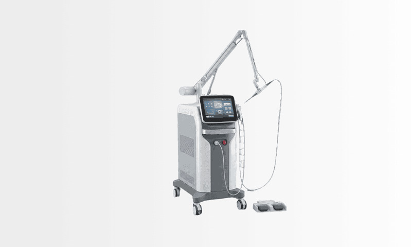
Technology supports every stage of this pathway: high-resolution CT and ultrasound for finding the stone and grading obstruction, endoscopic camera systems for direct visualisation, laser platforms for controlled fragmentation, fluoroscopic or ultrasound guidance for stent placement and drainage, and laboratory analysis of blood, urine and the stone itself to guide prevention afterwards.
The point of all of it is practical: a more accurate diagnosis, the least invasive treatment that will actually work, fewer unnecessary procedures, protected kidney function and a safer recovery. Technology is a means, not the plan itself.
Recovery After Treatment
Recovery depends on which route you took. After spontaneous passage, symptoms usually settle once the stone reaches the bladder or leaves the body. After SWL, expect some flank soreness, blood-tinged urine and the passage of fragments over days to weeks. After ureteroscopy, mild burning, urinary frequency, pink-tinged urine and stent-related discomfort are common for a short period.
Most patients are encouraged to drink fluids as advised, walk soon after the procedure, take prescribed medications as directed by the treating team, and avoid strenuous activity briefly. If a stent was placed, you will be told exactly how long it stays and when it comes out. Follow-up imaging confirms that the obstruction has resolved and no significant fragments remain, feeling better is a good sign, but it is not the same as confirmed clearance.
Clinicians treat certain developments after treatment as red flags requiring prompt reassessment: fever, chills, worsening pain, persistent vomiting, heavy bleeding, inability to urinate or severe stent discomfort. Your discharge instructions will spell out what applies to your specific situation.
Why Acting Early Matters
Not every ureteral stone is an emergency, but delay carries specific risks when obstruction, infection or reduced kidney function is in play. A blocked ureter raises pressure inside the kidney, and prolonged pressure can affect kidney function: a risk that rises sharply in people with a single kidney, existing kidney problems, diabetes or a suppressed immune system.
Pain itself is a reason to act. Repeated severe episodes lead to dehydration, vomiting, sleepless nights and repeated emergency visits. A stone that seems manageable on Monday can wedge into a narrower segment of the ureter by Friday and change the situation entirely.
The most serious scenario is the infected, obstructed kidney. Fever and chills alongside a known ureteral stone represent a genuine medical emergency in clinical terms, because antibiotics alone cannot work properly while infected urine sits trapped under pressure. Early drainage in this situation can be lifesaving and creates safe conditions for removing the stone later.
Early specialist assessment also prevents pointless waiting. Some stones have a realistic chance of passing; others never will because of their size, position or the anatomy around them. Knowing which category yours falls into lets you make an informed decision, especially when work commitments or other practical constraints are part of the calculation.
Benefits of Ureteral Stone Treatment
The specific benefits depend on the stone and the method used, but the overall aims are consistent: relieve symptoms, restore urine flow and reduce future risk.
| Benefit | What It Means for You |
|---|---|
| Pain relief | Removing the stone or helping it pass relieves the pressure and ureteral spasm behind the severe flank and groin pain. |
| Relief of obstruction | Treatment restores urine flow from kidney to bladder, protecting kidney function from pressure damage. |
| Lower infection risk | Treating the blockage appropriately reduces the risk of infection developing behind an obstructed kidney. |
| Minimally invasive options | Many stones are treated through the natural urinary passage or with external shock waves, usually avoiding open surgery. |
| Short recovery for most patients | Endoscopic and external approaches often allow same-day or short-stay treatment and a prompt return to daily activity. |
| Clear diagnosis and prevention planning | Imaging, stone analysis and laboratory testing identify why stones formed and guide steps to reduce recurrence. |
Recovery Timeline After Ureteral Stone Treatment
Recovery is individual, but the following timeline describes what many patients experience after the common treatments.
| Time Period | What Patients Can Expect |
|---|---|
| Day 1 | After ureteroscopy or shock wave treatment: mild discomfort, blood-tinged urine, urinary frequency or burning. You are monitored while anaesthesia or sedation wears off. |
| First week | Gradual return to light activities. If a stent is in place, bladder irritation or flank discomfort during urination may continue until it is removed. |
| First month | Follow-up may include stent removal, imaging to confirm drainage, review of the stone analysis and a prevention discussion. Energy and urinary comfort usually improve steadily. |
| Longer term | Recurrent stone formers begin a prevention plan: hydration targets, dietary changes, metabolic testing and, where indicated, medication prescribed by the treating doctor. |
What Influences a Good Outcome
Outcomes rest on four things: accurate diagnosis, appropriate timing, the right choice of treatment and proper follow-up. The stone itself is the first variable. Smaller stones pass more often than larger ones; stones in the lower ureter behave differently from stones near the kidney; and density matters, harder stones respond less predictably to shock waves and often suit ureteroscopic laser treatment better.
Your anatomy is the second variable. Narrowing, swelling, prior surgery, an enlarged prostate, congenital variations or kinking of the ureter can all affect both stone passage and surgical access. Infection status is the third and most decisive: bacteria behind an obstruction mean drainage and antibiotics come first, and everything else waits.
General health shapes decisions too. Patients with existing kidney disease, a single functioning kidney, diabetes, bleeding disorders, pregnancy, heart disease or immune suppression need additional precautions, and medication use, particularly blood thinners, influences timing and the safest method. None of this rules out treatment; it changes how treatment is sequenced.
The treating team’s judgement is the fourth variable, and it is not a small one. Ureteral stone care is a series of decisions: when to wait, when to intervene, when to drain urgently, and how to avoid ureteral injury or leftover fragments. Experienced urologists adjust the plan during a procedure based on what the endoscope actually shows, not just what the scan predicted.
Follow-up is part of the outcome, not an optional extra. A patient can feel entirely well while a silent partial obstruction persists, which is why imaging or clinical reassessment after treatment matters even when the pain is gone. And for recurrent stone formers, prevention is where the long-term result is won: more fluid is usually the foundation, but a proper plan may also involve reducing sodium, adjusting animal protein, correcting metabolic abnormalities found on testing, treating infections and, where the stone type justifies it, medication managed by the treating doctor.
Finally, your own participation counts. Taking medications as prescribed, attending the stent removal appointment, following activity guidance and reporting the warning symptoms your team described all contribute directly to a safe recovery. Arranging follow-up before you leave hospital, including a confirmed stent removal date, if one was placed, is one of the most practically important steps in the whole pathway.
Ureteral Stone Care at Acibadem
At Acibadem, ureteral stone care is delivered by urology teams experienced across the full range of stone situations, from uncomplicated stones suitable for observation to obstructed, infected systems needing urgent drainage. Complex cases involve collaboration among urologists, radiologists, anaesthesiologists, nephrologists, infectious disease specialists, emergency physicians and intensive care teams where needed. That multidisciplinary structure matters most for patients with infection, kidney disease, recurrent stones, multiple medical conditions or previous urinary tract surgery.
Diagnosis and treatment draw on the technologies described above: advanced cross-sectional imaging, ultrasound, endoscopic visualisation systems, laser lithotripsy platforms, fluoroscopic guidance and laboratory testing for stone analysis and metabolic evaluation. Which of these is used for an individual patient depends on the clinical indication: the tools are selected to support safe, effective care, not to add complexity for its own sake.
Treatment planning is deliberately individual, because two patients with stones of similar size can need entirely different approaches: one stone is dense, one system is infected, one stone sits near the bladder, one patient has a single functioning kidney. The clinical process weighs these details before recommending observation, ureteroscopy, shock wave treatment, drainage or staged care.
Making Sense of Your Options
A ureteral stone can disrupt life without warning, but the treatment decision should still be a considered one. The right approach depends on the stone’s size and position, the degree of blockage, the infection risk, your kidney function and your personal circumstances. Some stones can be safely monitored while they pass. Others should be treated promptly, and a few, the infected, obstructed ones, need drainage before anything else.
The useful question is rarely just “Can the stone be removed?” Almost every stone can be. The better questions are which approach is safest for this particular stone and this particular kidney, whether waiting is genuinely reasonable or merely convenient, and what the stone’s composition says about preventing the next one. Those answers come from imaging, laboratory results and specialist judgement, and they are worth having before the next wave of pain makes the decision for you.
Preparation
- Evaluation usually includes urine tests, blood tests and imaging such as ultrasound or CT to locate the stone and assess kidney function. Patients may be asked to stop certain blood thinners before intervention. Fasting is required if ureteroscopy or another procedure under anesthesia is planned.
Aftercare
- Drink fluids as advised and take prescribed pain relief or antibiotics if needed. A temporary ureteral stent may cause urgency or mild discomfort until removed. Follow-up imaging and metabolic evaluation may be recommended to reduce the risk of future stones.
Frequently Asked Questions
What affects the cost of ureteral stone treatment?
The main factors are stone size and location, severity of obstruction, infection risk, imaging needs, treatment method, anaesthesia, stent placement, hospital stay and follow-up requirements. Travel, interpreter support and accommodation can also affect the overall budget for international patients.
How can I get a personalised quote?
A personalised quote usually requires recent imaging, urine and blood test information, symptom history and details of any previous stone treatment. Acibadem International can review available medical records and arrange a free consultation to help estimate the most appropriate treatment pathway and package inclusions.
Is the least invasive option always the lowest cost?
Not necessarily. Observation or medication may have lower initial costs, but repeat visits, imaging or emergency care can add expense if the stone does not pass. A specialist will balance safety, likelihood of success and the total treatment plan.
Does a ureteral stent change the cost?
Yes, a stent can affect cost because it involves equipment, placement and later removal or follow-up. In some cases, stenting is necessary to relieve obstruction, protect kidney drainage or support healing after ureteroscopy.
What is typically included in an international patient package?
Package contents vary by hospital and clinical need, but may include specialist consultation, imaging review, the procedure, anaesthesia, standard hospital services, routine medications, interpreter support and transfers. It is important to confirm whether follow-up imaging, stent removal and extra tests are included.
Is this information medical or financial advice?
No. This is general educational information only. The safest treatment and the final cost can only be confirmed after assessment by a qualified urology specialist and review of your medical records.
Medically reviewed by the Acıbadem International Medical Board September 1, 2026
See our medical review board →
Update history
- PublishedJune 8, 2026
- Medical review approvedSeptember 1, 2026
- Last content updateSeptember 8, 2026
References2
- Kidney Stones medlineplus.gov
- Kidney stones nhs.uk
Trusted care for international patients
From the Acibadem Blog

Will a Ureteral Stone Pass on Its Own? Size, Position and When Intervention Is Planned
Many ureteral stones do pass without a procedure, and the odds depend mostly on size and position. Stones smaller than 4 mm pass on…

After Ureteral Stone Removal: Stent Discomfort, Fluids and the Metabolic Work-Up That Follows
A ureteral stent after stone removal is a soft, hollow tube that keeps the ureter open while swelling settles; it commonly causes urgency, flank…