Barrett Esophagus Treatment
Barrett esophagus is a precancerous change in the esophageal lining usually linked to chronic reflux. Care includes endoscopic diagnosis, surveillance, reflux control and ablation when dysplasia is found.
Quick answer
Barrett's esophagus is a change in the lining of the lower esophagus, usually caused by long-standing acid reflux. It is not cancer, but it raises the risk of esophageal adenocarcinoma. Care combines reflux control, surveillance endoscopy with biopsies, and, when precancerous cells (dysplasia) are found, endoscopic resection or ablation to remove or destroy the abnormal tissue.
Barrett’s Esophagus: What the Diagnosis Means
Barrett’s esophagus is a change in the lining of the lower esophagus, in which the normal squamous cells are replaced over time by cells more similar to intestinal tissue, usually because of repeated exposure to stomach acid and bile from chronic reflux. It is not cancer. It is classed as a precancerous condition because it raises the risk of esophageal adenocarcinoma, a serious cancer that is more treatable when found early. Managing Barrett’s esophagus means three things: controlling the reflux that caused the injury, monitoring the tissue at planned intervals, and treating abnormal cells when they appear, usually through the endoscope, without external incisions.
Most people first hear the term after an upper endoscopy performed for chronic heartburn, acid regurgitation, difficulty swallowing, chest discomfort, or a persistent throat sensation. Others are diagnosed during an evaluation for long-standing gastroesophageal reflux disease, usually shortened to GERD. The word “precancerous” is unsettling, and it is worth stating the balance plainly: in most people, Barrett’s esophagus does not progress to cancer. The purpose of structured care is to identify the smaller group of patients whose tissue is changing, and to act at a stage when endoscopic treatment is still an option.
That balance, a condition that is usually stable but occasionally dangerous, is why the diagnosis deserves a plan rather than either alarm or neglect. Careful endoscopic assessment, reliable pathology, honest risk stratification and a defined follow-up schedule turn an anxious diagnosis into a manageable one. A patient who knows exactly which category their Barrett tissue falls into, and what happens next, is in a fundamentally different position from one who has only heard the word “precancerous” and been left to search for answers.
A well-organised Barrett’s esophagus programme therefore answers the questions patients actually ask. Is the diagnosis accurate? Do I need treatment now, or monitoring? What does “dysplasia” mean? Can Barrett tissue be removed? How often will I need endoscopy? What happens if cancer is suspected? The sections below work through each of these in turn, following the pathway a patient actually travels: cause, diagnosis, risk category, treatment options, recovery and long-term follow-up.
What causes Barrett’s esophagus?
The main cause of Barrett’s esophagus is chronic gastroesophageal reflux, years of acid and bile washing up from the stomach into the lower esophagus. The esophageal lining is not built to withstand this exposure. Faced with repeated injury, the body replaces the normal lining with a tougher, intestinal-type lining. This process is called intestinal metaplasia, and it is the defining feature of Barrett’s esophagus under the microscope. The change is a defensive adaptation, but the new tissue is genetically less stable than the original lining, which is where the cancer risk comes from. Not everyone with reflux develops Barrett changes, and some people develop them after years of silent reflux they never noticed. Because chronic reflux is the root cause, understanding and treating reflux disease is a foundation of Barrett care rather than a side issue. Additional factors that make the condition more likely include age over 50, male sex, central obesity, a smoking history, a hiatal hernia, and a family history of Barrett’s esophagus or esophageal adenocarcinoma.
Barrett esophagus, Barretts esophagus, Barrett’s disease, one condition, many names
Barrett esophagus, Barretts esophagus and Barrett’s esophagus are the same condition; the spelling simply varies between medical reports, textbooks and search results. Some patients are told they have Barrett’s disease of the esophagus, or see the abbreviation BE on an endoscopy report. Spelling of the organ itself also varies, esophagus or oesophagus, Barrett’s change means the same thing in both traditions, and British sources write it as Barrett’s oesophagus. Whichever form appears on your report, the clinical questions are identical: is intestinal metaplasia confirmed, how long is the affected segment, and is dysplasia present?
What Barrett’s Esophagus Treatment Involves
Barrett’s esophagus treatment is not a single procedure applied to every patient. It is a coordinated care plan that may include accurate diagnosis, scheduled surveillance endoscopy, medical reflux control, lifestyle guidance, endoscopic therapy for dysplasia and, in selected cases, anti-reflux surgery or cancer-directed treatment. The right approach depends almost entirely on whether abnormal cells called dysplasia are present in the biopsies and, if so, how advanced those changes are.
Dysplasia is the microscopic finding that separates monitoring from treatment. It describes cells that have begun to change in ways associated with progression towards cancer, graded by how disordered they appear. The pathologist’s report will usually place your Barrett tissue in one of five categories:
- Non-dysplastic Barrett esophagus: Barrett tissue is present, but no precancerous cellular changes are seen under the microscope. Management usually means reflux control and surveillance at planned intervals.
- Indefinite for dysplasia: the biopsy shows changes that may be caused by inflammation rather than true dysplasia, and the pathologist cannot make a clear call. Reflux therapy is typically intensified and the endoscopy repeated after the lining has had time to heal.
- Low-grade dysplasia: early precancerous changes are present. Depending on confirmation by expert pathology review and patient-specific factors, either endoscopic treatment or close surveillance may be appropriate.
- High-grade dysplasia: more advanced precancerous changes with a higher risk of progression to cancer. Endoscopic treatment is commonly recommended when the patient is a suitable candidate.
- Early esophageal adenocarcinoma: cancer is present but may be limited to the superficial layers of the lining, where endoscopic therapy may still be possible in carefully selected cases.
Modern care aims to find and manage dysplasia before invasive cancer develops. Endoscopic mucosal resection can remove visible abnormal areas; ablation techniques can destroy remaining flat Barrett tissue so that healthier lining regrows. Both are performed through the mouth, preserving the esophagus in many patients who a generation ago would have faced major surgery. When cancer is suspected or confirmed, planning becomes more complex and typically involves multidisciplinary specialist boards deciding between endoscopic resection, esophageal surgery, chemotherapy, radiotherapy or combined approaches.
Can Barrett’s esophagus be reversed or cured?
Barrett tissue can often be removed or destroyed with endoscopic treatment, but doctors avoid the word “cure”, because the underlying tendency does not disappear. After successful resection and ablation, the visible Barrett segment is frequently replaced by regrown squamous lining, clinicians call this eradication rather than reversal. Recurrence is possible, which is why follow-up endoscopy continues even after treatment appears complete. For non-dysplastic Barrett’s esophagus, ablation is generally not recommended: the risk of the procedure is not justified when the tissue shows no precancerous change, and acid suppression alone does not make established Barrett tissue revert reliably. The honest summary is this: the abnormal lining can often be treated effectively, the cancer risk can be managed, and the condition itself is best understood as a long-term risk state that stays under observation.
Barrett’s Esophagus Symptoms and Who Needs Evaluation
Barrett’s esophagus symptoms are, in truth, mostly reflux symptoms: the Barrett change itself usually causes no sensation of its own. What patients feel is the chronic reflux behind it, frequent heartburn, acid regurgitation, a sour taste in the mouth, night-time reflux, chronic cough, hoarseness, repeated throat clearing, or the feeling of food coming back up after meals. This is why the condition is so often discovered incidentally, during an endoscopy arranged for reflux complaints rather than for Barrett’s specifically.
Some patients have little or no reflux discomfort yet still carry Barrett changes, particularly when reflux has been present silently for years. This silent group is one reason evaluation is considered for people with long-standing GERD even when symptoms are currently controlled, especially when other risk factors are present: age over 50, male sex, central obesity, a smoking history, a hiatal hernia, or a family history of Barrett’s esophagus or esophageal adenocarcinoma. Barrett’s esophagus is more common in men, but women develop it too and are evaluated on the same clinical grounds. Reflux-related cough and hoarseness can also overlap with airway conditions, so patients with a long cough history are sometimes investigated in parallel for pulmonary disease before reflux is confirmed as the driver.
How is Barrett’s esophagus diagnosed?
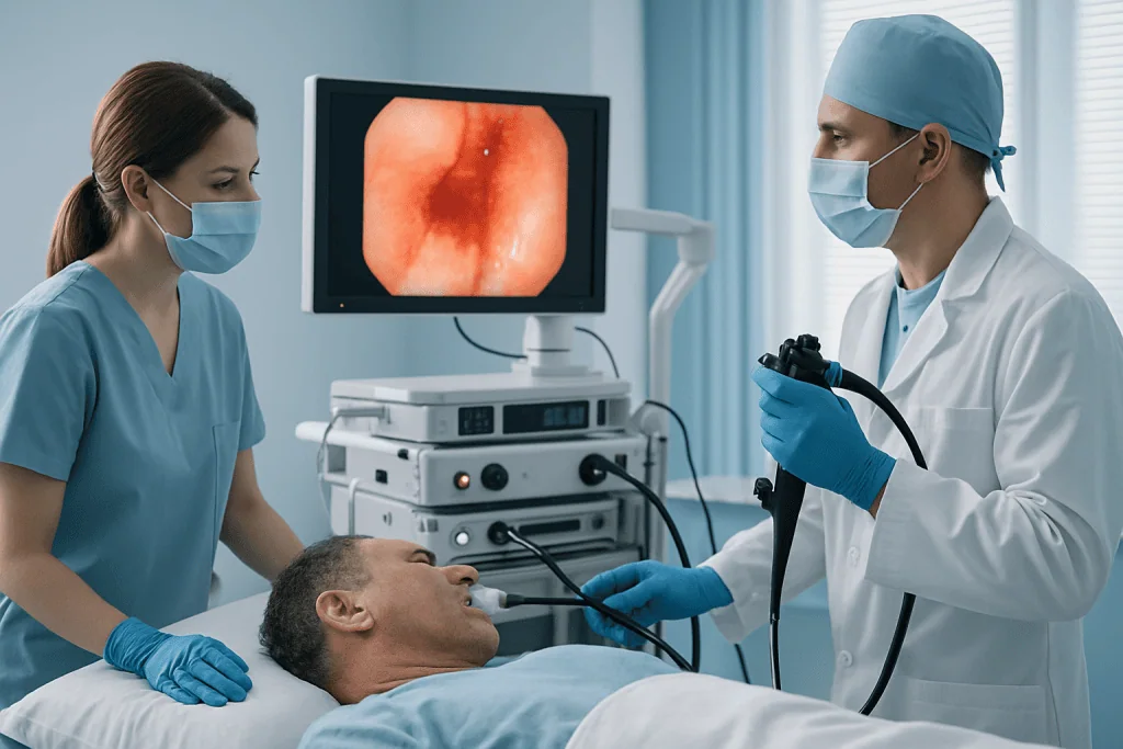
Barrett’s esophagus is diagnosed with upper gastrointestinal endoscopy and biopsy: there is no blood test or scan that can confirm it. During the procedure, a thin flexible instrument is passed through the mouth while you are sedated. The physician examines the esophagus, the junction between the esophagus and stomach, the stomach itself and the upper small intestine. If Barrett tissue is suspected, its length and appearance are documented, and biopsies are taken in a systematic pattern along the segment as well as from any visibly irregular areas. The tissue samples then go to pathology, where the diagnosis rests on two findings: confirmation of intestinal metaplasia, and the presence or absence of dysplasia. Both halves matter. The endoscopist’s eye determines where to sample and whether any area needs removal rather than biopsy; the pathologist’s assessment determines the risk category that drives every subsequent decision.
Because those microscopic distinctions are subtle, second opinions play a genuine role in Barrett care. When dysplasia has been reported, particularly low-grade dysplasia, which is the hardest category to grade consistently, review by experienced gastrointestinal pathologists helps confirm the finding before treatment decisions are made. This protects patients in both directions: it avoids ablating tissue that was actually inflamed rather than dysplastic, and it avoids delaying treatment when genuine dysplasia was under-called.
Active treatment rather than surveillance becomes the question when biopsies show confirmed dysplasia, when nodules or visible lesions appear within the Barrett segment, when early cancer is suspected, or when reflux remains uncontrolled despite medication. The decision is always individualised, weighing biopsy results, endoscopic findings, overall health, anatomy, patient preference and the availability of advanced endoscopic therapy.
Conditions the Barrett Care Pathway Addresses
Barrett’s esophagus care manages a spectrum of reflux-related and precancerous conditions, not just the Barrett segment itself. The associated problems influence both risk and recovery, and a plan that ignores them tends to fail.
Chronic GERD is the most common companion diagnosis. Ongoing acid and bile exposure keeps the lining inflamed, may contribute to progression, and makes the esophagus harder to assess accurately at endoscopy. Medical therapy, usually acid-suppressing medication prescribed and adjusted by the treating doctor, is the standard foundation, supported by lifestyle changes tailored to the individual patient’s eating pattern, weight, sleep habits and daily routine.
Hiatal hernia is another frequent association. Part of the stomach moves upward through the diaphragm, disrupting the natural barrier between stomach and esophagus and worsening reflux mechanics. Patients with severe reflux, a large hiatal hernia, or persistent symptoms despite medication may be evaluated for anti-reflux surgery. It is important to be precise here: surgery treats the reflux, not the Barrett tissue, and it does not replace surveillance.
Esophagitisinflammation or ulceration of the esophageal lining, complicates diagnosis because inflamed tissue can mimic dysplasia under the microscope. This is exactly why the “indefinite for dysplasia” category exists. The usual response is to intensify reflux therapy, allow the lining to heal, and repeat the endoscopy so the pathologist can assess calm tissue rather than inflamed tissue.
Dysplasia and early cancer are where the therapeutic side of the pathway engages fully. Endoscopic mucosal resection removes raised or suspicious areas so pathology can examine them completely; ablation then treats the remaining flat Barrett tissue. For many patients with dysplasia or very early cancer confined to the superficial layers, this sequence treats the abnormality while preserving the esophagus. More advanced cancer requires a different plan and may involve esophageal surgery, systemic therapy, radiotherapy or combined care decided in a multidisciplinary setting.
How Barrett’s Esophagus Treatment Is Performed
In practice, care moves through a defined sequence. The details vary by patient, but the typical pathway looks like this:
- Review of history, prior endoscopy reports, biopsy results, medications and symptoms.
- High-quality upper endoscopy with systematic biopsies and documentation of the Barrett segment.
- Pathology assessment, and, where dysplasia is reported, expert confirmation of the grade.
- Assignment of a risk category: non-dysplastic, indefinite, low-grade, high-grade, or early cancer.
- Treatment matched to that category: surveillance and reflux control, endoscopic resection, ablation, or multidisciplinary cancer planning.
- Follow-up endoscopy at intervals set by the diagnosis and the response to treatment.
Preparation and diagnostic planning
The first step is a careful review of your history, previous endoscopy reports, biopsy results, medication use and symptoms. Specialists assessing a case that began elsewhere will usually want to see prior endoscopy images, pathology slides or tissue blocks when available, laboratory results, imaging studies and a current medication list, with these, the team can decide whether repeat endoscopy, pathology review, imaging or additional testing is genuinely needed, and avoid repeating investigations that were done well the first time.
Before an endoscopy, you will be asked not to eat or drink for a set period. Blood thinners, diabetes medications and certain other drugs sometimes need temporary adjustment around the procedure; those decisions belong to your treating doctor, made before the day itself, never to the patient alone. The clinical team also reviews allergies, anaesthesia history, heart and lung conditions and any prior reactions to sedation.
The diagnostic examination itself has become considerably more detailed than a quick look. The endoscopist documents the length and appearance of the Barrett segment and inspects it for subtle abnormalities, nodules, ulcers, depressions, irregular mucosal patterns, colour changes. Enhanced imaging modes, magnification and targeted inspection techniques help identify areas that need focused biopsy or removal rather than routine sampling. The point of this thoroughness is simple: early dysplasia and early cancer can be nearly invisible on a casual examination, and the entire strategy depends on not missing them.
The endoscopy and biopsy process
During upper endoscopy, sedation keeps the procedure comfortable for most patients. The endoscope passes through the mouth into the esophagus, and the physician examines the lining, the gastroesophageal junction and the stomach. Where Barrett’s esophagus is suspected or already known, biopsies follow a structured protocol along the segment, plus targeted samples from any visible lesion.
The examination itself is usually brief; the full visit takes longer because of preparation, sedation, monitoring and recovery. Most patients go home or back to their accommodation the same day with an escort. A mild sore throat, bloating or fatigue from sedation can occur and usually settles quickly.
The biopsy results set the direction. No dysplasia means the focus stays on reflux control and scheduled surveillance. Reported dysplasia usually triggers confirmation by experienced pathology review, because the grade determines whether ablation, resection or close follow-up is the right next step.
Endoscopic resection for visible lesions
If a raised, nodular or otherwise suspicious area is seen, endoscopic resection may be performed. Unlike a small biopsy, resection removes the whole abnormal area as a larger specimen, which lets the pathologist see how deep any abnormal cells extend and, if cancer is present, whether it appears limited to the superficial layers. The procedure is therefore both diagnostic and therapeutic at once.
Endoscopic mucosal resection or related techniques are chosen according to the lesion’s appearance and location. The physician lifts or isolates the abnormal area and removes it through the endoscope, with no external incision. If pathology confirms the lesion was fully removed and limited to an early stage, endoscopic treatment of the remaining Barrett tissue usually follows. If deeper invasion or high-risk features are found, the case moves to multidisciplinary review to consider additional treatment: this staged, evidence-first approach is what prevents both undertreatment and unnecessary surgery.
Ablation for dysplastic Barrett tissue
Ablation destroys the abnormal Barrett lining in a controlled way so that healthier squamous lining can grow back during healing. It is commonly considered for confirmed low-grade dysplasia, for high-grade dysplasia, and after resection of certain early lesions. The technique chosen depends on the length of the Barrett segment, anatomy, prior treatments and physician judgement; thermal ablation applies controlled energy at a carefully selected depth, and focal techniques can treat residual islands of Barrett tissue in later sessions.
Everything happens through the endoscope. Several sessions are often needed, because the esophagus must heal between treatments and residual areas may require additional therapy, patients planning care, especially from abroad, should build this staged rhythm into their expectations rather than assuming a single visit.
After ablation, acid-suppressing medication is usually prescribed to protect the healing lining. Temporary chest discomfort, swallowing discomfort, nausea or sensitivity with eating can occur. Diet typically advances gradually from liquids or soft foods back to normal, following the physician’s instructions. Follow-up endoscopy then assesses healing, confirms whether visible Barrett tissue has been eradicated, and continues surveillance, because recurrence can occur even after an apparently complete result.
Reflux control and long-term management
Reflux control is the permanent backbone of Barrett’s esophagus care. Proton pump inhibitors or other acid-suppressing medications are commonly prescribed, with timing, dose and duration decided by the treating doctor according to symptoms, endoscopic findings and whether resection or ablation has been performed. Individualised lifestyle guidance sits alongside medication: avoiding late meals, elevating the head of the bed for night-time symptoms, reducing foods that clearly trigger your own reflux, stopping tobacco, and moving towards a healthier weight where relevant.
For selected patients, severe reflux, regurgitation despite medication, medication intolerance, or a significant hiatal hernia, further evaluation may include esophageal function testing, pH monitoring or imaging, and anti-reflux surgery may be considered where the expected benefits outweigh the risks. That decision is made carefully and with one caveat repeated deliberately: surgery corrects reflux mechanics, but the Barrett tissue still needs its surveillance schedule afterwards.
What to eat if you have Barrett’s esophagus?
No diet reverses Barrett’s esophagus, so honest dietary advice aims at reducing reflux rather than treating the tissue. Practical measures many patients find useful include smaller meals, finishing dinner well before lying down, and limiting personal trigger foods, commonly fatty or fried meals, alcohol, coffee, chocolate, mint and heavily spiced dishes, though triggers vary widely between individuals and there is no universal forbidden list. Weight reduction, where appropriate, tends to help reflux more than any single food rule. After resection or ablation, the situation is different and temporary: your care team will specify a soft or liquid diet for a defined healing period before you return to normal eating. Dietary guidance works best when it is tailored to your own symptom pattern rather than copied from a generic list.
Why Acting Early Matters
Barrett’s esophagus develops slowly, and many patients live for years without progression. The value of early action lies in identifying the smaller group at higher risk before invasive cancer develops. Esophageal adenocarcinoma is difficult to treat when discovered late; early dysplasia, by contrast, can often be managed through the endoscope. That asymmetry is the entire logic of surveillance.
Delay carries specific costs. Persistent reflux inflammation worsens symptoms, feeds esophagitis, and makes pathology harder to interpret. In a patient with unconfirmed or untreated dysplasia, long gaps between evaluations raise the chance that more advanced changes are found at the next look. Doctors also recognise a set of alarm features that change the pace and priority of investigation: progressive difficulty swallowing, unexplained weight loss, vomiting blood, black stools, persistent chest pain, and anaemia.
Acting early does not always mean having an immediate procedure. Sometimes the most appropriate action is confirming the diagnosis, optimising reflux therapy, and repeating the endoscopy at the right moment. What matters is eliminating uncertainty: knowing your risk category, what should happen next, and when follow-up is due.
Benefits of Barrett’s Esophagus Treatment and Surveillance
The benefits of structured Barrett care come from four things working together: accurate diagnosis, risk-based monitoring, reflux control, and timely treatment when precancerous changes appear.
| Benefit | What It Means for You |
|---|---|
| Clarifies your true risk | Careful endoscopy and pathology review establish whether Barrett tissue is non-dysplastic, dysplastic, or suspicious for early cancer: the fact that determines everything else. |
| Finds concerning changes earlier | Surveillance is designed to detect dysplasia or early cancer at a stage when less invasive treatment may still be possible. |
| Treats dysplasia without external incisions | Endoscopic resection and ablation treat abnormal lining through the mouth, avoiding open surgery in appropriate cases. |
| Improves reflux control | Medication, lifestyle guidance and selected surgical evaluation reduce ongoing injury from acid and bile reflux. |
| Supports long-term monitoring | A defined follow-up plan tells you when repeat endoscopy is due and which symptoms should prompt earlier review. |
Recovery Timeline After Endoscopy, Resection or Ablation
Recovery depends on whether you had diagnostic endoscopy only, tissue resection, ablation, or an additional reflux procedure. Most patients return to light activity quickly after endoscopic care; the table below sets out the usual pattern.
| Time Period | What Patients Can Expect |
|---|---|
| Day 1 | After diagnostic endoscopy: mild throat irritation, bloating, or drowsiness from sedation. After ablation or resection: chest discomfort and swallowing sensitivity are more noticeable, and the diet instructions matter. |
| First week | Routine daily activities usually resume, avoiding strenuous exercise if advised. Soft foods or a gradual diet progression may be recommended after therapeutic procedures. |
| First month | The esophagus continues to heal. Acid-suppressing medication is usually continued as prescribed. The care team reviews pathology and schedules the next treatment or surveillance step. |
| Longer term | Follow-up endoscopy at intervals set by the diagnosis and treatment response. Some patients need further ablation sessions or ongoing surveillance even after visible Barrett tissue has cleared. |
Factors That Influence Outcomes
A good result in Barrett’s esophagus care depends on identifiable medical and technical factors, and it is worth knowing them because several are within reach of the patient and the care team.
Diagnostic accuracy comes first. The presence and grade of dysplasia drive every treatment decision, so experienced endoscopic assessment and reliable pathology interpretation are central. Where dysplasia is suspected, confirmation prevents unnecessary ablation in some patients and prevents dangerous delay in others.
The Barrett segment itself matters. Shorter segments are generally easier to treat and monitor; longer segments demand more extensive surveillance and often multiple ablation sessions. Visible lesions, nodularity, ulceration or scarring change the technical plan. If early cancer is found, the depth of invasion and the pathology features determine whether endoscopic therapy is sufficient or additional treatment should be considered.
Reflux control shapes healing. Ongoing reflux symptoms, a large hiatal hernia, obesity, tobacco use, or inconsistent use of prescribed medication mean more inflammation, a greater chance of persistent or recurrent Barrett tissue, and less reliable surveillance. Optimising medical therapy and lifestyle factors supports healing after ablation and makes each follow-up examination more informative.
Overall health frames the plan. Heart disease, lung disease, bleeding risk, anticoagulant use, prior esophageal surgery and anaesthesia risk all affect procedure planning. In older patients or those with significant medical conditions, the balance between surveillance, treatment and quality of life deserves an explicit, unhurried discussion rather than a default pathway.
Technical expertise is not interchangeable. Barrett endoscopy requires detailed inspection, systematic biopsy, recognition of subtle lesions and familiarity with the full range of therapeutic options. Endoscopic resection and ablation are highly operator-dependent. Care is stronger where gastroenterologists, pathologists, surgeons, radiologists and oncology specialists can discuss complex findings together and adapt the plan as new information arrives.
Finally, follow-up is part of the treatment. Even after successful eradication of dysplasia or visible Barrett tissue, surveillance usually continues, because Barrett’s esophagus is a chronic risk condition rather than a one-time event. Patients who understand their follow-up schedule, medication plan and warning symptoms stay engaged in their own care, and engaged patients are the ones whose surveillance actually happens on time.
Living With Barrett’s Esophagus
For most people, life with Barrett’s esophagus looks remarkably ordinary: reflux management day to day, an endoscopy at planned intervals, and attention to the general health measures, weight, tobacco, meal habits, that reduce reflux burden. The condition does not restrict travel, work or exercise in itself. Two questions dominate patients’ searches, and both deserve straight answers.
Can Barrett’s esophagus kill you?
Barrett’s esophagus itself is not fatal and is not cancer; the concern is that, in a minority of patients, it can progress over time to esophageal adenocarcinoma, which is a life-threatening disease when found late. That is precisely the risk surveillance exists to manage: regular endoscopy is designed to catch dysplasia or early cancer at a stage when endoscopic treatment remains possible. The realistic framing is that Barrett’s esophagus is a marker of elevated risk to be monitored, not a terminal diagnosis, and that skipping surveillance is the decision that genuinely increases danger.
How long can a person live with Barrett’s esophagus?
Many people live with Barrett’s esophagus for decades, and for most patients life expectancy is shaped far more by their general health, heart, lungs, weight, smoking status, than by the Barrett segment itself. The condition usually remains stable, and structured surveillance is what keeps it that way in practical terms: it does not change the tissue, but it ensures that if the tissue changes, the response comes early. Patients whose dysplasia is found and treated endoscopically typically keep their esophagus and return to normal life with a follow-up schedule attached.
Barrett’s Esophagus Care at Acibadem
Barrett’s esophagus sits at the intersection of gastroenterology, pathology, reflux management, advanced endoscopy and, when necessary, cancer care. At Acibadem, these strands are organised into one pathway: patients are evaluated by physicians experienced in digestive diseases and therapeutic endoscopy, with access to multidisciplinary boards for complex cases. When biopsy findings suggest high-grade dysplasia or early cancer, discussion among the relevant specialists determines whether endoscopic treatment, surgery, oncology care or additional staging is the appropriate next step, rather than leaving that judgement to a single reading of a single report. The same gastroenterology teams manage the broader range of digestive conditions, from Crohn disease to celiac disease, which matters when reflux symptoms turn out to have company.
Diagnostic and endoscopic technology is used the way this condition demands: high-definition imaging, enhanced mucosal assessment, structured biopsy protocols and close pathology correlation, so that the plan rests on both what the physician sees during the procedure and what the tissue shows under the microscope. Treatment planning is personalised because the right option genuinely differs between patients: a person with non-dysplastic short-segment Barrett needs reflux control and surveillance; a patient with confirmed low-grade dysplasia weighs ablation against close monitoring; a visible lesion needs resection before any ablation decision; suspected cancer needs staging and tumour-board review. These distinctions take time and careful explanation, and they are the substance of the consultation rather than an afterthought.
For patients travelling from abroad, Acibadem’s international patient services coordinate appointments, medical record transfer, interpretation and hospital admission, and support communication with the clinical teams in the patient’s own language. This coordination is particularly relevant to Barrett care because treatment may involve staged procedures: knowing in advance whether a visit is for diagnosis, therapeutic endoscopy, pathology review or a second opinion changes how a trip is planned. Where ongoing surveillance will continue in the patient’s home country, the care plan is documented clearly so that local physicians can carry it forward without gaps.
Moving Forward With Clarity
Barrett’s esophagus deserves careful attention, but it does not mean cancer is inevitable. Most patients are managed safely with reflux control and scheduled surveillance; where dysplasia appears, endoscopic therapies can often deal with it while the problem is still early and the esophagus can be preserved. The single most useful step for any patient is understanding their exact diagnosis, the risk category, the length of the segment, the pathology grade, and following a plan matched to it. When endoscopic findings, pathology, reflux history and overall health are brought together in one place, the result is a plan that is medically sound, honestly explained, and practical to live with.
Preparation
- Your doctor may request previous endoscopy and biopsy reports, medication lists and reflux history. You may need to stop blood thinners before endoscopic biopsy or ablation. Fasting is usually required for several hours before the procedure.
Aftercare
- Mild throat discomfort, bloating or chest burning can occur after endoscopy or ablation. Follow acid-suppressing medication and diet instructions carefully. Regular surveillance endoscopy is important to monitor dysplasia or cancer risk.
Frequently Asked Questions
What affects the cost of Barrett esophagus care?
The main factors are the purpose of care, endoscopy and biopsy needs, pathology review, dysplasia status, whether ablation or resection is required, anesthesia, hospital setting, specialist fees and follow-up planning.
How can I get a personalised quote?
You can request a free consultation and share your endoscopy reports, biopsy results, medication list and any imaging or reflux test results. A specialist team can then review your case and prepare a personalised plan and quote.
Is ablation always needed for Barrett esophagus?
No. Many patients require surveillance and reflux control only. Ablation is usually considered when dysplasia is confirmed or when a specialist determines that endoscopic eradication therapy is appropriate.
Will pathology results change the treatment cost?
Yes. Biopsy findings are central to Barrett esophagus management. If dysplasia or a visible lesion is found, additional procedures such as mucosal resection, ablation or closer follow-up may be recommended.
Are travel and language services included?
International patient services may help coordinate appointments, translation, hospital navigation and travel-related planning. What is included should be confirmed in the individual quote.
Is this information medical or financial advice?
No. This is general educational information. Diagnosis, treatment suitability and cost planning should be discussed with a qualified specialist through a personalised consultation.
Medically reviewed by the Acıbadem International Medical Board August 31, 2026
See our medical review board →
Update history
- PublishedJune 8, 2026
- Medical review approvedAugust 31, 2026
- Last content updateSeptember 8, 2026
References1
- Barrett's Esophagus my.clevelandclinic.org
Trusted care for international patients
From the Acibadem Blog

After Barrett’s Ablation: Throat Discomfort, Diet Steps and When Eating Feels Normal Again
Recovery after Barrett's ablation usually means a sore throat and chest discomfort for a few days, a diet that moves from cool liquids to…