Endometrial Cancer Treatment
Endometrial cancer is a cancer of the uterine lining, most often treated with surgery and, when needed, radiation, chemotherapy, or targeted therapies tailored to stage and health.
Quick answer
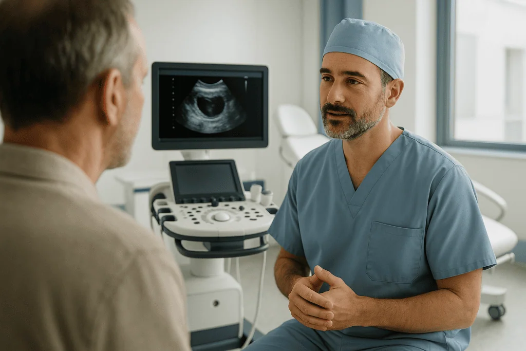
Endometrial cancer begins in the endometrium, the inner lining of the uterus, and most often affects women after menopause. It usually announces itself early through abnormal vaginal bleeding. Diagnosis rests on a tissue biopsy, and treatment typically centres on surgery to remove the uterus, with radiation, chemotherapy, immunotherapy, targeted therapy or hormone therapy added when the tumour's stage, grade or molecular profile calls for it.
What Is Endometrial Cancer?
Endometrial cancer is cancer that begins in the endometrium, the inner lining of the uterus. It develops when cells of that lining acquire genetic changes that let them grow abnormally, form a tumour, and, if left untreated, invade the muscle of the uterus and spread beyond it. Most people diagnosed with endometrial cancer are past menopause, although the disease can occur earlier, and it is often found while it is still confined to the uterus, at a point where treatment is most straightforward.
That early detection is not luck. Endometrial cancer tends to announce itself through abnormal vaginal bleeding before it has spread, which gives you and your doctors a genuine head start over many other cancers. It is one of the most common cancers of the female reproductive system in many countries, and when it is diagnosed early, many patients can be treated with surgery alone. Others need additional therapy, radiation, chemotherapy, immunotherapy, targeted therapy or hormone therapy, depending on the cancer’s type, stage, molecular profile and the patient’s overall health. The pattern matters: this is a disease that usually signals its presence early, and a disease whose treatment can be tailored precisely once the right diagnostic steps have been completed.
It helps to understand what the endometrium actually does. During the reproductive years, the lining of the uterus builds up under the influence of oestrogen in the first half of each menstrual cycle, matures under the influence of progesterone in the second half, and is shed as a period if pregnancy does not occur. This constant cycle of growth and shedding is normal and tightly controlled. Endometrial cancer arises when that control fails, when cells of the lining keep growing without the usual signals to stop, mature or shed. After menopause, the lining should become thin and quiet, which is exactly why any bleeding at that stage of life is treated as a signal worth investigating rather than a nuisance to be tolerated.
A diagnosis brings many questions at once. You may be trying to understand how serious the cancer is, whether the uterus must be removed, whether chemotherapy or radiation will be needed, and how treatment may affect your body, hormones, sexuality, fertility and everyday life. You may also be wondering what the unfamiliar words on a pathology report mean, endometrioid, serous, grade, myometrial invasion, mismatch repair, and how long the road from diagnosis through treatment to recovery usually takes. This page walks through each of those questions in order, from symptoms and causes through diagnosis, staging, treatment options and recovery, and it is deliberately honest about what medicine can and cannot say before your own results are in.
Treatment matters because endometrial cancer can progress from the inner lining into the uterine muscle, the cervix, the lymph nodes, the abdomen or distant organs. Modern care is not simply about removing the uterus. It is about defining the exact type of cancer, determining how far it has spread, estimating the risk of recurrence, and building a plan that treats the disease effectively while avoiding unnecessary treatment wherever possible, because overtreatment carries its own costs in side effects and quality of life. At Acibadem, patients with suspected or confirmed endometrial cancer are evaluated through a coordinated pathway within the wider oncology and cancer treatment programme, drawing on gynaecological oncology, radiology, pathology, medical oncology, radiation oncology, nuclear medicine, anaesthesiology, fertility specialists where appropriate, and supportive care teams.
Is endometrial cancer the same as uterine cancer?
Almost, but not exactly, endometrial cancer and uterine cancer are often used interchangeably because most cancers of the uterus start in the endometrium. Strictly speaking, uterine cancer is the broader term: it covers any cancer arising anywhere in the uterus, including rarer cancers of the uterine muscle and supporting connective tissue. Endometrial cancer refers specifically to cancer of the lining. In everyday conversation, and in much of what you will read online, the two terms point at the same disease; in a pathology report, the distinction becomes real, because a cancer of the muscle wall is a different disease with a different treatment pathway. Both terms are also distinct from cervical cancer, which begins in the cervix, the lower neck of the uterus, behaves differently, has different causes, and is diagnosed and treated along a separate pathway with its own screening tests. When you read your own report, the word that matters most is not uterine versus endometrial; it is the precise subtype the pathologist has identified, because that word drives everything that follows.
Cancerous uterine tumors: the two main groups
Cancerous uterine tumors fall into two broad groups: carcinomas, which begin in the endometrial lining and account for the large majority of cases, and sarcomas, which begin in the muscle wall or connective tissue of the uterus. This page deals with carcinomas, endometrial cancer in the strict sense. Uterine sarcomas are managed differently, usually within a dedicated sarcoma pathway, because their biology, staging system and treatment choices are not the same; the surgical decisions differ, the role of radiation differs, and the drugs used differ. Occasionally a single tumour contains both carcinoma and sarcoma elements; this is called carcinosarcoma, and despite the name it is treated as an aggressive form of endometrial cancer rather than as a sarcoma, because its behaviour follows the carcinoma component. If your report mentions carcinosarcoma, expect the team to recommend more thorough staging and, in many cases, treatment beyond surgery alone.
How fast does endometrial cancer spread?
There is no single answer, because speed depends on the grade and subtype of the tumour rather than on the diagnosis alone. Low-grade endometrioid cancers, the most common form, often grow slowly and can remain confined to the uterus for a long time, sometimes developing from precancerous changes over a period of years. High-grade cancers, and subtypes such as serous carcinoma, clear cell carcinoma and carcinosarcoma, can behave far more aggressively, invading the uterine muscle earlier and spreading sooner. It also helps to know the routes the disease can take. Endometrial cancer can grow directly through the uterine wall towards the outer surface of the uterus and the cervix; it can travel through lymphatic channels to the pelvic and para-aortic lymph nodes; it can seed across the lining of the abdomen, particularly with serous subtypes; and, less commonly, it can travel through the bloodstream to distant organs such as the lungs or liver. You cannot judge the speed or route of a cancer from symptoms alone, and neither can your doctor by examination. That is precisely why grading, imaging, surgical staging and molecular profiling exist: they replace guesswork with measurement, and they are the reason two patients with the same diagnosis can honestly be given very different advice.
Endometrial Cancer Symptoms
The most common endometrial cancer symptoms involve abnormal vaginal bleeding. In women after menopause, that means any bleeding at all, even light spotting, even a single episode, even bleeding that stops on its own. In women who have not reached menopause, warning signs include unusually heavy periods, periods that last longer than they used to, bleeding between periods, or bleeding that departs from a long-established pattern. If you have been searching for uterus lining cancer symptoms, this is the same list: the endometrium is the lining of the uterus, so the two phrases describe one disease seen from two directions.
What is the red flag for endometrial cancer?
Postmenopausal bleeding is the single clearest red flag. It does not matter how light it is, how brief it is, or how long ago your periods stopped, bleeding after menopause always deserves prompt medical evaluation. Most postmenopausal bleeding turns out to have a benign cause, such as thinning of the vaginal or uterine tissue, a polyp, or a hormonal effect from a medicine. But because endometrial cancer so reliably produces this symptom at an early, treatable stage, doctors treat it as a signal that must be investigated rather than watched. The investigation itself is usually simple, an examination, an ultrasound and, where indicated, a small tissue sample, and in the majority of cases it ends with reassurance. The point of taking the symptom seriously is not to assume the worst; it is to close the question quickly, one way or the other.
What are usually the first signs of endometrial cancer?
For most patients, the first sign is a change in bleeding: postmenopausal spotting, heavier or longer periods, or bleeding between cycles. Some notice a watery, pink or blood-tinged vaginal discharge before any frank bleeding appears, and this discharge deserves the same attention as bleeding itself. Patients who later describe their first signs often mention exactly these things: a small amount of spotting they almost dismissed, a discharge that seemed unusual, a period that would not settle. Other symptoms tend to arrive later, particularly as disease becomes more advanced. These can include pelvic pain or a feeling of pressure, pain during intercourse, unexplained weight loss, abdominal swelling or bloating, difficulty or discomfort passing urine, and changes in bowel habits. The later symptoms are less specific, which is another reason the bleeding pattern carries so much diagnostic weight: it is the earliest and most reliable signal the disease gives.
Uterine cancer symptoms overlap heavily with those of benign conditions such as fibroids, polyps, infection and hormonal disturbance, which is why symptoms alone never make the diagnosis, tissue does. The honest message is twofold. First, most abnormal bleeding is not cancer, and most women investigated for it will hear exactly that. Second, the only way to know is to have it evaluated, and evaluation is straightforward: an examination, usually an ultrasound, and where indicated a small tissue sample taken from the lining. Persistent or unusual symptoms deserve assessment even when each individual symptom seems minor, because it is the pattern and persistence, not the drama of any single episode, that carries meaning.
Can endometrial cancer be found before symptoms appear?
There is no routine screening test for endometrial cancer in the general population, in the way that cervical screening exists for cervical cancer or mammography for breast cancer. A cervical smear test is not designed to detect endometrial cancer and cannot be relied on to rule it out, although occasionally abnormal endometrial cells are noticed on one incidentally. Some cancers are found by chance, during investigation of another problem, on imaging done for a different reason, or in the uterus removed for what was thought to be a benign condition. For women at substantially increased inherited risk, such as those with Lynch syndrome, doctors may discuss individualised surveillance or risk-reducing options; that conversation belongs to a genetics and gynaecology team who know the family history in detail. For everyone else, awareness of the bleeding signal is, in practice, the screening tool, which is why it is worth taking seriously.
Causes and Risk Factors
What causes endometrial cancer?
Endometrial cancer is caused by genetic changes in the cells of the uterine lining that allow them to grow without normal control. In most individual cases, no single triggering event can be identified, but the strongest known driver for the common endometrioid subtype is prolonged exposure of the endometrium to oestrogen that is not balanced by progesterone. Oestrogen stimulates the lining to grow; progesterone normally keeps that growth in check and pushes the cells towards maturation. When the balance tilts towards oestrogen over years, the lining can thicken abnormally, develop precancerous changes known as atypical hyperplasia, and eventually progress to cancer. The rarer, more aggressive subtypes, serous and clear cell carcinoma among them, follow a different biology that is less dependent on hormones, which is one reason they can arise in women without the classic risk profile and why they are treated differently once found.
Several factors are known to increase risk, and it is worth understanding why each one appears on the list rather than simply memorising it:
- Obesity: fat tissue converts other hormones into oestrogen, raising the lifetime exposure of the endometrium. This effect continues after menopause, when fat tissue becomes the body’s main source of oestrogen, which is part of the reason the disease is most common in later life.
- Diabetes and metabolic conditions: these frequently accompany the hormonal and metabolic environment that favours endometrial changes, and they also matter later, because they influence surgical planning and recovery.
- Polycystic ovary syndrome (PCOS): irregular or absent ovulation means long stretches of oestrogen exposure without the progesterone that normally follows ovulation, leaving the lining stimulated but unopposed.
- Reproductive history: never having been pregnant, an early first period or a late menopause all extend the total number of years the endometrium spends under oestrogen’s influence.
- Older age: most endometrial cancers are diagnosed after menopause, and risk accumulates with the years of hormonal exposure behind it.
- Oestrogen-only hormone therapy: in women who still have a uterus, unopposed oestrogen stimulates the lining, which is why combined regimens exist; any question about hormone therapy belongs to the prescribing doctor, who weighs it against the reasons the treatment was started.
- Tamoxifen: this medicine, used in the care of breast cancer, has an oestrogen-like effect on the uterine lining even though it blocks oestrogen in breast tissue. Any decision about it belongs entirely to the treating oncologist, but women taking it are followed with this awareness, and new bleeding on tamoxifen is always evaluated.
- Lynch syndrome: an inherited condition affecting DNA repair that raises the risk of endometrial cancer as well as colon cancer and ovarian cancer. A family pattern of these cancers, or a diagnosis at a young age, may prompt genetic assessment, and modern tumour testing often flags the possibility even when no one suspected it.
- Prior pelvic radiation: radiation to the pelvis for another condition can, years later, contribute to the development of uterine tumours, which is one reason a full treatment history matters at diagnosis.
Two honest caveats belong here. Having risk factors does not mean you will develop endometrial cancer, and some patients who develop it have no identifiable risk factor at all. Risk factors explain patterns across populations; they do not decide individual fate, and they should never be read as blame. If you recognise yourself in the list above, the practical takeaway is heightened awareness of the bleeding signal, not fear.
Can the risk of endometrial cancer be reduced?
Some of the factors above can be influenced and some cannot. Age, reproductive history and inherited conditions are fixed; body weight, metabolic health and physical activity are, at least partly, within reach, and maintaining a healthy weight is associated with lower endometrial cancer risk because it reduces the background oestrogen exposure described above. Certain hormonal factors, including combined hormonal regimens and pregnancy, are associated with lower risk, but these are never decisions to make for cancer prevention alone; they belong to broader conversations with your own doctor about your health as a whole. For women found to have precancerous changes of the lining, treating those changes properly is itself a form of prevention, which is one reason atypical hyperplasia is taken seriously rather than watched indefinitely. What no lifestyle measure can do is eliminate risk, which is why the symptom awareness described earlier remains the most dependable safeguard for everyone.
Diagnosis: How Endometrial Cancer Is Confirmed
Diagnosis follows a logical sequence, and understanding it helps you see why each step exists. Imaging raises or lowers suspicion; only tissue confirms cancer; and staging tests then map how far confirmed disease extends. Skipping steps saves no time in the end, because treatment cannot be planned properly without the information each step provides. A typical pathway looks like this:
- Gynaecological examination and history: the doctor reviews your symptoms, bleeding pattern, medications, family history and personal risk factors, and performs a pelvic examination. This conversation shapes everything after it, so detail helps, when the bleeding started, how it has behaved, what medicines you take, what surgeries you have had.
- Transvaginal ultrasound: a probe placed in the vagina gives a close, clear view of the uterus. It measures the thickness of the uterine lining and looks for polyps, fibroids, fluid or other findings. The scan takes minutes and requires no special preparation.
- Endometrial sampling: the decisive step. This may be an office endometrial biopsy, in which a thin flexible tube collects cells from the lining, or hysteroscopy with dilation and curettage, in which the physician views the uterine cavity directly through a slender camera and obtains tissue under vision. Hysteroscopy is often chosen when an office biopsy is inconclusive, when a focal lesion such as a polyp needs targeted sampling, or when the cervical canal is narrow.
- Pathology review: a pathologist experienced in gynaecological cancers examines the tissue under the microscope and confirms whether cancer is present, identifies its subtype, and assigns a grade describing how abnormal the cells look and, by implication, how they are likely to behave.
- Staging investigations: once cancer is confirmed, pelvic, abdominal and chest imaging, ultrasound, MRI, CT or PET/CT depending on the case, together with laboratory tests help estimate the extent of disease before treatment planning. MRI is particularly useful for judging how deeply a tumour has grown into the uterine muscle and whether the cervix is involved; CT and PET/CT look further afield.
- Molecular and immunohistochemical testing: in selected cases, tumour testing for features such as mismatch repair status, microsatellite instability, p53 expression, POLE mutation status and hormone receptors refines the diagnosis, sharpens the risk assessment and directly guides therapy choices, especially for advanced or recurrent disease.
Imaging does not replace surgical staging in many patients, but it provides a map that supports safer and more accurate planning: it tells the surgeon what to expect, flags findings that would change the operation, and identifies patients whose disease needs a different first step altogether. Expert pathology review matters just as much. The difference between a low-grade endometrioid tumour and a serous carcinoma changes the entire treatment plan, the extent of surgery, the role of chemotherapy, the follow-up schedule, so second review of biopsy slides by a specialist gynaecological pathologist is common practice and entirely reasonable to expect.
What does an endometrial biopsy involve?
An office endometrial biopsy is a brief outpatient procedure: a thin, flexible tube is passed through the cervix into the uterine cavity, and gentle suction collects a small sample of the lining. No incision is made and no general anaesthetic is needed for most patients. Cramping during and shortly after the procedure is common, similar to strong period pain for some women, and light spotting for a day or two afterwards is normal. The sample goes to the laboratory, and results typically follow within days. If the biopsy cannot be completed, for example because the cervical canal is tight, or if the result does not match the clinical picture, hysteroscopy with dilation and curettage under anaesthesia is the next step. It is worth knowing that a negative office biopsy in the face of continuing symptoms is not the end of the pathway; persistent bleeding after a benign biopsy still warrants the more thorough look that hysteroscopy provides, because a small sample can miss a focal lesion.
What percentage of endometrial thickness is cancer?
There is no percentage of endometrial thickness that equals cancer, and the question rests on a common misunderstanding worth untangling. Endometrial thickness is measured on ultrasound in millimetres, not as a percentage, and thickness is a screening signal rather than a diagnosis. A thin lining after menopause is generally reassuring; a thickened lining, or any postmenopausal bleeding regardless of the measurement, prompts tissue sampling. Plenty of thickened linings turn out to be benign, polyps, hyperplasia without atypia, hormonal effects, or simply a measurement taken at an angle that exaggerates the true thickness. And no ultrasound measurement, however carefully taken, can prove or exclude cancer on its own; only a biopsy examined by a pathologist can do that. Before menopause the measurement is even less decisive, because the lining naturally varies through the cycle. If your report mentions a thickened endometrium, the correct reading is not that cancer is present, but that sampling is worthwhile: a threshold for looking closer, not a verdict.
Does fluid in the endometrial cavity mean cancer?
Usually not. A small amount of fluid in the endometrial cavity is a common finding, particularly after menopause, and it most often reflects benign causes such as narrowing of the cervical canal or thinning of the tissues, which lets normal secretions pool inside the uterus rather than drain. Fluid becomes more meaningful when it appears alongside other findings, a thickened or irregular lining, abnormal bleeding, or a visible mass, and in those situations doctors will usually recommend sampling the lining rather than relying on the scan alone. Fluid by itself, with a thin regular lining and no symptoms, is frequently just observed or rechecked. As with thickness, the principle holds: the scan raises questions; tissue answers them. If your report mentions fluid and you are unsure what it means in your case, the useful question for your doctor is not whether fluid can ever mean cancer, but what the whole picture, lining, symptoms, history, suggests should happen next.
How is endometrial cancer staged?
Endometrial cancer is staged surgically and pathologically: the definitive stage comes from examining the tissue removed at operation, supported by the imaging done beforehand. In broad terms, stage I means the cancer is confined to the body of the uterus; stage II means it has grown into the supporting tissue of the cervix; stage III means it has reached nearby structures such as the outer surface of the uterus, the ovaries, the vagina or the pelvic and para-aortic lymph nodes; and stage IV means it involves the lining of the bladder or bowel, or has spread to distant organs. Within these broad bands sit finer subdivisions that describe exactly how deep and how far the disease extends. Modern staging also weighs grade and, increasingly, molecular findings, which is why two tumours of the same anatomical extent can end up in different risk categories with different treatment recommendations: a POLE-mutated tumour and a p53-abnormal tumour of identical size are not the same disease in practice. The stage is not a verdict. It is the coordinate system the team uses to choose treatment and follow-up with precision, and it is the reason your final treatment recommendation may only be settled after surgery, when the complete pathological picture is in hand.
Who May Need Treatment for Endometrial Cancer
Most patients who need treatment first sought medical care because of abnormal bleeding, were investigated along the pathway above, and received a biopsy result confirming cancer or a high-risk precancerous change. Treatment is generally indicated for patients who have:
- A biopsy showing endometrial carcinoma, or a precancerous condition with high-risk features such as atypical hyperplasia.
- Persistent postmenopausal bleeding with abnormal endometrial findings, even when initial sampling has been inconclusive.
- Imaging or pathology suggesting cancer that may extend beyond the uterus, where the sequence and combination of treatments needs specialist planning.
- A recurrence after previous endometrial cancer treatment, where prior surgery, prior radiation and the site of recurrence all shape what can be offered.
- A medical condition that makes standard surgery more complex and requires specialised planning, anaesthetic assessment or an alternative to operation.
- A wish to preserve fertility in a carefully selected early cancer, which requires expert counselling, strict eligibility assessment and close monitoring throughout.
Because endometrial cancer often affects patients in later adulthood, treatment decisions also have to respect the rest of your health. Heart disease, diabetes, obesity, blood clot risk, respiratory conditions, kidney function, previous surgeries and current medications all shape what a safe plan looks like, not as obstacles, but as facts the plan must be built around. A good treatment plan is not the most aggressive one available; it is the one that treats the cancer appropriately while respecting your overall health and your own priorities. Sometimes that means optimising a medical condition before surgery so the operation itself is safer. Sometimes it means choosing radiation instead of an operation for a patient in whom anaesthesia carries real danger. Sometimes it means accepting a slightly longer preparation period in exchange for a markedly safer treatment. These are individual decisions, made with full information and genuine discussion, not defaults applied to everyone with the same diagnosis.
Conditions and Indications Addressed
Endometrial cancer care covers a range of tumour types and patient situations, and the plan differs meaningfully between them. The most common form is endometrioid adenocarcinoma, which is often associated with oestrogen exposure, frequently diagnosed at an early stage, and graded from low to high according to how abnormal the cells appear. Other subtypes, serous carcinoma, clear cell carcinoma, carcinosarcoma and undifferentiated carcinoma, tend to behave more aggressively regardless of how early they are found, and they usually require more extensive staging and additional treatment even when they appear confined to the uterus. Knowing the subtype is therefore not academic detail; it is the first fork in the road of the entire treatment plan.
Treatment planning also addresses precancerous changes in the uterine lining, called atypical endometrial hyperplasia or endometrial intraepithelial neoplasia. These conditions matter for two reasons: they can coexist with a cancer that the biopsy happened to miss, and they can progress to cancer over time if untreated. For patients who have completed childbearing, surgery is usually recommended, because it removes both the precancerous tissue and the uncertainty about what might lie beside it. For carefully selected younger patients who strongly wish to preserve fertility, hormone-based management may be considered, but only after thorough evaluation and frank counselling about the risks, the need for repeated surveillance biopsies to confirm the treatment is working, and realistic pregnancy planning afterwards.
In practice, treatment may be indicated for:
- Early-stage disease limited to the uterus: often treated with surgery, with any additional therapy decided by the risk factors found at final pathology rather than assumed in advance.
- Higher-risk uterine-confined disease: may require radiation, chemotherapy, or both after surgery, depending on grade, subtype, depth of invasion and molecular findings.
- Cancer involving lymph nodes or nearby pelvic structures: usually managed with a combined approach that may include surgery, chemotherapy and radiation, sequenced according to the individual case.
- Advanced or metastatic disease: treated with systemic therapy as the backbone and, in selected cases, surgery or radiation for symptom control or disease reduction.
- Recurrent endometrial cancer: managed according to where the cancer has returned, what treatments were used before, the tumour’s molecular findings and the patient’s general health, recurrence in a previously untreated site offers different options from recurrence in an irradiated field.
- Patients who cannot safely undergo major surgery: may be offered radiation as primary treatment, hormone therapy, or other individualised approaches designed around their medical limits.
The goal throughout is not only to treat the visible cancer but to reduce the risk of recurrence where that risk is significant, and, equally, to spare patients treatment they do not need. Both halves of that goal depend on precise staging, accurate pathology and discussion among specialists who manage gynaecological cancers regularly, because the judgement calls in this disease sit exactly at the boundary between doing enough and doing too much.
How Endometrial Cancer Treatment Is Performed
Endometrial cancer treatment is a personalised medical and surgical plan built around the biology of your specific tumour and the realities of your health. It may include one or more of the following, alone or in sequence:
- Surgery: the foundation of treatment for many patients, used to remove the uterus and establish the true extent of disease at the same time.
- Radiation therapy: used after surgery for selected patients to reduce the risk of the cancer returning in the pelvis or vaginal area, or as primary treatment when surgery is not possible.
- Chemotherapy: often recommended for higher-risk cancers, advanced-stage disease, or aggressive subtypes, and given in planned cycles.
- Immunotherapy: may be used for recurrent or advanced disease, particularly when tumour testing suggests the immune system is likely to respond.
- Targeted therapy: drugs aimed at specific cancer pathways, sometimes combined with immunotherapy or used in defined molecular settings.
- Hormone therapy: occasionally used for low-grade, hormone-sensitive cancers, particularly in medically fragile patients or carefully selected younger patients who wish to preserve fertility.
Preparation and diagnostic planning
Before treatment begins, the team reviews the biopsy results, medical history, medications, prior surgeries, imaging studies and any existing pathology slides or reports. This early review frequently identifies missing tests, clarifies how urgent treatment genuinely is, and allows the team to sketch the likely treatment pathway before any irreversible decision is made. It is also the point at which surprises are cheapest to handle: a subtype that needs re-review, an imaging finding that changes the surgical plan, a medical condition that needs attention first.
Preoperative evaluation typically includes blood tests, anaesthesia assessment, imaging where indicated, and screening for conditions that could affect surgery or recovery. Patients with diabetes, high blood pressure, heart disease, obesity or a history of blood clots may need additional preparation, sometimes a cardiology review, sometimes adjustments to the anaesthetic plan, sometimes measures to reduce clot risk around the time of surgery. Every medicine you take, including blood thinners, hormone treatments and supplements, is reviewed by the surgical and anaesthesia teams, and any adjustment before surgery is decided and directed by the treating doctors, never by a checklist or by the patient acting alone. Practical preparation matters too: understanding what the day of surgery will look like, arranging support at home for the first days afterwards, and knowing what questions remain open until final pathology.
For younger patients hoping to preserve fertility, preparation looks different. The diagnosis must be confirmed with particular care, usually with hysteroscopic assessment and expert pathology review; the cancer must genuinely appear low grade and confined to the lining on imaging; and you must understand that conservative treatment demands strict follow-up with repeated sampling of the endometrium to confirm the disease is responding. A fertility specialist is usually involved early, because the realistic timeline for attempting pregnancy, and the plan for what happens afterwards, sit at the centre of every decision. This pathway is genuinely available for the right candidates, and genuinely unsafe for the wrong ones, which is why the selection process is deliberately strict.
Surgery for endometrial cancer
For many patients, the operation is a total hysterectomy, removal of the uterus and cervix, with bilateral salpingo-oophorectomy, meaning removal of both fallopian tubes and ovaries. This removes the primary site of disease and provides the tissue needed for accurate staging, which is why the operation is both treatment and diagnosis at once. In selected premenopausal patients with very low-risk disease, preserving the ovaries may be discussed to avoid immediate surgical menopause, but that decision requires careful specialist evaluation of the trade-offs between hormonal health and oncological safety.
Lymph node assessment is a central part of staging. Many patients have sentinel lymph node mapping, a technique in which a tracer dye injected into the cervix travels along the same lymphatic channels a cancer cell would take, identifying the first lymph nodes most likely to receive drainage from the uterus. Those sentinel nodes are removed and examined closely. If they are free of cancer, more extensive lymph node removal can often be avoided, which lowers the risk of lymphoedema, chronic leg swelling, and other complications while still providing the staging information the team needs. Other patients require removal of additional pelvic or para-aortic nodes, depending on tumour type, imaging findings and what the surgeon sees during the operation. In broad strokes, the operation proceeds like this:
- Anaesthesia is given and the surgical field prepared.
- The surgeon accesses the pelvis, through several small incisions in laparoscopic or robotic-assisted surgery, or a single larger incision in open surgery.
- The abdomen and pelvis are inspected systematically for any sign of disease beyond the uterus, and washings may be taken for examination.
- Sentinel lymph nodes are mapped and removed where this technique is used, or fuller node dissection is performed where indicated.
- The uterus, cervix, fallopian tubes and (usually) ovaries are removed intact, without cutting through the tumour.
- All removed tissue goes to pathology, whose report, usually available within days to weeks, will shape everything that follows.
Minimally invasive approaches, laparoscopic or robotic-assisted, use small incisions and magnified visualisation, and for many suitable patients they mean less postoperative pain, a shorter hospital stay and a faster return to normal activity than open surgery. Robotic assistance can be particularly helpful in patients with a higher body weight, where the instruments’ range of movement and the stable camera view make precise work easier. But open surgery remains the safer or more effective choice in certain situations: a very large uterus that cannot be removed intact through small incisions, extensive prior surgery with internal scarring, advanced disease requiring wider access, or complex anatomy. The right approach is the one that removes the cancer properly, not the one with the smallest scars, and a surgeon who explains why they recommend one route over another is doing exactly what they should.
The operation commonly takes several hours, though the exact duration depends on the extent of staging, prior abdominal surgery, body build and whether additional procedures are needed. Most patients stay in hospital briefly after minimally invasive surgery; open surgery usually means a longer stay. Throughout the admission, the care team monitors pain, bleeding, urination, bowel function, mobility, and signs of blood clots or infection, and early walking is actively encouraged because it protects against clots and speeds the return of normal bowel function.
Radiation therapy
Radiation therapy may be recommended after surgery for patients whose final pathology shows a meaningful risk of recurrence in the pelvis or at the vaginal cuff, the top of the vagina where the uterus was removed, and it can also serve as the main treatment when surgery is not safe because of other medical conditions. Two forms are common. Vaginal brachytherapy delivers radiation from inside, through an applicator placed in the vagina, concentrating the dose exactly where recurrence is most likely in selected patients while sparing surrounding organs; each session is short and the course is typically completed in a few visits. External beam radiation therapy directs carefully planned radiation from outside the body to the pelvis or other target areas, and is typically delivered in brief daily sessions over several weeks. Modern planning uses imaging-based techniques to shape the radiation fields around the treatment area while limiting exposure to the bladder, bowel, rectum and other nearby tissues as far as possible. Side effects depend on the technique and target: brachytherapy mainly affects local vaginal tissue, while external beam treatment can cause temporary bowel or bladder irritation and fatigue during the course. The recommendation, which form, whether both, or neither, depends on stage, grade, lymph node status, surgical findings and any prior treatments, and it is one of the decisions most worth discussing in detail with the radiation oncology team.
Chemotherapy, immunotherapy, targeted therapy and hormone therapy
Chemotherapy may be used after surgery for high-risk cancers or advanced-stage disease, and as the main treatment when disease has spread beyond what surgery and radiation can address. It circulates through the bloodstream to treat cancer cells that may have travelled beyond the uterus, and it is typically given in cycles, with recovery time between infusions so the body can rebuild. Side effects vary between patients and between drugs, but can include fatigue, nausea, lowered blood counts, hair loss, numbness or tingling in the hands and feet, and increased infection risk. Supportive medications and close monitoring help many patients complete their planned course more safely, side effects are managed actively, with dose adjustments, protective drugs and practical advice, not simply endured.
Immunotherapy and targeted therapy have become increasingly important for selected patients with advanced or recurrent endometrial cancer, and they are the main reason molecular testing of the tumour has moved from optional extra to routine step in many centres. Tumour testing may reveal features that make immunotherapy more likely to help, such as mismatch repair deficiency or microsatellite instability, findings that describe how well the tumour repairs its own DNA and, indirectly, how visible it is to the immune system. Some targeted drugs are combined with immunotherapy or used in specific molecular settings. These treatments require careful monitoring for immune-related side effects: blood pressure changes, thyroid changes, liver inflammation, bowel symptoms, skin reactions and others. The monitoring schedule is part of the treatment, not an optional extra, and patients on these drugs are asked to report new symptoms promptly precisely because early recognition keeps side effects manageable.
Hormone therapy may be considered for low-grade, hormone receptor-positive cancers, for recurrent disease with favourable biology, or as fertility-sparing treatment in carefully selected early cases. Options include progestin-based therapy, given as tablets or through a hormone-releasing intrauterine device, and other endocrine approaches. It works by opposing the oestrogen stimulation that drives these particular tumours, which is why it only makes sense for cancers whose biology is genuinely hormone-dependent. It is not suitable for every patient, and it always comes with a structured follow-up plan involving repeated sampling of the lining, because the treatment only makes sense if its effect is verified rather than assumed.
Recovery process after treatment
Recovery depends on what treatment you received. After minimally invasive surgery, many patients begin walking the same day or the next day and gradually resume light activities over the first week. Fatigue is normal and often lasts longer than patients expect; internal healing continues long after small incisions look closed, which is why patients are usually advised to avoid heavy lifting, sexual intercourse and placing anything in the vagina until the surgeon confirms healing is complete. After open surgery, recovery takes longer, because the abdominal incision and deeper tissues need more time; extra support for mobility, wound care and blood clot prevention is common, and the return to ordinary activity is more gradual. Whatever the surgical route, you leave hospital with clear instructions on pain control, diet, bowel function, incision care, warning signs and follow-up appointments, and with a named point of contact for questions in the first weeks.
If you had not yet reached menopause and both ovaries were removed, menopause begins with surgery rather than gradually. Hot flushes, sleep disturbance, mood changes and vaginal dryness can appear within days, and they deserve the same active management as any surgical symptom. Whether and how these symptoms are treated, including any question of hormone replacement, depends on the type of cancer you had and belongs to a conversation with your treating team, because the right answer genuinely differs from patient to patient and from subtype to subtype. Bone and heart health also enter longer-term planning after early loss of ovarian function, which is one more reason follow-up visits cover more than recurrence checks.
If radiation, chemotherapy or other systemic therapy follows surgery, recovery becomes a longer pathway rather than a single postoperative period. Some patients keep working and maintain daily routines during treatment with adjustments; others need considerably more rest, and neither response is a failure. The team adapts supportive care continuously according to symptoms, laboratory results and how the treatment is tolerated, anti-nausea strategies, blood count monitoring, nutrition support, physiotherapy where mobility has suffered, and psychological support where the weight of the diagnosis lands hardest. Recovery from cancer treatment is rarely a straight line, and a realistic plan accounts for the uneven weeks as well as the good ones.
Why Acting Early Matters
Endometrial cancer usually announces itself through abnormal bleeding, especially after menopause. Acting on that signal early gives the medical team the best opportunity to diagnose the cancer while it is still limited to the uterus, when surgery alone, or surgery with less intensive additional therapy, is more often sufficient. Delay allows the tumour time to invade more deeply into the uterine muscle, reach lymph nodes, or spread to the abdomen or distant organs. Delay can also make surgery itself more complex: advanced disease may require more extensive procedures, longer recovery, and combined treatment with chemotherapy and radiation. For younger patients, the window for fertility-preserving management can close if the cancer progresses or develops higher-risk features while unaddressed.
Acting early does not mean rushing into treatment without understanding it. It means moving efficiently through the right diagnostic steps, tissue diagnosis, pathology review, imaging where needed, assessment of overall health, and specialist discussion, so that the decision you eventually make rests on complete information rather than on fear or momentum. In most cases there is time to gather that information properly, and a team that encourages your questions is a team planning well.
Is endometrial cancer curable?
Doctors are careful with the word, and so is this page, but the honest answer is encouraging: endometrial cancer found while it is confined to the uterus is often treated with surgery performed with the intention of removing the disease completely, and many patients in that situation need no further therapy afterwards, only structured follow-up. Whether that applies to you depends on the stage, grade, subtype and molecular features of your particular tumour, which is exactly what the diagnostic pathway is designed to establish before anyone makes predictions. Higher-risk and advanced disease can still be treated meaningfully, with combinations of surgery, radiation and systemic therapy aimed at controlling the cancer and reducing the chance of it returning, and the range of options for advanced disease has widened as molecular testing and immunotherapy have matured. No responsible clinician promises an outcome before staging is complete; a responsible clinician tells you what the pathology shows, what it means for your options, and how confident they can honestly be.
What is the survival rate of endometrial cancer?
Published survival rates are population averages, grouped by stage and subtype, and this page deliberately does not print them: a single number cannot describe your situation, and quoting one out of context misleads more than it informs. What can be said honestly is this: outlook is generally more favourable when the cancer is confined to the uterus, low grade, and of the common endometrioid subtype, and less favourable with deeper invasion, lymph node involvement, aggressive subtypes or distant spread. Molecular features shift the picture further in both directions, some findings identify tumours that behave better than their appearance suggests, others the reverse. The meaningful conversation about prognosis happens with your own treatment team, after staging and pathology are complete, because only then do the population statistics acquire any relevance to you personally, and only then can they be translated into what actually matters: what treatment is recommended, what follow-up will look like, and what you can reasonably expect.
Benefits of Endometrial Cancer Treatment
The benefits of treatment depend on the stage and biology of the cancer, but the overall aim is constant: remove or control the disease while preserving health and quality of life as far as possible. The table below summarises what each element of a well-built plan is actually for.
| Benefit | What It Means for You |
|---|---|
| Removal of the primary cancer | Surgery can remove the uterus, where most endometrial cancers begin, and provides the most accurate staging information available. |
| Personalised risk assessment | Pathology, imaging, lymph node evaluation and molecular testing together determine whether additional treatment is needed, or can be safely avoided. |
| Lower risk of recurrence in selected patients | Radiation, chemotherapy, immunotherapy, targeted therapy or hormone therapy may be recommended when the risk of the cancer returning is significant. |
| Less invasive surgical options when appropriate | Minimally invasive surgery may reduce pain, hospital stay and recovery time for suitable patients. |
| Symptom relief | Treatment may stop abnormal bleeding, reduce pelvic symptoms and improve comfort, especially when the tumour is causing the symptoms. |
| A clear follow-up plan | After treatment, structured surveillance helps detect concerning changes and supports long-term recovery. |
Recovery Timeline After Endometrial Cancer Treatment
Recovery is individual, and the pace depends on the surgical approach, any additional therapy and your health going in, but most patients find it useful to see the usual milestones laid out plainly, so that normal fatigue is not mistaken for a setback and genuine warning signs are not mistaken for normal fatigue.
| Time Period | What Patients Can Expect |
|---|---|
| Day 1 | Monitoring of pain control, bleeding, urination, nausea and early mobility. Walking is encouraged as soon as it is safe, to reduce the risk of blood clots. |
| First week | Fatigue, mild abdominal discomfort, gas pain and light vaginal spotting are common. Walking increases gradually; incision and medication instructions are followed at home. |
| First month | Energy usually improves. The final pathology report is reviewed, and the team explains whether observation alone or additional treatment is recommended. |
| During additional therapy | If radiation, chemotherapy, immunotherapy, targeted therapy or hormone therapy is needed, the schedule and side effects are monitored closely with supportive care. |
| Longer term | Follow-up visits focus on recurrence surveillance, management of treatment effects, sexual health, menopause symptoms, emotional wellbeing and general health. |
Factors That Influence Outcomes and a Good Result
Outcomes in endometrial cancer are shaped by several factors, and it is worth understanding them because they explain why two patients with the same diagnosis can receive quite different plans, and why neither plan is wrong. Stage is among the most important: cancers limited to the uterus generally carry a more favourable outlook than cancers that have reached lymph nodes or distant organs. Tumour grade matters too, low-grade tumours tend to grow slowly and stay put longer, while high-grade tumours behave more aggressively and are more likely to need treatment beyond surgery.
The subtype is another key factor. Endometrioid cancers often respond differently from serous, clear cell, carcinosarcoma and other high-risk subtypes, both to surgery-based strategies and to drugs. Molecular findings refine the picture further: mismatch repair status, p53 expression, POLE mutation status, hormone receptor expression and other tumour features can move a cancer into a higher or lower risk category and directly influence whether systemic therapy is recommended, and which kind. This is why a modern pathology report reads longer than it once did, each extra line narrows the uncertainty about how your particular tumour is likely to behave.
Surgical quality and accurate staging carry real weight. Removing the uterus intact and safely, assessing lymph nodes appropriately, avoiding preventable complications and obtaining a complete pathology evaluation all contribute to a strong plan. For selected patients, sentinel lymph node mapping offers reliable staging information while reducing the need for broader lymph node removal: a genuine improvement in both accuracy and quality of life, since extensive node dissection carries a lasting risk of leg swelling. Experience matters here in a practical sense: teams that manage gynaecological cancers regularly make these judgement calls constantly, and the judgement calls are where outcomes are quietly won or lost.
Your overall health shapes the result as much as the tumour does. Diabetes, obesity, smoking, heart disease, kidney disease, prior pelvic radiation and blood clot risk can affect surgery, wound healing, treatment tolerance and recovery. Optimising these factors before and during treatment may reduce complications; nutrition, physical activity within your limits, careful anaesthesia planning and coordinated management of existing medicines by the treating doctors all play their part. None of this requires perfection: it requires the treatment plan to be built around your actual health rather than an idealised version of it.
Adherence to follow-up is essential and often underestimated. Endometrial cancer surveillance usually consists of regular clinical visits and symptom review, more frequent in the early years after treatment, then gradually spaced out, with imaging or laboratory tests added when symptoms, examination findings, tumour type or prior stage make them appropriate. New vaginal bleeding, pelvic pain, persistent cough, unexplained weight loss, abdominal swelling, or changes in bowel or bladder function are the signals your follow-up team will ask about, and they are worth reporting between visits rather than saving for the next appointment. Follow-up is also where the non-cancer consequences of treatment are managed: menopausal symptoms, bone health, lymphoedema if node surgery was extensive, and the slow rebuilding of energy and confidence.
Finally, emotional and sexual health should not be treated as afterthoughts. Hysterectomy, removal of the ovaries, radiation and systemic therapy can affect body image, intimacy, menopausal symptoms and mood, and these effects are common enough that asking about them should be routine, not awkward. A high-quality cancer programme addresses them openly: counselling, pelvic floor support, menopause management and sexual health guidance are legitimate parts of recovery, and asking for them is part of getting well, not a detour from it. Partners often carry their own share of worry, and involving them in conversations about recovery, where you want them involved, tends to help both of you.
How Acibadem Coordinates Endometrial Cancer Care
Endometrial cancer care involves several specialties, multiple test results, and decisions that can change after final pathology, which is why coordination matters as much as any single treatment. At Acibadem, patients with endometrial cancer are evaluated by physicians experienced in gynaecological oncology and related cancer specialties, and cases may be discussed in multidisciplinary tumour boards, where surgeons, medical oncologists, radiation oncologists, radiologists, pathologists, nuclear medicine physicians and other experts review the diagnosis and treatment options together. This collaborative review is particularly valuable when a case is complex, when the tumour has high-risk features, or when the plan changes after surgery reveals more than imaging predicted: the situations in which a single perspective, however skilled, benefits from challenge and confirmation.
Diagnostic pathways may include advanced imaging, detailed pathology review, minimally invasive surgical planning, sentinel lymph node techniques where appropriate, modern radiation planning, and molecular testing to guide therapy. The purpose of this technology is not to add complexity; it is to improve the accuracy of staging, support precise treatment delivery, reduce avoidable harm, and help the team match therapy to the biology of your tumour rather than to an average patient who does not exist. Coordination extends past the clinical decisions too: sequencing appointments sensibly, making sure each specialist works from the same complete file, and keeping you informed of what each result means for the plan.
Not every patient needs the same depth of review, and a second opinion has a legitimate place in endometrial cancer care. It typically involves independent review of the biopsy slides, imaging and any operative notes, and it can confirm the original plan, identify additional testing that would refine it, or explain why a different approach is reasonable. Second opinions tend to be most useful where genuine judgement calls exist: the extent of lymph node surgery, fertility preservation, radiation recommendations, chemotherapy, immunotherapy, or the management of recurrent disease, because those are precisely the decisions where two well-trained teams can reasonably differ, and where hearing the reasoning twice leaves you more confident in whichever path you take.
Moving Forward with Clarity
Endometrial cancer is a serious diagnosis, but it is also one of the more structured cancers to evaluate and treat: the disease usually declares itself early, the diagnostic pathway is well defined, and the treatment decisions follow logically from the findings. The single most important early step is establishing the exact type and stage of disease, because everything else follows from that, whether surgery alone is likely to be sufficient, whether additional therapy is needed to reduce risk, and what follow-up should look like once treatment ends.
Specialist review works best when it draws on complete information. In practice, that means biopsy reports, pathology slides where available, imaging results, operative notes from any previous surgery, a current medication list and a summary of medical history: the raw material from which an accurate plan is built. With those pieces in place, a gynaecological oncology team can define the disease precisely, weigh the options honestly, and set out a pathway that covers not only the treatment itself but recovery, surveillance, and continuity of care after treatment ends. The distance between uncertainty and a clear plan is usually shorter than it feels on the day of diagnosis, and the plan, once made, tends to replace the hardest part of this experience, which is not knowing, with something you can actually act on.
Preparation
- Evaluation usually includes pelvic examination, biopsy review, imaging, blood tests, and staging assessment. Your team reviews medical history, medications, anesthesia risks, and fertility or menopause-related concerns. You may be asked to stop certain blood thinners and fast before surgery.
Aftercare
- After surgery, follow wound care, activity, and medication instructions carefully, and attend scheduled oncology visits. Further treatment such as radiation, chemotherapy, immunotherapy, or targeted therapy may be recommended based on pathology results. Report fever, heavy bleeding, severe pain, leg swelling, or breathing difficulty promptly.
Frequently Asked Questions
What affects the cost of endometrial cancer treatment?
The final cost depends on the confirmed diagnosis, tumour features, surgery type, lymph node assessment, hospital stay, pathology, imaging, and whether radiotherapy, chemotherapy, immunotherapy, targeted therapy, or hormonal treatment is needed.
How can I get a personalised quote from Acibadem?
You can request a complimentary consultation and share available medical reports, biopsy results, imaging, and previous treatment notes. A specialist team can then review your case and prepare a personalised treatment plan and quote.
Is surgery always required for endometrial cancer?
Surgery is a common main treatment when the cancer is operable, but it is not the only option. Some patients may need radiation, systemic therapy, hormonal treatment, or supportive care depending on specialist assessment.
Are travel and interpreter services included in the treatment cost?
In Turkey, international patient packages may include coordination, interpreter support, transfers, and assistance with accommodation planning. The exact inclusions should be confirmed in the written quote before travel.
Will I need treatment after surgery?
Some patients need no further treatment after surgery, while others may require radiation, chemotherapy, immunotherapy, targeted therapy, or hormonal therapy. This decision is based on pathology results and multidisciplinary specialist review.
Is this information medical or financial advice?
No. This is general educational information. A gynaecologic oncology specialist and the international patient team can provide personalised medical guidance and a tailored quote after reviewing your records.
Medically reviewed by the Acıbadem International Medical Board August 30, 2026
See our medical review board →
Update history
- PublishedJune 6, 2026
- Medical review approvedAugust 30, 2026
- Last content updateSeptember 8, 2026
References2
- Endometrial Cancer Treatment (PDQ) – Patient Version cancer.gov
- Uterine Cancer medlineplus.gov
Trusted care for international patients
From the Acibadem Blog

Life After Endometrial Cancer Treatment: Surgical Menopause, Energy and Long-Term Care
Menopause after endometrial cancer surgery happens when both ovaries are removed along with the uterus, so estrogen falls within days rather than over years.…

Recovering From Endometrial Cancer Surgery: Activity, Wound Care and Follow-Up Timing
Endometrial cancer surgery recovery time is usually measured in weeks, not days. Most people leave the hospital within one to five days, feel steadier…
How Endometrial Cancer Treatment Is Planned: Surgery First, Then What the Pathology Says
Endometrial cancer treatment is usually planned in two steps. Most people have surgery first, typically a hysterectomy with removal of the ovaries and fallopian…