Brain Tumors Treatment
Brain tumor treatment may include surgery, radiosurgery, radiotherapy, chemotherapy, or targeted therapies, planned according to tumor type, location, grade, and the patient’s neurological condition.
Quick answer
A brain tumor is an abnormal growth of cells in or around the brain. Some are benign, some malignant, and some spread from cancers elsewhere in the body. Treatment depends on the tumor's type, location and behaviour, and may involve observation, surgery, stereotactic radiosurgery, radiotherapy, chemotherapy or targeted drugs, often in a planned sequence designed to control the tumor while protecting brain function.
What is a brain tumor?
A brain tumor is an abnormal growth of cells in the brain or in the structures immediately around it: the membranes that cover the brain, the cranial nerves, the pituitary region or the skull base. Brain tumor treatment refers to the medical, surgical and radiation-based therapies used to manage these growths, and it is relevant to anyone whose scan shows a tumor, whose symptoms suggest one, or whose existing cancer has spread to the brain. It is not one procedure. It is a planned pathway that may include observation, surgery, stereotactic radiosurgery, fractionated radiotherapy, chemotherapy, targeted therapy, immunotherapy in selected cases, rehabilitation, or a combination of these.
Not all brain tumors behave the same way. Some are benign, meaning they do not behave like cancer, yet even a benign tumor can cause serious harm if it presses on areas that control movement, speech, vision or hormone balance. Others are malignant, meaning they grow aggressively or infiltrate healthy brain tissue. Tumors may also be primary, beginning in the brain or its surrounding structures, or metastatic, meaning they have travelled to the brain from a cancer elsewhere in the body, most often the lung, breast, skin, kidney or bowel. These categories matter because they lead to genuinely different treatment plans, not just different labels.
A tumor’s size matters, but it is rarely the deciding factor. What shapes the plan is the exact location, the growth pattern, the biological behaviour, the relationship to critical brain pathways, and whether the tumor began in the brain or arrived there. Two tumors that look almost identical on a scan may require very different treatment once advanced imaging, pathology and molecular testing are complete. That is why a diagnosis made from a single image is a starting point, never a conclusion, and why experienced centres review each case in a multidisciplinary setting before committing to a plan.
What is the difference between a brain tumor and brain cancer?
Brain cancer means a malignant brain tumor: a growth that invades brain tissue, tends to recur and can behave aggressively. Not every brain tumor is brain cancer. Meningiomas, most pituitary adenomas and vestibular schwannomas, for example, are usually benign: they do not spread through the body and many grow slowly. A metastatic brain tumor sits in a third category: it is cancer, but the cancer of the organ it came from, and its treatment is guided partly by that original diagnosis. Pathologists classify tumors along a spectrum from slow-growing to aggressive, and that classification, combined with molecular markers, does more to determine treatment than the word ‘tumor’ or ‘cancer’ alone.
What causes brain tumors?
In most cases, no single cause of a brain tumor can be identified. Primary brain tumors develop when cells in or around the brain acquire changes in their DNA that let them grow without normal control, and for the great majority of patients, nothing they did or failed to do explains why that happened. A small number of recognised risk factors exist: previous therapeutic radiation to the head, rare inherited syndromes such as neurofibromatosis, tuberous sclerosis, Li-Fraumeni syndrome and von Hippel-Lindau disease, and a weakened immune system, which raises the likelihood of central nervous system lymphoma. Age plays a role too, with different tumor types tending to appear at different stages of life.
When people ask what the main cause of a brain tumor is, the honest answer splits in two. For metastatic tumors, the cause is clear: an existing cancer elsewhere in the body has released cells that settled in the brain. For primary tumors, research has not established a main cause, and everyday exposures that worry many patients, including mobile phone use, have not been shown to cause brain tumors. Family history matters only in the rare inherited syndromes; most patients with a brain tumor have no affected relatives.
Brain tumor symptoms
Brain tumor symptoms depend far more on where a tumor sits than on what type it is. The brain has no spare space, so a growing mass can cause trouble in two ways: by pressing on or irritating the region around it, and by raising pressure inside the skull, through its own bulk, through swelling in surrounding tissue, or by blocking the circulation of cerebrospinal fluid. A small tumor in a critical area can cause obvious symptoms early, while a larger tumor in a quieter region may grow for a long time before anyone notices. This is why symptom lists are useful but never diagnostic on their own.
What are the first warning signs of a brain tumor?
There is no single first warning sign that reliably identifies a brain tumor. What clinicians look for is a pattern of new, persistent or progressive neurological change rather than any one symptom in isolation. The presentations that most often lead to a scan include:
- Headaches that are new, changing in character, or steadily worsening, particularly if worse in the morning or when lying down
- A first seizure in adulthood
- Nausea or vomiting linked to raised pressure rather than a stomach problem
- Weakness, clumsiness or numbness affecting one side of the body
- Difficulty finding words, speaking or understanding speech
- Changes in vision or hearing, including double vision or visual field loss
- Dizziness, unsteadiness or balance problems
- Memory or concentration changes noticed by the person or their family
- Personality or behaviour changes
- Hormonal symptoms, such as unexplained changes in menstruation, growth or lactation
- Unexplained, persistent fatigue
Some patients experience subtle changes that progress over weeks or months and are easy to attribute to stress, age or lack of sleep. Others present urgently after a seizure or a sudden neurological decline. Both routes to diagnosis are common, and neither says anything definitive about how serious the underlying tumor is.
What does a brain tumor feel like?
Most brain tumors do not produce a distinctive sensation, and many cause no pain at all in their early stages. When headaches occur, they are often described as a dull, persistent pressure rather than a sharp pain, sometimes worse on waking, when coughing, or when bending forward, and sometimes accompanied by nausea. Just as often, what a patient ‘feels’ is not a physical sensation but a change in function: a hand that no longer types accurately, a word that will not come, a personality that family members say has shifted. Some patients later describe having felt vaguely unwell or ‘not themselves’ for months before diagnosis; others felt entirely normal until a seizure occurred.
It helps to keep perspective. Headache is one of the most common symptoms in medicine, and the overwhelming majority of headaches are not caused by a tumor. What matters clinically is change: a headache pattern that is new for you, that progresses, or that arrives alongside neurological symptoms, rather than the presence of headache itself.
How do you know if you have a brain tumor?
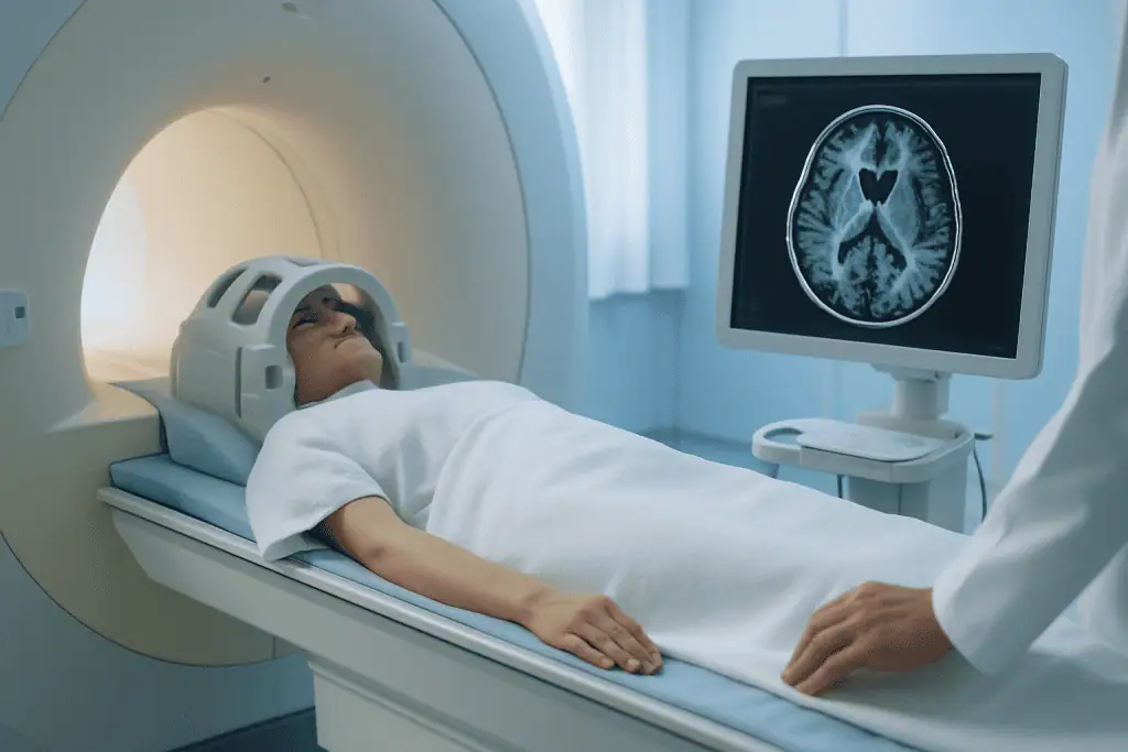
The only way to know whether you have a brain tumor is imaging, no combination of symptoms can confirm or exclude one. Magnetic resonance imaging of the brain with contrast is the central test: it shows the tumor’s location, size, enhancement pattern, surrounding swelling, relationship to functional areas, and whether there are multiple lesions. Computed tomography is used in urgent settings, particularly to detect bleeding, calcification or pressure effects quickly. Even then, a scan usually shows that a tumor exists and suggests what it might be; the definitive diagnosis often depends on pathology after a biopsy or operation.
People searching for accounts of how someone knew they had a brain tumor find remarkably varied stories: a driving-test eye check that revealed swollen optic nerves, a seizure at work, months of misattributed fatigue, a scan ordered for something unrelated. That variability is precisely the point. Brain tumors announce themselves in many ways or not at all, which is why unexplained, progressive neurological symptoms are investigated rather than assumed to be benign or assumed to be sinister.
Brain cancer symptoms: are they different from benign tumor symptoms?
Brain cancer symptoms are broadly the same as the symptoms caused by benign brain tumors, because symptoms arise from pressure, swelling and location, not from whether a tumor is malignant. A benign meningioma pressing on the motor cortex can cause more dramatic weakness than a small malignant tumor in a quieter region. The differences tend to lie in tempo: aggressive tumors more often cause symptoms that progress over weeks rather than years, and they more often provoke significant swelling. But tempo alone cannot separate the two, which is why imaging and pathology, not symptom patterns, determine the diagnosis.
Signs of head tumors beyond the brain itself
Signs of head tumors arising outside the brain, in the skull base, sinuses, nasopharynx or surrounding soft tissues, can overlap with brain tumor symptoms because these growths sit close to cranial nerves and can extend inward. Persistent one-sided hearing loss, facial numbness or weakness, double vision, swallowing difficulty or a change in voice can point toward the skull base or head and neck region rather than the brain proper. These conditions are assessed and treated differently, often involving head cancer specialists alongside neurosurgeons. Careful imaging usually distinguishes where a growth originates, which in turn determines which team leads treatment.
Who may need brain tumor evaluation and treatment
Patients arrive at brain tumor evaluation by three main routes: symptoms that prompt a scan, a tumor found incidentally on imaging done for another reason, or a known cancer that has spread to the brain. Each route raises different questions. A symptomatic tumor asks how urgently pressure and neurological risk must be addressed. An incidental finding asks whether treatment is needed at all or whether structured observation is safer. A metastasis asks how brain-directed treatment fits into the wider cancer plan.
Formal diagnosis begins with a neurological examination and imaging. Contrast-enhanced MRI is the cornerstone for most suspected brain tumors, mapping the lesion’s anatomy in detail. CT is valuable when speed matters or when bone involvement, bleeding or calcification need assessment. Depending on what the initial scans show, advanced imaging may follow: functional MRI to locate speech and movement areas, diffusion tensor imaging to map the white matter pathways that carry signals between brain regions, MR spectroscopy to analyse the tissue’s chemical signature, and perfusion imaging to assess blood flow within the tumor. These studies refine both the likely diagnosis and the safety of any planned intervention.
If the tumor may be metastatic, imaging of the chest, abdomen and pelvis, or whole-body imaging, may be recommended to find the primary cancer or assess how widespread the disease is. For particular tumor types, blood tests, hormone studies, visual field testing, spinal imaging or cerebrospinal fluid analysis add essential information. The final diagnosis usually rests on pathology after biopsy or surgery, and increasingly on molecular and genetic tumor markers, because these markers can change both the expected behaviour of a tumor and the treatments considered appropriate for it.
Patients typically seek specialist care when a scan suggests a brain tumor, when symptoms are progressing, when a previously treated tumor has returned, or when they want a second opinion before committing to surgery or radiation. Second opinions are particularly common, and particularly reasonable, when a tumor lies near critical structures such as the speech areas, motor pathways, brainstem, optic nerves, pituitary gland or skull base, or when different doctors have recommended different approaches. Patients with brain metastases often need care coordinated between neurosurgery, radiation oncology, medical oncology and the team already treating the original cancer.
Conditions and indications brain tumor treatment addresses
Brain tumor treatment covers a broad range of diagnoses, each with its own behaviour and standard approach. Primary brain tumors include gliomas such as astrocytoma, oligodendroglioma and glioblastoma; meningiomas arising from the membranes around the brain; schwannomas involving the cranial nerves, most commonly the vestibular nerve; pituitary adenomas; ependymomas; medulloblastomas and other embryonal tumors; central nervous system lymphomas; craniopharyngiomas; pineal region tumors; and rare skull base or ventricular tumors. Naming the tumor precisely matters, because the plan for a meningioma bears little resemblance to the plan for a glioma or a lymphoma.
Metastatic brain tumors are among the most common brain tumors in adults. They occur when cancer cells from another organ travel to the brain, frequently from lung cancer, breast cancer, melanoma, kidney cancer or colorectal cancer. Treatment may involve surgery, stereotactic radiosurgery, partial-brain or whole-brain radiotherapy in selected situations, systemic cancer therapy, or combinations of these. The decision rests on the number of lesions, their size, the symptoms they cause, the cancer type and its molecular profile, how well disease outside the brain is controlled, and what treatment has already been given.
Benign tumors may still require treatment. A meningioma, vestibular schwannoma, pituitary adenoma, craniopharyngioma or low-grade glioma can grow, compress the brain, affect cranial nerves, cause seizures, disturb hormones, block cerebrospinal fluid circulation, or threaten vision, hearing, movement or cognition. For small, stable, asymptomatic tumors, observation with scheduled scans is often a legitimate first choice, but that decision should rest on careful assessment of the specific tumor, not on general reassurance that benign means harmless.
High-grade tumors usually require combined treatment. Aggressive gliomas, for instance, are typically managed with maximal safe surgical removal followed by radiotherapy and chemotherapy, adapted according to pathology and molecular features. Some tumors demand urgent intervention because of mass effect, swelling, hydrocephalus or rapidly worsening neurological symptoms. Others allow time for planned evaluation, second opinion and a deliberately sequenced strategy. Distinguishing between the two is one of the most important judgements a specialist team makes.
Treatment also addresses the complications that travel with brain tumors: seizures, cerebral oedema, raised intracranial pressure, hydrocephalus, neurological deficits, endocrine imbalance, pain, fatigue, and cognitive or emotional effects. Managing these is part of comprehensive care rather than an afterthought, because good symptom control strongly influences safety, comfort and how well a patient tolerates the rest of treatment.
How brain tumor treatment is performed
Every pathway is individual, but most follow a recognisable sequence. Understanding that sequence makes the process less opaque and helps you ask better questions at each stage.
- Record and imaging reviewexisting scans, reports and any pathology are assessed to establish what is known and what is missing.
- Diagnostic completionadditional imaging, functional mapping, hormone testing or biopsy fill the gaps.
- Multidisciplinary planningthe case is discussed by the relevant specialists and a sequenced plan is agreed.
- Treatmentsurgery, radiosurgery, radiotherapy, drug therapy or observation, in the agreed order.
- Recovery and surveillancerehabilitation where needed, followed by scheduled MRI monitoring over the long term.
The process begins with a detailed review of medical history, neurological symptoms, prior imaging, pathology if available, and any cancer history. Specialist centres usually ask to see MRI images, CT scans, pathology reports, operative notes, radiation records, medication lists and recent laboratory results before a patient travels, where that is possible. This allows the team to judge urgency, plan any additional testing in advance, and make the visit itself focused rather than exploratory.
Diagnostic confirmation and treatment planning come next. A neuroradiologist and the treating physicians review the imaging to understand the tumor’s anatomy and behaviour. If surgery may be needed, advanced MRI techniques help map functional brain areas and important nerve pathways. For tumors near speech or motor regions, planning may include functional imaging, tract mapping, neuropsychological assessment, and discussion of awake brain mapping where appropriate. For pituitary or skull base tumors, evaluation typically adds hormone testing, visual field examination and focused skull base imaging. None of this is delay for its own sake: it is the information that makes the eventual treatment safer.
Complex cases are discussed in a multidisciplinary tumor board. Neurosurgeons, radiation oncologists, medical oncologists, neuroradiologists, pathologists, neurologists, endocrinologists and rehabilitation specialists contribute depending on the tumor. This format exists for a simple reason: no single specialty sees the whole picture, and treatment decisions made from one perspective alone are more likely to be poorly sequenced. Board review aligns the plan with evidence-based protocols while adapting it to the individual patient’s neurological condition and priorities.
If surgery is recommended, preparation includes anaesthesia evaluation, a medication review by the treating team, including planning around blood-thinning drugs, seizure prevention where indicated, steroid treatment for swelling where needed, and a clear explanation of the risks specific to the tumor’s location. You will be guided on fasting, admission and what to expect afterwards. The surgical goal is defined in advance: remove the tumor completely when that is safe, remove as much as possible when total removal would risk neurological harm, or take a biopsy when establishing the diagnosis is the primary aim. That distinction, maximal safe resection rather than removal at any cost, is central to modern neurosurgery.
During the operation itself, the neurosurgeon uses image-guided navigation built from your MRI or CT scans to reach the tumor accurately. Microsurgical technique allows work through small corridors under high magnification. In selected cases, intraoperative ultrasound, fluorescence-guided visualisation, neurophysiological monitoring, endoscopy or awake mapping support safe removal. Awake mapping is used for tumors near language or movement areas, because the team can test function during parts of the operation and adjust accordingly. Not every patient needs these techniques; they are chosen according to the tumor’s location and the specific risks involved, not applied as a standard package.
Operating time varies widely: a biopsy is comparatively short, while complex skull base, deep-seated or functional-area operations can take several hours. Afterwards, you are monitored closely in a recovery or intensive care setting, and a postoperative MRI or CT is usually performed to assess the result and check for complications. The team tracks strength, speech, vision, alertness, pain, nausea, seizure activity and wound healing. Length of hospital stay depends on the operation, your neurological condition and whether early rehabilitation is needed.
Radiation-based treatment may follow surgery, replace it, or address recurrent disease. Stereotactic radiosurgery delivers a high dose of tightly focused radiation to a defined target in one or a few sessions, often used for small metastases, vestibular schwannomas, meningiomas and other selected lesions. Despite the name, it involves no incision. Fractionated radiotherapy spreads treatment over multiple sessions, which is safer for larger tumors or lesions close to sensitive structures such as the optic nerves or brainstem. Planning uses fused MRI and CT images to shape the dose around the tumor while sparing normal tissue as far as possible, and immobilisation masks keep the head stable and the targeting consistent from session to session.
Drug treatment depends entirely on the diagnosis. Some gliomas are treated with oral chemotherapy given during and after radiotherapy. Central nervous system lymphoma, medulloblastoma, germ cell tumors and certain recurrent tumors follow their own specific chemotherapy protocols. Targeted therapies are considered when molecular testing identifies alterations that a specific medicine can address, or when the original cancer that spread to the brain carries actionable markers. Immunotherapy plays a role in selected metastatic cancers, depending on cancer type and treatment history. These systemic decisions sit within the broader framework of oncology and cancer treatment, coordinated with whatever therapy the body’s disease requires.
Recovery is highly individual. Some patients feel noticeably better soon after pressure on the brain is relieved. Others need time for swelling to settle, medications to be adjusted, or structured neurological rehabilitation. Physiotherapy, occupational therapy, speech and swallowing therapy, cognitive rehabilitation, psychological support, seizure management and endocrine follow-up may all form part of recovery, depending on what the tumor and its treatment affected. Follow-up MRI scans are scheduled to monitor healing, detect any recurrence early and guide further treatment if needed.
Why acting early matters
Not every brain tumor is an emergency, but timely evaluation matters. Delay can allow a tumor to grow, worsen swelling or seizures, and cause neurological deficits that become harder to reverse. A tumor near the optic pathways may threaten vision. A lesion blocking cerebrospinal fluid flow can cause hydrocephalus and rising pressure. A tumor in or near the motor cortex, speech centres, cerebellum, brainstem or cranial nerves may progressively affect movement, communication, balance, swallowing, hearing or facial function. Time changes what is at stake.
Early specialist assessment also prevents unnecessary or badly sequenced treatment, which is a real risk, not a theoretical one. Some tumors should not be operated on before proper endocrine testing or cancer staging. Some metastases are better treated with radiosurgery, while others need surgery first because of their size, the swelling they cause, or diagnostic uncertainty. Some low-grade tumors can be monitored safely, but observation should be structured, with defined imaging intervals and explicit criteria for stepping in, rather than an open-ended wait.
Acting promptly also preserves options. Surgery may be safer before a tumor becomes larger or more symptomatic. Radiation planning is more focused when disease is limited. Systemic therapy works best when it is chosen on molecular evidence and coordinated with the rest of the patient’s cancer care. For anyone considering treatment abroad, early communication of scans and reports allows a specialist centre to clarify how urgent the situation is, and whether travel should happen immediately or after further testing at home.
Benefits of brain tumor treatment
The realistic benefits depend on the tumor type, its location and the treatment used, but well-planned care can improve both disease control and daily functioning.
| Benefit | What It Means for You |
|---|---|
| Tumor control | Treatment may remove, shrink, stabilise or slow the tumor, depending on its biology and the therapies used. |
| Protection of neurological function | Careful planning aims to treat the tumor while preserving speech, movement, vision, memory, balance and independence as far as possible. |
| Relief of pressure and symptoms | Surgery, medication, radiation or combined treatment may reduce headaches, seizures, weakness, swelling or hydrocephalus. |
| Accurate diagnosis | Biopsy or surgical pathology, supported by molecular testing where indicated, clarifies the tumor type and directs the most appropriate therapy. |
| Personalised treatment sequence | A coordinated plan determines whether surgery, radiosurgery, radiotherapy, medication, rehabilitation or observation should come first, and what follows. |
Recovery timeline after brain tumor treatment
Recovery varies by diagnosis and treatment type, but the following gives a general sense of what many patients experience.
| Time Period | What Patients Can Expect |
|---|---|
| Day 1 | After surgery, close monitoring of neurological function, pain, swelling, seizures and early complications. After radiosurgery or radiotherapy sessions, most patients return to normal activities sooner, though fatigue or mild headache can occur. |
| First Week | Hospital discharge may follow within days after less complex surgery; larger operations may need longer monitoring or inpatient rehabilitation. Medication adjustments and wound care are reviewed before discharge. |
| First Month | Energy gradually improves. Follow-up imaging or the final pathology report guides the next step, radiotherapy, chemotherapy, targeted therapy or observation. Rehabilitation continues if speech, strength, balance or cognition were affected. |
| During Additional Therapy | Radiotherapy and chemotherapy schedules depend on tumor type. Fatigue, hair changes in the treated area, scalp sensitivity, nausea, blood count changes or steroid-related effects can occur and are monitored by the care team. |
| Longer Term | Regular MRI surveillance is important for years. Some patients return to work and travel; others need ongoing neurological, endocrine, oncology or rehabilitation support depending on the tumor and treatment received. |
What influences outcomes after brain tumor treatment
Outcomes in brain tumor care depend on many factors, and no responsible medical team predicts an individual result from imaging alone. Tumor type comes first. A benign meningioma, a low-grade glioma, a pituitary adenoma, a solitary brain metastasis and a high-grade glioma behave differently and warrant different expectations. Grade, molecular markers, growth rate and response to any previous treatment all shape what is realistic, which is why the honest answer to most prognosis questions begins with the pathology report, not the scan.
Location matters just as much. A small tumor in the brainstem can be more challenging than a larger tumor in a less sensitive region. Tumors near speech, movement, vision or memory structures, cranial nerves or major blood vessels demand especially careful planning. Whether a tumor can be removed safely depends not only on surgical skill but on the tumor’s borders, how far it infiltrates normal tissue, its blood supply and its relationship to functional pathways.
Your condition at the start of treatment matters too. Patients who are walking, speaking, eating and functioning well before treatment usually have a different recovery path from those who already have severe weakness, confusion, frequent seizures or raised intracranial pressure. Age, general health, immune status, heart and lung function, diabetes, prior radiation, previous surgery and active cancer elsewhere in the body all influence both what treatments are offered and how recovery unfolds.
Accurate pathology and molecular testing can change the plan entirely. In modern neuro-oncology, specific markers classify gliomas, indicate likely behaviour and guide chemotherapy or targeted treatment decisions. For metastatic tumors, molecular profiling of the primary cancer can steer systemic therapy in ways that help control disease in both the brain and the body. This is why tissue handling, expert pathology review and communication between specialists are not administrative details: they are clinically decisive.
Finally, a good result is not defined only by what the scan shows. It includes neurological preservation, symptom control, a safe recovery, clarity of diagnosis, an appropriate follow-up plan and quality of life. For some patients, the best outcome is complete removal. For others, it is a deliberate partial removal followed by radiotherapy, chosen to avoid unacceptable neurological risk. For others still, the best decision is structured monitoring rather than immediate intervention. Strong plans are individual, realistic and revisited as new information arrives.
Can people survive a brain tumor?
Yes, many people survive a brain tumor, and some live for decades after diagnosis. Survival depends heavily on the tumor type: many benign tumors, once treated or stabilised, never threaten life at all, and some are simply monitored for years without ever needing intervention. Malignant tumors vary widely, from types that respond well to combined treatment to aggressive gliomas where treatment aims to extend life and protect function rather than eliminate disease permanently. No general statement fits every diagnosis, which is why survival questions are best answered by a specialist who has seen your specific pathology and imaging, not by averages that may not describe your situation.
Can I live a normal life with a brain tumor?
Many people do live full, working, independent lives with or after a brain tumor, particularly those with small benign tumors under observation, tumors that were removed completely, or lesions controlled with focused radiation. Others live well with adjustments: medication for seizures, temporary driving restrictions after a seizure, rehabilitation for a specific deficit, or hormone replacement after pituitary treatment. Some tumors and treatments do leave lasting effects on energy, cognition or movement that require ongoing support. What life looks like afterwards depends on the tumor type, its location, the treatment needed and how your brain recovers, and it is a question worth asking your team directly, in terms specific to your diagnosis.
Brain tumor care at Acibadem for international patients
International patients come to Acibadem for careful second opinions, coordinated treatment planning, and access to neurosurgical and oncology services within a hospital group experienced in caring for patients from abroad. Brain tumor care can involve several specialties within a short period, and patients benefit when imaging, surgical assessment, pathology, radiation planning, medical oncology, intensive care, rehabilitation and follow-up are organised around a single clinical plan rather than scattered across disconnected appointments.
Complex cases are reviewed by multidisciplinary teams that can include neurosurgery, radiation oncology, medical oncology, neuroradiology, pathology, neurology, endocrinology and rehabilitation medicine, with tumor board discussion for decisions that should not rest on one perspective. Technology supports this work, advanced MRI and CT, functional imaging and tract mapping, image-guided navigation, microsurgical visualisation, intraoperative monitoring, precision radiation planning, and molecular pathology, but its value lies in how it is matched to the individual case, not in the equipment list itself.
That matching is the point. A patient with a small vestibular schwannoma may be best served by observation, radiosurgery or microsurgery, depending on hearing, balance symptoms, tumor size and documented growth. A patient with a glioma may need maximal safe resection followed by radiotherapy and chemotherapy shaped by pathology. A patient with brain metastases needs coordination between existing cancer records, systemic therapy options, radiosurgery and possibly surgery for a symptomatic lesion. There is no single standard pathway: the right plan depends on details.
For patients travelling from Europe, the Middle East, Africa, the Americas or elsewhere, Acibadem’s international patient services help with medical record transfer, appointment coordination, interpretation, admission guidance and communication with the clinical team throughout the stay. If you are weighing up travelling for neurosurgical treatment, it is worth asking the team to walk you through how a typical care pathway is organised, from first record review through to follow-up after returning home, so that each stage is clear before any decision is made.
What a specialist review usually involves
A thorough specialist review draws on specific materials: the MRI or CT images themselves rather than only the written reports, the radiology reports, pathology results if a biopsy or operation has already taken place, details of any previous cancer treatment, a current medication list, and a clear account of symptoms and how they have changed over time. With this information, a specialist team can usually say whether the working diagnosis is well supported, whether further testing would change the plan, and how urgent any treatment genuinely is.
Second opinions are a normal part of brain tumor care, not a sign of distrust. They are most valuable before irreversible steps, surgery, radiation or the start of systemic therapy, and particularly when a tumor sits near critical structures or when two doctors have recommended different approaches. A good second opinion either confirms the plan, which brings confidence, or improves it, which is the entire purpose.
Whatever team you work with, some questions consistently produce useful answers:
- What do you believe this tumor is, and how confident are you before pathology?
- What happens if we monitor rather than treat now, and how would monitoring be structured?
- If surgery is proposed, what is the goal, complete removal, maximal safe removal, or biopsy?
- What are the specific neurological risks given this tumor’s location?
- What treatment is likely to follow surgery, and what does the full sequence look like?
- How will follow-up be organised, and for how long?
Brain tumor care is complex, but decisions do not need to be made in the dark. A careful consultation should leave you understanding what the tumor is likely to be, what each option involves, what each option may mean for neurological function and recovery, and how the whole sequence of care fits together, whether treatment happens close to home or abroad.
Our Specialists Explain
 Treating Selected Brain Tumors Without an Incision at Acibadem
Preparation
- Evaluation usually includes neurological examination, MRI or other imaging, laboratory tests, and pathology review when available. The care team assesses tumor location, grade, symptoms, and overall health to plan surgery, radiosurgery, radiation, or systemic therapy. Patients may be advised to adjust medications and fast before procedures requiring anesthesia.
Aftercare
- After treatment, neurological status, wound healing, and possible side effects are closely monitored. Follow-up imaging and oncology or neurosurgery visits help assess response and detect recurrence. Rehabilitation, seizure control, steroid tapering, or supportive therapies may be recommended according to individual needs.
Frequently Asked Questions
What mainly affects the cost of brain tumor treatment?
Cost is influenced by tumor type and location, required imaging, whether biopsy or surgery is needed, pathology and molecular testing, ICU or ward stay, radiotherapy or drug treatment, and the level of follow-up or rehabilitation required.
How can I get a personalised quote from Acibadem?
You can request a free consultation by sharing recent imaging, medical reports, pathology results if available, current symptoms and previous treatment history. The international patient team can coordinate specialist review and provide a personalised treatment plan and quote.
Is a package price always possible for brain tumor care?
A package may be possible after medical review, but complex cases may need itemised planning because the final approach can change after specialist assessment, updated imaging, surgery findings or pathology results.
Can travel, translation and accommodation be included?
International patient services may help coordinate interpreter support, airport transfers, accommodation guidance and appointment scheduling. What is included should be confirmed in the written quote before travel.
Why do quotes differ between hospitals or countries?
Quotes may differ because hospitals use different billing models, technologies, specialist teams, inpatient pathways, medication policies and follow-up arrangements. Comparing what is included is as important as comparing the headline treatment plan.
Medically reviewed by the Acıbadem International Medical Board August 30, 2026
See our medical review board →
Update history
- PublishedJune 5, 2026
- Medical review approvedAugust 30, 2026
- Last content updateSeptember 8, 2026
References3
- Adult Central Nervous System Tumors Treatment (PDQ) – Patient Version cancer.gov
- Brain tumours (malignant) nhs.uk
- Brain Tumors medlineplus.gov
Trusted care for international patients
Compare options, costs and recovery
From the Acibadem Blog
Seeking a Second Opinion on a Brain Tumor Plan: What to Bring and Which Questions to Ask
A brain tumor second opinion is a review of your scans, pathology and proposed plan by a separate neuro-oncology team. Bring the actual imaging…