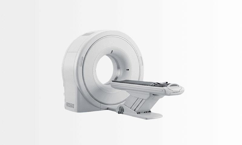
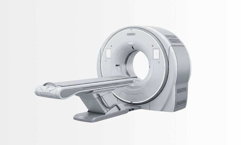
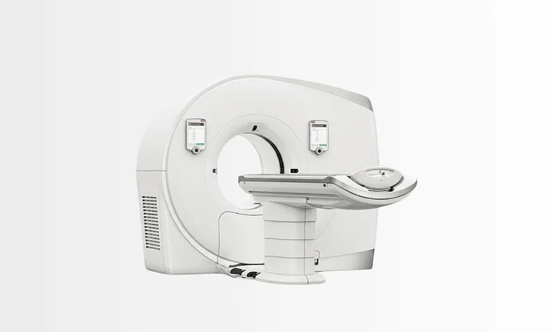
Coronary CT Angiography
Coronary CT Angiography is a non-invasive heart imaging test that uses CT scanning and contrast dye to visualize coronary arteries and assess narrowing or blockage.
Quick answer
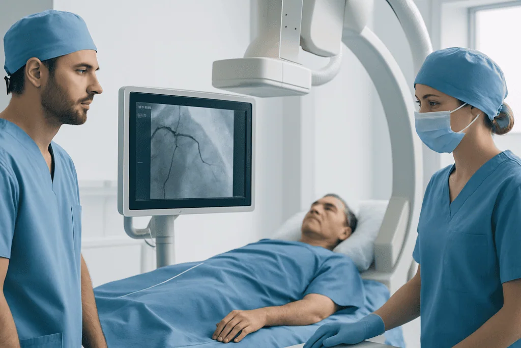
Coronary CT angiography is a non-invasive heart scan that uses a CT scanner and an intravenous contrast dye to image the coronary arteries. It shows whether plaque is narrowing or blocking the vessels that supply the heart muscle, without a catheter entering the body. The scanning itself takes minutes, and most people go home the same day and resume normal activities almost immediately.
Coronary CT angiography: a non-invasive angiogram of the heart
Coronary CT angiography is a non-invasive imaging test that uses a computed tomography scanner and an intravenous contrast dye to produce detailed pictures of the coronary arteries: the vessels that supply oxygen-rich blood to the heart muscle. It works as an angiogram of the heart without any catheter being passed into the body, and it exists to answer one central question: are those arteries narrowed or blocked? It is used mainly in people with chest symptoms or cardiac risk factors whose diagnosis is not yet settled.
Hearing that you may need a heart test can be unsettling, especially if you have chest discomfort, shortness of breath, a strong family history of heart disease, or a stress test that raised more questions than it answered. Most people in that position want to know three things: is an artery blocked, am I at risk of a heart attack, and will the next step involve an invasive procedure? Those concerns are reasonable. When the heart is involved, uncertainty feels urgent, and vague reassurance is not enough.
For many patients, the practical appeal of this test is simple. Because it is a non-invasive angiogram, it produces detailed images of the coronary arteries without instruments being threaded into the heart. That can make the diagnostic process faster and less demanding, while still giving cardiologists the anatomical detail they need to judge whether symptoms are likely to be caused by coronary artery disease.
Just as importantly, the scan helps guard against both under-treatment and over-treatment. A normal or low-risk result may spare you an invasive test you never needed. A concerning result identifies the need for closer evaluation, medication, lifestyle change or a further cardiac procedure. The value of the study is therefore not only in the images themselves; it is in moving you from uncertainty towards an informed plan.
What coronary CT angiography involves
Coronary CT angiography is a specialised examination that combines computed tomography with an intravenous contrast dye to create highly detailed images of the coronary arteries. The scanner takes many rapid cross-sectional pictures of the heart; advanced software then reconstructs them so that radiologists and cardiologists can examine both the artery walls and the open channel inside each vessel. The whole study is built around a single anatomical question, which is why the images are acquired, processed and read quite differently from those of a routine chest CT.
The test is used primarily to look for atherosclerosis: the gradual build-up of fatty, fibrous and sometimes calcified plaque within the artery wall. It can show whether plaque is present, where it sits, and whether it appears to be narrowing the vessel enough to limit blood flow. In many cases it also characterises the type and extent of plaque, which adds useful information to your overall risk assessment, because soft, calcified and mixed deposits carry different implications and shape treatment differently.
Unlike a routine CT of the chest, coronary CT angiography must be timed to the cardiac cycle. The heart never stops moving, so image acquisition is synchronised with your heartbeat using ECG leads attached during the scan. If your heart rate is fast, the team may give medication to slow and steady it, because a calmer rhythm produces sharper pictures. The aim throughout is to capture the coronary arteries at the moment they are most still.
It is important to understand that this is a diagnostic test, not a treatment. The scan does not open a blocked artery, and it does not replace emergency care for someone in the middle of an active heart attack. What it provides is a detailed, non-invasive map of the coronary circulation that guides the next decision. Depending on the findings, that next step may be reassurance, preventive cardiology, additional functional testing, invasive coronary angiography, or treatment with medication or revascularisation.
In selected patients, physicians may add further CT-based analysis to gauge overall plaque burden, or to estimate whether a particular narrowing is likely to be restricting blood flow in a functionally meaningful way. Whether that extra analysis is worthwhile depends on your symptoms, your history and the precise clinical question being asked.
Why is CT coronary angiography done?
CT coronary angiography is done to establish whether the coronary arteries are narrowed or blocked, without resorting to an invasive procedure. Cardiologists order it most often when symptoms such as chest pain or breathlessness could plausibly be cardiac, when accumulated risk factors make coronary disease a realistic possibility, or when an earlier test, a treadmill test, stress imaging study or echocardiogram, has produced findings that do not fully explain the picture. It is also used to identify congenital anomalies of the coronary arteries and, in carefully selected cases, to add anatomical detail in people with known disease.
Can CT coronary angiography detect blockage?
Yes, detecting blockage and narrowing is precisely what the test is designed to do. A well-performed scan shows where plaque has formed, whether it is calcified or soft, and how far it encroaches on the channel that carries blood. There are honest limits, though. Heavy calcification can exaggerate how severe a narrowing looks, very small vessels are harder to judge, and the scan shows anatomy rather than blood flow. A borderline narrowing therefore sometimes needs a functional test, or an invasive study, to settle how much it actually matters to the heart muscle downstream.
What does a CT angiogram show?
A CT angiogram shows the contrast-filled channel of a blood vessel together with the vessel wall around it. Applied to the heart, that means each of the main coronary branches can be traced from origin to tip, with narrowings, plaque deposits, unusual anatomical courses and, after previous treatment, stents and bypass grafts all visible to a trained reader. Because the images are volumetric, the reporting specialist can rotate and slice them in any plane, examining a suspicious segment from several angles before drawing a conclusion.
Angiogram, angiography and related terms, explained
The vocabulary around this test causes genuine confusion, because several overlapping words are used loosely, sometimes even in referral letters. It is worth separating them, since the differences affect what you would actually experience on the day.
What is angiography?
Angiography is the general name for any imaging technique that visualises blood vessels, usually with the help of a contrast agent. It is applied across the body, to arteries in the brain, lungs, legs and kidneys, and even to the small vessels of the retina in eye angiography. When the word appears on its own in a cardiology setting, it almost always refers to imaging of the coronary arteries, by one route or another.
CT angiogram and angiography: how do the two differ?
The terms CT angiogram and angiography describe the same goal reached by different routes: both produce pictures of blood vessels, but the CT version acquires them from outside the body using a scanner and an injection into an arm vein, while conventional catheter angiography injects contrast directly into the artery being studied through a thin tube. The catheter route gives real-time moving images and allows treatment during the same session; the CT route avoids arterial puncture altogether. Which is more appropriate depends largely on how likely you are to need treatment on the spot.
Is a coronary angiogram the same as coronary angiography?
In everyday use, a coronary angiogram and angiography of the coronary arteries mean the same thing: imaging of the arteries that supply the heart muscle. Strictly speaking, angiography names the technique and an angiogram names the picture it produces, but hospitals, referral letters and search results use the two interchangeably. What matters when reading your own paperwork is the prefix: the letters CT in front of either word signal the non-invasive scan; without them, the catheter-based test is usually what is meant.
How does the catheter-based angiogram procedure compare?
The angiogram procedure most people picture: a thin catheter guided from the wrist or groin up to the heart, contrast injected directly into the coronary arteries, images captured under X-ray, remains the reference standard for measuring narrowings precisely, and it carries one decisive advantage: if a significant blockage is found, it can often be treated during the same session with balloon dilation and coronary stent applications. It is, however, an invasive procedure, with its own preparation, a period of bed rest afterwards and a small procedural risk of its own. Coronary CT angiography sits earlier in the pathway: its job is to clarify whether that invasive step is genuinely needed at all.
Who may need coronary CT angiography
The scan is most often considered when symptoms could reflect reduced blood flow to the heart but the diagnosis remains uncertain. Typical triggers include chest pain or pressure, particularly on exertion; unexplained shortness of breath; discomfort radiating to the arm, jaw, neck or back; falling exercise tolerance; or symptoms that are not classic for angina yet still raise concern because of age or risk profile.
Some patients are referred because they sit at an intermediate likelihood of coronary artery disease. They have no established diagnosis, but enough risk factors have accumulated to make imaging sensible: high blood pressure, diabetes, raised cholesterol, a smoking history, obesity, a sedentary lifestyle, or a family history of premature heart disease. Others arrive after a treadmill test, stress imaging study or echocardiogram has produced results that are inconclusive, or that simply do not match how they actually feel.
The scan can also be used in people with known coronary disease when additional anatomical detail is needed, although not everyone with established disease is a good candidate. Extensive calcification, previously implanted stents, bypass grafts, an irregular heart rhythm or difficulty holding the breath can all limit image clarity, and in those situations another diagnostic route may serve better. Test selection is always individualised.
The pathway begins with a careful clinical assessment rather than the scanner. A cardiologist reviews your symptoms, personal and family history, current medications and risk profile; a physical examination, ECG and blood tests are often part of the work-up. Kidney function is checked because the contrast dye is cleared through the kidneys. You will also be asked about previous reactions to contrast material, asthma, thyroid disease and any possibility of pregnancy, since each of these changes how, or whether, the scan is performed.
The question the test tries to answer is deliberately narrow: is there meaningful coronary narrowing, and if so, where and how extensive is it? For many patients, an angiogram obtained by CT answers that question efficiently. For others: those in an acute emergency, or those very likely to need treatment during the same procedure: a different pathway makes more sense. The deciding factors are the likelihood of disease, the urgency of the situation and how much a non-invasive anatomical answer would actually change the next step.
The conditions and situations it helps evaluate
Coronary CT angiography is most strongly associated with the evaluation of coronary artery disease, but its clinical use is broader than a single diagnosis. It can contribute to several related assessments:
- Suspected coronary artery disease: detecting narrowing or blockage in the coronary arteries in patients with chest pain or related symptoms.
- Stable angina evaluation: determining whether exertional chest symptoms correspond to plaque build-up and a reduced coronary artery diameter.
- Assessment after inconclusive cardiac testing: adding anatomical detail when earlier tests suggested a possible problem without resolving it.
- Evaluation of coronary anomalies: identifying unusual origins or courses of the coronary arteries, which can be clinically significant.
- Risk stratification in selected patients: understanding the presence and distribution of plaque, even when severe obstruction is not obvious.
- Planning further cardiology care: helping decide whether medication, preventive care, invasive angiography or another intervention should follow.
The test is not usually the right choice in an emergency. When symptoms strongly suggest an acute heart attack, physicians prioritise emergency evaluation, blood tests, ECG monitoring and rapid invasive management where needed. The strength of coronary CT angiography lies in carefully selected patients for whom a detailed, non-invasive view of the coronary arteries will meaningfully shape care.
How is CT coronary angiography done?
The process begins before the scan itself, because image quality in cardiac CT depends heavily on timing, heart rate and the safe use of contrast. You are usually asked to avoid caffeine for a period beforehand, since it raises the heart rate, and you may be asked to fast for several hours. The care team reviews your current medications in advance and tells you exactly how to handle them on the day: that decision always belongs to your treating doctor, not to a website. On the day itself, the sequence typically runs as follows:
- Arrival checks. A nurse or technologist confirms your blood pressure, pulse and recent medical history, and reviews the safety questions about kidneys, allergies and pregnancy one final time.
- Intravenous line. A small cannula is placed, usually in the arm, so contrast dye can be given during the scan.
- ECG leads. Sticky electrodes are attached to the chest so image capture can be synchronised with your heartbeat.
- Heart-rate preparation. If your heart is beating faster than ideal, the team may give medication to slow it, and in some cases a medicine that widens the coronary arteries is used shortly before acquisition so the vessels show up more clearly.
- Positioning and breath-hold practice. You lie on the CT table, which moves through the scanner opening; the technologist rehearses the short breath-hold with you.
- Contrast and acquisition. The dye is injected through the cannula, a warm sensation or metallic taste is common and passes quickly, and the scanner collects its data within seconds.
- Observation. You are watched briefly afterwards, particularly if heart-rate medication was used, the cannula is removed, and drinking fluids is usually encouraged to help clear the contrast.
From a technology standpoint, the study relies on high-speed CT imaging, ECG synchronisation and reconstruction software that turns a moving heart into still, readable images. Modern systems are designed to capture the coronary arteries with high spatial and temporal resolution while managing radiation exposure thoughtfully; the imaging team adjusts the protocol to each patient wherever possible, balancing image quality against dose. Where kidney function is reduced, the team also weighs the benefit of the scan against the contrast load, adjusting hydration and contrast volume or considering an alternative test, one more reason the pre-scan assessment is never a formality. Post-processing tools then let physicians review each artery from multiple angles and distinguish calcified from non-calcified plaque.
The interpretation phase matters as much as the scan. A radiologist with cardiovascular imaging expertise, often working with a cardiologist, evaluates the images and prepares a report addressing whether plaque is present, where narrowings sit, how severe they appear and whether further assessment or treatment is advisable. The report is never read in isolation: it is weighed against your symptoms, examination findings, risk factors and previous results, and complex cases can be reviewed in a specialist board setting before a recommendation is made.
How long does a CT coronary angiography take?
The image acquisition itself takes only seconds, and the time on the scanner table is usually a matter of minutes. The overall visit is longer: registration, placement of the intravenous line and ECG leads, heart-rate control where needed, the scan, and a short period of observation afterwards all add up. It is sensible to plan for a visit measured in hours rather than minutes, and your care team can tell you what to expect for your specific appointment. Most people leave the same day and return to their usual routine almost immediately.
What should you not do before an angiogram?
Before a CT-based angiogram, the standard advice is to avoid caffeine, coffee, tea, energy drinks and some soft drinks, for the window your team specifies, because it pushes the heart rate up and blurs the images. You are usually asked not to eat for several hours beforehand; the exact length of that fast varies between centres and between patients, so treat the written instructions you receive as the final word rather than any general rule. Do not make any changes to your own medication: your treating doctor reviews everything you take and tells you precisely what to do. Finally, do not withhold information, kidney problems, previous contrast reactions, asthma, thyroid disease and any possibility of pregnancy all need to be declared, because they directly affect how the scan is planned.
Why acting early can make a difference
Coronary artery disease often develops gradually, and in its early stages it may cause no symptoms at all. Yet plaque can progress over time, narrowing the arteries or becoming unstable. When that happens, the first sign may be worsening angina, shrinking exercise tolerance, an emergency admission or a heart attack. That is why timely evaluation matters.
When chest symptoms are dismissed or testing is delayed, opportunities for prevention can be missed. A patient with early or moderate disease may benefit from cholesterol management, blood pressure control, smoking cessation support, diabetes optimisation and targeted lifestyle changes, measures that may reduce the chance of future cardiac events. If significant narrowing is present, early diagnosis lets physicians decide whether further testing or intervention is needed before the condition produces more serious consequences.
Delay also prolongs uncertainty. People limit physical activity, avoid travel or live with ongoing anxiety simply because they do not know whether their symptoms are dangerous. Not every chest symptom comes from a blocked artery, but it is usually better to clarify the cause than to assume it is benign, and coronary CT angiography can be a valuable part of that clarification in appropriately selected patients.
Potential benefits of coronary CT angiography
For the right patient, the scan offers several practical and clinical advantages.
| Benefit | What It Means for You |
|---|---|
| Non-invasive evaluation | The coronary arteries can be visualised without placing a catheter into the heart, which may reduce procedural burden and recovery needs. |
| Detailed anatomical information | The test can show where plaque is located, whether narrowing is present, and how extensive coronary artery disease may be. |
| Helpful triage after symptoms or unclear test results | It can help determine whether symptoms are likely related to coronary disease and whether you may need medication, more testing, or an invasive procedure. |
| Faster clarification for many patients | A normal or low-risk study may help rule out significant coronary blockage and reduce uncertainty more quickly. |
| Support for preventive heart care | Even when severe obstruction is not found, identifying plaque can guide risk-factor treatment and long-term cardiovascular prevention. |
Is CT coronary angiography better than other heart tests?
It is better for some purposes and not for others: there is no universally superior test. For people with a low-to-intermediate likelihood of disease, the CT approach is often the more sensible first step, because a clearly normal scan makes significant blockage unlikely without any arterial puncture. The catheter-based study remains preferable when the probability of disease is high and treatment during the same session is realistic, and it allows pressure measurements inside the artery that CT cannot make directly. Functional tests, stress echo, stress MRI, nuclear imaging, answer a different question again: not what the arteries look like, but how well blood actually flows under load. A good cardiologist chooses among these based on your probability of disease, your symptoms and what the answer would change.
Recovery: what to expect afterwards
Recovery from coronary CT angiography is usually brief, because the test involves no surgery and no arterial catheterisation. The exact experience depends on whether heart-rate medication or other medicines were used on the day.
| Time Period | What Patients Can Expect |
|---|---|
| Day 1 | Most patients complete the scan and go home the same day. Mild temporary sensations from contrast or medication may occur but usually pass quickly. |
| First Week | Normal routines are typically resumed immediately or within a day. The main focus is reviewing results and understanding the next step in care. |
| First Month | If plaque or narrowing is found, follow-up may include cardiology consultation, medication changes, risk-factor management, or additional testing. |
| Longer Term | The long-term impact depends on the findings. Some patients need only reassurance and prevention, while others may move on to more active cardiac treatment planning. |
Is an angiogram very painful?
Most people find a CT-based angiogram easy to tolerate: there is no arterial puncture, so discomfort is usually limited to the needle used for the intravenous line and the brief warm flush that the contrast produces. The catheter-based version is performed under local anaesthetic at the wrist or groin; patients typically describe pressure or an odd sensation rather than sharp pain, though experiences differ and some soreness at the access point afterwards is common. In both cases the team monitors your comfort throughout and can respond if something feels wrong.
How long is bed rest after an angiogram?
After a CT angiogram, no bed rest is normally required at all: a short period of observation is usually the only delay before you leave. After a catheter-based angiogram, a period of lying flat or resting is standard while the arterial puncture site seals. Its length varies with the access route, wrist access generally allows earlier mobilisation than groin access, and with the closure method used, so the treating team sets it individually rather than by a fixed rule.
How long does recovery after an angiogram take?
Following coronary CT angiography, most people resume their usual routine the same day unless their physician advises otherwise; drinking extra fluids to help clear the contrast is often the only instruction. After a catheter-based angiogram, teams commonly advise taking things gently for a short period and avoiding heavy lifting while the access site heals, with the exact timeframe depending on the access route, the closure technique and your individual circumstances. If a long journey or a flight is planned soon after a catheter-based study, it is worth asking the treating team when travelling is sensible, because the answer depends on the access site and how well it is healing.
How much does a coronary CT angiography cost?
There is no single honest answer, because the cost depends on where the scan is performed and on what the quoted figure actually includes. Several drivers explain why quotes for what sounds like the same test can differ substantially:
- What the package covers: some quotes include the pre-scan cardiology consultation, ECG, blood tests and the specialist report; others cover only the scan itself.
- Additional imaging: a coronary calcium score is sometimes performed alongside the angiographic study and may or may not be bundled in.
- Medication and consumables: contrast dye, heart-rate medication and monitoring all form part of the true cost.
- Interpretation and follow-up: review by a cardiovascular imaging specialist, and a results consultation afterwards, may be priced separately.
- Country, city and institution: healthcare pricing varies widely between health systems and between hospitals within them.
When comparing quotes, the useful discipline is to compare like with like: confirm exactly which elements are included, whether the reporting is done by a specialist in cardiac imaging, and what happens, and what it costs, if the images need to be repeated or a follow-up consultation is required. Insurance and national health coverage rules also differ, so what you personally pay can bear little relation to the headline figure.
What influences image quality, interpretation and outcomes
When patients ask how successful this test is, the most accurate answer is that it is generally a highly useful study in appropriately selected patients, particularly for evaluating suspected coronary artery disease outside of an acute emergency. Like any test, its value depends on the quality of the images, the clinical context and how the results are used.
Several factors shape image quality. A lower, regular heart rate produces clearer pictures, while significant arrhythmias make scanning harder. Heavy coronary calcification can limit how precisely a narrowing is measured, because calcium creates imaging artefacts. Body habitus, the ability to hold the breath briefly, and previously implanted stents or bypass grafts can also affect what the reader can confidently say.
Preparation matters too. Following the instructions on fasting, caffeine and medication improves the chances of a smooth, single-pass examination, and accurate disclosure of kidney disease, contrast allergy, asthma, thyroid problems or possible pregnancy is essential for safety.
Interpretation quality depends on experience. Coronary CT angiography produces complex data that need careful review by specialists trained in cardiac imaging, and a strong programme does more than issue a scan report: it integrates the findings with your history, symptoms, laboratory results and earlier studies. That integration is what turns pictures into decisions.
Finally, a good result is not defined only by normal arteries. Often the most useful outcome is clarity: sometimes that means reassurance, sometimes it means starting preventive treatment, and sometimes it means moving promptly to invasive angiography or revascularisation because a significant blockage has been found. The best outcome is a more accurate, more timely decision than would otherwise have been possible.
Cardiac imaging at Acibadem: how the pathway is organised
Choosing where to have cardiac imaging involves more than choosing a scanner. The scan is most valuable when it sits inside a complete cardiology pathway, assessment before, expert reading after, and a clear route to whatever comes next.
At Acibadem, coronary CT angiography is typically integrated into a structured diagnostic process involving cardiologists, radiologists, cardiac imaging specialists and nurses, with interventional cardiology and cardiovascular surgery teams available when the findings call for them. That multidisciplinary structure matters because the significance of a result is rarely obvious from images alone: a moderate narrowing in one patient may warrant medication and observation, while the same finding in another, with different symptoms and risk factors, may prompt more urgent evaluation. Clinical judgement carries the decision.
Technology plays its part. Image speed, acquisition timing, ECG-based synchronisation, radiation management strategies and reconstruction software all influence how useful a study turns out to be, and protocols are tailored to each patient’s heart rhythm, body profile and clinical question wherever possible.
Continuity is another practical consideration. A coronary CT angiogram is often the beginning of a conversation rather than the end of one. Some patients seek a second opinion after earlier testing left the picture unclear; others go on to need cardiology consultation, invasive angiography or an assessment for coronary artery bypass surgery if significant disease is found. Within one network, imaging findings can be reviewed in context rather than in isolation, including through multidisciplinary specialist discussion when a case is complex, so the step that follows the scan is grounded in the whole clinical picture rather than a single report.
Most importantly, decisions remain individual. Not every patient with chest symptoms needs this scan, and not every abnormal result leads to the same next step. The aim is to match the test, and the plan that follows it, to the person in front of the physician, using evidence-based protocols and experienced judgement.
Understanding your results and what may follow
Reports from coronary CT angiography tend to fall into recognisable patterns, and knowing them in advance makes the conversation with your cardiologist easier to follow. Clearly normal arteries generally point away from coronary disease as the cause of symptoms, and attention turns to other explanations. Plaque without significant obstruction usually shifts the focus to prevention, cholesterol, blood pressure, smoking, diabetes and activity, because the finding changes long-term risk even when it does not require a procedure. Narrowing that appears significant typically leads either to functional testing, to confirm whether blood flow is genuinely restricted, or to invasive angiography, where treatment can follow in the same setting if needed.
Whichever category your result falls into, the scan has done its job when the next step is clearer than it was before. For some people that means the relief of a settled answer; for others it means treatment starting earlier than it otherwise would have. Understanding the state of your coronary arteries, precisely, and without unnecessary intervention, is one of the more useful things modern cardiology can offer, and it is the entire purpose of this test.
Preparation
- You may be asked to avoid food, caffeine, and smoking for several hours before the scan. Kidney function, allergy history, and current medications are reviewed because contrast dye is used. In some cases, heart rate-lowering medication may be given before the test to improve image quality.
Aftercare
- Most patients can return home shortly after the scan and resume normal activities the same day. Drinking extra fluids may help clear the contrast dye unless your doctor advises otherwise. Seek medical advice if you develop rash, shortness of breath, or unusual symptoms after contrast exposure.
Frequently Asked Questions
What is coronary CT angiography and what does it show?
Coronary CT angiography is a noninvasive heart imaging test that uses CT scanning and contrast dye to create detailed pictures of the coronary arteries. It helps doctors look for narrowing, plaque buildup, congenital vessel differences, and other signs of coronary artery disease. The scan can also provide information about nearby heart structures. At Acibadem, radiology and cardiology specialists review the images together when needed to support an accurate, personalized assessment.
Why might my doctor recommend a coronary CT angiography?
Your doctor may recommend coronary CT angiography if you have chest pain, shortness of breath, an abnormal stress test, or risk factors such as diabetes, smoking, high blood pressure, or a family history of heart disease. It is often used to evaluate suspected coronary artery disease in a quick, noninvasive way. It can also help clarify uncertain findings from other tests. Acibadem specialists decide whether this scan is the most suitable option for your situation.
How should I prepare for a coronary CT angiography scan?
Preparation usually includes avoiding caffeine for several hours before the scan and following instructions about eating, drinking, and medications. You may be asked not to smoke beforehand, because nicotine can affect heart rate. Some patients receive medication to slow the heart for clearer images. Tell your doctor if you are pregnant, have asthma, kidney disease, thyroid problems, or a contrast allergy. Acibadem teams provide detailed preparation guidance based on your medical history.
Is coronary CT angiography painful or invasive?
Coronary CT angiography is generally not painful and is considered noninvasive because it does not require catheters inside the heart arteries. You will have an IV line placed so contrast dye can be injected, which may cause a brief warm sensation or metallic taste. During the scan, you lie still and may be asked to hold your breath for a few seconds. Most patients find the procedure fast and well tolerated.
How long does a coronary CT angiography take?
The actual image acquisition is very quick and often takes only a few seconds, but the full appointment usually lasts longer to allow for registration, preparation, heart rate monitoring, IV placement, and post-scan observation if needed. In many cases, the visit may take around 30 minutes to an hour. Timing can vary depending on your heart rhythm and whether medication is needed beforehand. Acibadem staff explain each step so international patients know what to expect.
Is the contrast dye used in coronary CT angiography safe?
The contrast dye used in coronary CT angiography is commonly used and safe for most patients, but it can pose risks in people with certain allergies, kidney problems, or thyroid conditions. Reactions are usually mild, such as itching or nausea, but more serious reactions can occur rarely. That is why your medical history and blood tests may be reviewed before the scan. Acibadem specialists assess these factors carefully to plan the safest approach for you.
How much radiation is involved in coronary CT angiography?
Coronary CT angiography uses X-rays, so there is some radiation exposure. Modern CT technology and optimized imaging protocols are designed to keep the dose as low as reasonably possible while maintaining image quality. The exact amount varies based on your body type, heart rate, and scan technique. For many patients, the diagnostic value outweighs the risk, especially when the test may help avoid unnecessary invasive procedures. Your Acibadem team can explain the benefit-risk balance in your case.
Can coronary CT angiography detect blocked arteries accurately?
Coronary CT angiography is very useful for identifying plaque and showing whether coronary arteries appear narrowed or blocked. It is especially helpful for ruling out significant coronary artery disease in selected patients. However, it does not replace every other heart test, and in some cases heavy calcification, fast heart rate, or irregular rhythm can affect image quality. If needed, Acibadem specialists may recommend additional testing to confirm the findings and guide treatment planning.
What happens after a coronary CT angiography and when will I get the results?
After the scan, you can usually return to normal activities unless your doctor gives different instructions. Drinking fluids may be advised to help clear the contrast dye. A radiologist analyzes the images, and the results are then discussed with your cardiologist or referring doctor. Timing varies, but many patients receive a report within a short period. At Acibadem, your results are reviewed in the context of your symptoms and risk factors for a personalized assessment.
Can international patients travel to Turkey for coronary CT angiography at Acibadem?
Yes, international patients often travel to Turkey for coronary CT angiography, especially when they want timely access to advanced imaging and specialist evaluation. Before travel, you may be asked to share previous reports, blood test results, and a list of medications. This helps the team plan your scan efficiently and safely. Acibadem supports international patients with appointment coordination and personalized assessment, making the process smoother from pre-arrival preparation to follow-up after imaging.
Medically reviewed by the Acıbadem International Medical Board September 1, 2026
See our medical review board →
Update history
- PublishedJuly 19, 2026
- Medical review approvedSeptember 1, 2026
- Last content updateSeptember 1, 2026
From the Acibadem Blog

After Coronary CT Angiography: Fluids, Contrast Dye and the Symptoms Worth a Call
After coronary CT angiography, most people notice only a warm flush, a metallic taste or a brief urge to urinate, and these pass within…