Melanoma Skin Cancer Treatment
Melanoma skin cancer care focuses on accurate diagnosis, staging, and personalized treatment such as surgery, immunotherapy, targeted therapy or radiation, delivered by multidisciplinary oncology teams.
Quick answer
Melanoma is a skin cancer that develops in melanocytes, the pigment-producing cells, and can spread if not treated early. Treatment depends on stage: early melanoma is usually removed surgically with a margin of healthy skin, sometimes with a sentinel lymph node biopsy. Higher-risk or advanced melanoma may also involve immunotherapy, targeted therapy or radiotherapy, planned by a multidisciplinary team.
Melanoma: What It Is and Why It Deserves Prompt, Careful Attention
Melanoma is a cancer that begins in melanocytes, the pigment-producing cells of the skin. It most often appears as a new dark spot or an existing mole that changes in size, shape or colour, and its first treatment is almost always surgical removal. Melanoma matters more than the other common forms of skin cancer because it can spread to lymph nodes and distant organs if it is not detected in time. Found early and completely removed, it often needs no treatment beyond the operation itself.
People arrive at a melanoma diagnosis by different routes. Many first notice a changing mole, or a dark spot that looks different from every other mark on their skin. Others are diagnosed after a routine skin check, a biopsy taken for reassurance, or an evaluation of enlarged lymph nodes. Whatever the route, the diagnosis tends to raise the same urgent questions: has it spread, do I need surgery, will I need immunotherapy or targeted therapy, how quickly should treatment begin, and how can I be sure the plan is right?
These are fair questions, and this page answers them as directly as the evidence allows. Melanoma is a serious diagnosis precisely because of its capacity to spread. At the same time, treatment has changed substantially in recent years. Immunotherapy, targeted therapy, refined surgical techniques, radiotherapy in selected situations and careful imaging-based staging now allow physicians to personalise care far more precisely than in the past, including for disease that has already travelled beyond the skin.
Melanoma decisions often turn on small but critical details: tumour thickness, ulceration, margin status, lymph node involvement, genetic mutations, previous treatments and your overall health. That is why melanoma care at Acibadem is organised around accurate diagnosis, careful staging and individualised planning. Dermatology, surgical oncology, medical oncology, radiation oncology, pathology, radiology, nuclear medicine and reconstructive teams may all be involved, depending on the stage and location of the disease.
Is melanoma skin cancer?
Yes. Melanoma is a form of skin cancer: the most serious of the common types, because of its ability to spread. Melanoma skin cancer differs from basal cell and squamous cell carcinoma, which grow locally and only rarely travel to distant organs. Melanoma cancer cells retain the natural ability of melanocytes to migrate, which is part of the reason the disease can move through lymphatic channels and the bloodstream once it grows deep enough into the skin. If you are reading about the broader family of skin tumours, including the far more common non-melanoma types, the skin cancer page covers those separately.
What causes melanoma skin cancer?
The single most important cause of melanoma skin cancer is ultraviolet (UV) radiation, from the sun and from tanning beds. UV light damages the DNA of melanocytes, and blistering sunburns, particularly in childhood, are strongly linked to melanoma later in life. Intermittent, intense exposure appears to matter more for melanoma than the steady, cumulative exposure associated with other skin cancers, which is why melanoma is often seen on skin that is burned occasionally rather than tanned constantly.
UV exposure is not the whole story. Risk is also higher in people with fair skin, light eyes and red or blond hair; in people with many moles or atypical (dysplastic) moles; in those with a personal or family history of melanoma; and in people whose immune system is suppressed by illness or by medication prescribed for other conditions. Melanoma also occurs in people with darker skin tones, where it more often develops on the palms, soles, nail beds and mucous membranes, sites with little or no sun exposure. Rarely, melanoma arises in the eye. It is uncommon in children, although it does occur and is managed within specialised paediatric cancer programmes. The practical conclusion: sun protection genuinely lowers risk, but no skin type is exempt and no body site can be skipped during a proper skin examination.
Melanoma Symptoms and Warning Signs
Melanoma symptoms usually begin at the surface of the skin, which is one of this disease’s few mercies: unlike cancers of internal organs, melanoma can often be seen before it causes any other problem. The difficulty is that early melanoma frequently causes no discomfort at all, so detection depends on looking, not on waiting to feel something.
What are 5 signs of melanoma?
The five classic signs of melanoma are summarised by the ABCDE pattern, which describes how a suspicious lesion differs from an ordinary mole:
- A: Asymmetry: one half of the spot does not match the other half in shape.
- B: Border: the edges are irregular, ragged, notched or blurred rather than smooth and even.
- C: Colour: the colour is uneven, with shades of brown, black, tan, red, white or blue within the same lesion.
- D: Diameter: the spot is larger than most moles, bigger than a pencil eraser is a common reference, or is clearly growing.
- E: Evolution: the lesion is changing over weeks or months in size, shape, colour, elevation or symptoms.
A sixth, informal sign deserves equal weight: the “ugly duckling”. A mole that simply looks different from all your other moles warrants assessment even if it fails to tick any of the ABCDE boxes. Melanomas do not read textbooks, and experienced dermatologists take the odd one out seriously.
What does melanoma skin cancer look like?
Most melanomas look like an irregular dark spot: uneven in outline, varied in colour and different from the moles around it. Some are flat and subtle; others are raised and nodular. A minority are lightly pigmented or almost skin-coloured, so-called amelanotic melanomas, which makes them easy to dismiss as a harmless bump. Melanoma can also appear as a dark streak under a fingernail or toenail, a pigmented patch on the palm or sole, a changing spot on the scalp hidden by hair, or a lesion on mucosal surfaces. Bleeding, crusting, a sore that does not heal, rapid growth and new pain are all features that should prompt evaluation. Because melanoma varies so much from person to person, examination by an experienced clinician matters more than any checklist.
What does first stage melanoma look like?
First stage melanoma is usually small, flat or only slightly raised, and often looks like nothing more than an unusual mole. At this stage the tumour is thin, confined to the upper layers of the skin, and the visual clues are subtle: mild asymmetry, a slightly irregular border, a mix of brown tones, or simply the fact that the spot was not there a year ago. Melanoma in situ, the earliest form, is limited to the outermost layer of skin and can be even harder to distinguish by eye. This is exactly why early melanoma is so often found during deliberate skin checks rather than because it caused symptoms, and why changes over time are more telling than appearance on any single day.
Does melanoma skin cancer itch?
Sometimes. Some melanomas itch, tingle, feel tender or bleed, and a mole that develops a new sensation deserves attention. But many melanomas, including dangerous ones, cause no sensation at all, and plenty of harmless skin conditions itch persistently. Itching alone neither confirms nor excludes melanoma. Treat sensation as one signal among several: an itch in a mole that is also changing in size, shape or colour carries far more weight than an itch in skin that otherwise looks normal.
What does skin cancer look like?
Skin cancer can look like several different things, because it is not one disease. Basal cell carcinoma often appears as a pearly or waxy bump, a flat flesh-coloured patch, or a sore that heals and reopens. Squamous cell carcinoma tends to form a firm red nodule or a rough, scaly, crusted patch. Melanoma, by contrast, is usually pigmented: a new dark spot or a changing mole with the irregular features described above. Knowing the difference matters, because the non-melanoma cancers grow locally while melanoma can spread, but the practical rule is the same for all of them: a skin change that is new, growing or refusing to heal should be examined rather than watched indefinitely.
Should you compare your mole with skin cancer pictures?
Skin cancer pictures are a reasonable way to learn the general patterns, what asymmetry, irregular borders and colour variation actually look like, but they are a poor way to reassure yourself about a specific lesion. Melanoma has a wide visual range, and published images tend to show classic examples. Some genuine melanomas look mild; some alarming-looking moles are entirely benign. Photographs are useful for one specific job: documenting your own skin over time, so that change becomes visible. For deciding whether a particular spot is dangerous, dermoscopy in a clinic outperforms any comparison you can make at home.
Who May Need Melanoma Care
Anyone with a suspicious skin lesion, a newly confirmed melanoma, a previous melanoma requiring follow-up, or melanoma that has spread or returned may need specialised melanoma care. The stereotype, fair skin, a history of sunburns, describes the highest-risk group but not the only one. Melanoma affects people with darker skin tones as well, and in those patients it more often appears in less sun-exposed areas: the palms, soles, nail beds, mucous membranes and, rarely, the eye. Because presentation varies so widely, evaluation by clinicians who see melanoma regularly is important.
Many patients also need melanoma care when a biopsy has already been performed elsewhere and they want the diagnosis confirmed or the treatment plan checked. In these situations, review of the original pathology slides can be decisive. Small differences in reported thickness or ulceration can change the stage, and a change in stage can change everything that follows, margins, lymph node evaluation, systemic therapy discussions. A second opinion in melanoma is not a luxury; it is a routine and reasonable part of careful care.
Patients with advanced disease come to melanoma care for a different reason: melanoma has been found in lymph nodes, lungs, liver, brain, bone or other organs. Here, planning typically includes molecular testing of the tumour, assessment of symptoms and general fitness, and a structured discussion of systemic therapy options. Some patients need treatment to begin promptly because the disease is causing symptoms; others have time to complete staging thoroughly and weigh the alternatives. Distinguishing between the two is itself a clinical judgement, and it is one of the first things an experienced team will make.
How Melanoma Is Diagnosed and Staged
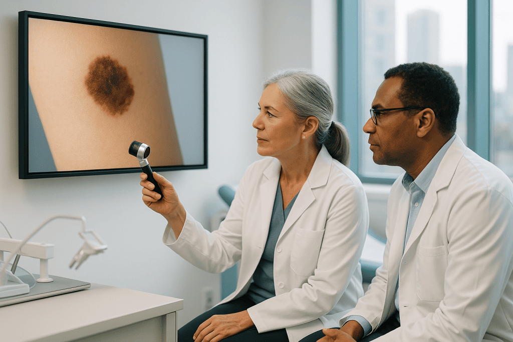
Diagnosis usually begins with a skin examination. Dermatologists use dermoscopy, a magnified, illuminated examination of the skin, to distinguish suspicious lesions from benign moles with far greater accuracy than the naked eye. For patients with many moles, digital skin imaging can map the whole skin surface so that new or changing lesions stand out at follow-up visits.
If melanoma is suspected, the lesion is removed or biopsied and examined by a pathologist. The pathology report is the foundation of every decision that follows. It typically records Breslow thickness (how deep the melanoma has grown, measured in millimetres), ulceration, mitotic activity, margin status and melanoma subtype. Breslow thickness and ulceration are the two features that most strongly shape the initial stage, which is why an incomplete or superficial biopsy can make accurate staging genuinely difficult.
Staging then extends outward from the skin as the risk demands. Thin, low-risk melanomas may need no investigation beyond the excision itself. Thicker or higher-risk tumours may call for lymph node ultrasound, sentinel lymph node biopsy, CT, PET-CT or MRI, depending on the clinical picture. For advanced disease, molecular testing of the tumour, most importantly for BRAF mutations, identifies whether targeted medicines are an option. When a biopsy was performed at another centre, pathology review is often recommended before finalising the plan, for the simple reason that the entire staging system rests on those few reported measurements.
How fast does melanoma skin cancer spread?
There is no single timetable; the honest answer is that it depends on the type of melanoma. Some melanomas, such as lentigo maligna on chronically sun-damaged skin, can remain confined to the surface for years. Superficial spreading melanoma, the most common type, typically grows outward across the skin for months before growing downward. Nodular melanoma is the exception that drives the urgency in melanoma care: it grows vertically from the outset and can thicken meaningfully within weeks to months. Because depth of invasion is what allows melanoma to reach lymphatic vessels and the bloodstream, speed of growth translates directly into risk. This is why a lesion that is visibly changing over weeks is treated as a different problem from one that has looked identical for a decade, and why evaluation should not wait on the assumption that all melanomas move slowly.
Conditions and Indications Addressed by Melanoma Treatment
Melanoma care covers a wide clinical range, from a thin tumour confined to the top layers of skin to metastatic disease involving distant organs. Each situation calls for a different intensity of treatment and follow-up:
- Melanoma in situ: melanoma cells are limited to the outermost layer of the skin. Treatment is usually surgical removal with appropriate margins, and nothing more.
- Early invasive melanoma: the tumour has entered deeper layers of the skin but shows no clear evidence of spread. Surgery is the main treatment, and sentinel lymph node biopsy may be considered depending on thickness and other features.
- Melanoma with higher-risk pathology: greater thickness, ulceration or high mitotic activity raises the risk of recurrence and influences staging, lymph node assessment and discussions about adjuvant therapy.
- Regional lymph node involvement: melanoma has reached nearby lymph nodes. Treatment may combine surgery, immunotherapy, targeted therapy and, in selected cases, radiotherapy.
- Locally advanced melanoma: the tumour has grown extensively in the skin or nearby tissue, or in-transit metastases have appeared between the original site and the lymph nodes. These cases need multidisciplinary planning and may involve systemic therapy before or after local treatment.
- Metastatic melanoma: the disease has spread to distant organs. Immunotherapy, targeted therapy, radiotherapy, surgery for selected lesions and supportive care may all play a role depending on the pattern of disease.
- Recurrent melanoma: melanoma has returned after previous treatment. Management depends on where it recurs, what therapies were used before and what current staging shows.
- Special-site melanoma: tumours under the nail, on the palms or soles, in mucosal areas or in the eye require diagnostic and surgical strategies adapted to those locations.
The purpose of matching treatment to stage and biology is to avoid two opposite mistakes: undertreatment, which allows the disease to progress, and overtreatment, which adds side effects without adding benefit. Most of the effort in a good melanoma programme goes into getting this match right before anything irreversible is done.
How Melanoma Treatment Is Performed
Treatment begins with preparation, not with an operation. Before a plan is finalised, the team reviews the biopsy report, dermoscopy records or photographs where available, prior imaging, laboratory results and your medical history. If the biopsy was performed elsewhere, pathology review may be recommended first. For most patients, the pathway then follows a recognisable sequence:
- The biopsy confirms the diagnosis and defines the tumour’s key features.
- Pathology and, where needed, imaging and lymph node evaluation establish the stage.
- A multidisciplinary tumour board reviews cases with complex staging or multiple reasonable options.
- Local treatment, usually surgery, removes the disease that can be removed.
- Systemic therapy or radiotherapy is added where the risk profile or the extent of disease justifies it, followed by structured surveillance.
Wide local excision
Wide local excision is the standard operation for melanoma. It removes the biopsy site together with a measured rim of normal-looking skin, because melanoma cells can extend microscopically beyond what the eye can see. The recommended margin depends on the tumour’s Breslow thickness and follows international guideline-based protocols, commonly in the range of half a centimetre to two centimetres. The procedure may be done under local anaesthesia, sedation or general anaesthesia, depending on the size and site of the lesion and whether lymph node surgery is planned at the same time. For melanomas in cosmetically or functionally sensitive areas, the face, hands, feet, or near joints, planning may involve reconstructive techniques to close the wound while preserving movement and appearance as far as possible.
Sentinel lymph node biopsy
A sentinel lymph node biopsy determines whether melanoma cells have reached the first lymph node or nodes that drain the tumour site. It is generally discussed when the melanoma is thicker than roughly 0.8 millimetres or carries other higher-risk features such as ulceration. A tracer technique maps the lymphatic drainage, usually with imaging before surgery and specialised detection tools in the operating theatre, and the sentinel node is removed through a small incision for pathological examination. If melanoma cells are found, the stage changes, and with it the conversation about further treatment. The procedure gives staging information; it is not itself a therapy, and understanding that distinction helps patients weigh it calmly.
Immunotherapy
Immunotherapy is one of the central treatments for advanced or high-risk melanoma. These medicines work by releasing natural brakes on the immune system so that immune cells can recognise and attack melanoma. They are typically given intravenously at regular intervals, either after surgery to reduce the risk of recurrence in higher-risk disease (adjuvant therapy) or as the main treatment for melanoma that has spread. Responses vary honestly and widely: some patients have strong, durable responses; others do not respond, or develop immune-related side effects affecting the skin, bowel, liver, lungs, thyroid or other glands. These effects are manageable when caught early, which is why monitoring and prompt reporting of new symptoms to the treating team are built into every immunotherapy schedule.
Targeted therapy
Targeted therapy is used when the tumour carries specific genetic changes, most often a mutation in the BRAF gene. These oral medicines block the signalling pathways that drive melanoma cell growth, and in tumours with the right mutation they can shrink disease relatively quickly, which matters when symptoms need prompt control. The choice between targeted therapy and immunotherapy, or the sequence in which to use them, depends on mutation status, the pace and burden of disease, side-effect profiles and durability considerations. It is one of the genuinely individual decisions in melanoma care, and a reasonable specialist will be able to explain why one order suits your situation better than the other.
Radiotherapy
Radiotherapy is not the main treatment for early melanoma, but it has clear roles in selected settings: treating melanoma that has spread to the brain or bones, controlling symptoms such as pain or bleeding, and reducing the risk of local recurrence after surgery in specific lymph node situations. Modern radiotherapy planning uses detailed imaging to shape the dose tightly around the target while limiting exposure to surrounding healthy tissue, which has made these focused uses considerably more precise than in earlier decades.
Technology supports melanoma care at every stage. Dermoscopy and digital skin imaging assist lesion evaluation and long-term monitoring of high-risk skin. High-resolution ultrasound assesses lymph node regions. CT, MRI and PET-CT stage higher-risk disease and measure treatment response. Nuclear medicine techniques guide sentinel node mapping. Pathology combines microscopic assessment with immunohistochemistry and molecular testing to sharpen the diagnosis and identify treatable mutations. In theatres and radiotherapy units, imaging-guided planning helps clinicians treat the intended target while protecting function and surrounding tissue.
How long treatment takes varies as much as the disease itself. A wide local excision alone may be completed as a same-day procedure, with recovery measured in days to weeks depending on the wound. Sentinel lymph node biopsy adds operative time and closer postoperative observation. Immunotherapy or targeted therapy may continue for months or longer, depending on the setting and response. Radiotherapy may run over a short or extended schedule depending on the indication. A good care team explains not only what is recommended but how long each phase should take and what follow-up will look like afterwards.
Recovery begins immediately after local treatment and continues through surveillance. After surgery, you receive instructions on wound care, activity limits, signs of infection and suture removal. If lymph nodes were removed, the team watches for fluid collections, numbness, stiffness and, less commonly, lymphoedema. During systemic therapy, recovery is less about an incision and more about managing fatigue, skin changes, digestive symptoms or hormone effects. Follow-up visits and imaging are scheduled according to stage, treatment type and recurrence risk, within the framework of a broader oncology programme rather than as isolated appointments.
Why Acting Early Matters
Melanoma is very treatable when found early, and considerably harder to control once it grows deep into the skin or spreads to lymph nodes and distant organs. Time matters because depth of invasion and the presence of spread are the dominant factors in staging. A small lesion removed completely at an early stage may need nothing further. The same lesion, left to progress before diagnosis, may require wider surgery, lymph node evaluation, systemic therapy or radiotherapy.
Each kind of delay carries its own cost. Delaying evaluation of a changing mole allows the tumour to thicken. Delaying definitive excision after a confirmed diagnosis prolongs uncertainty and can complicate surgical planning. Delaying staging in higher-risk melanoma can miss lymph node or distant disease that would change the treatment entirely. None of this means panic; it means the sensible default in melanoma is sooner rather than later.
Acting early is not the same as rushing. The best melanoma care is both timely and precise, and a short period spent confirming pathology, completing staging and reviewing the case in a multidisciplinary setting is time well used. The goal is straightforward: avoid unnecessary delay while making sure the first major treatment decision is the right one, because in melanoma the first decision shapes all the others.
Can melanoma skin cancer kill you?
Yes, it can, melanoma is the skin cancer responsible for most skin cancer deaths, because of its ability to spread to vital organs. That is the plain answer, and it is exactly why this disease is treated with the seriousness described on this page. But the risk is not uniform. Melanoma caught while it is thin and confined to the skin is usually removed completely with surgery, and for many patients that is the end of the medical story apart from follow-up. The danger rises with depth, ulceration and spread, which is why the entire logic of melanoma care is built around finding it early and staging it accurately.
Can you live a long life after having melanoma?
Yes. Many people treated for melanoma, particularly early-stage melanoma, go on to live long lives, and even in advanced disease, modern immunotherapy and targeted therapy have given a meaningful number of patients durable disease control that was not possible a generation ago. Living well after melanoma does come with obligations: people who have had one melanoma carry a higher risk of developing another, so lifelong sun protection, regular self-examination and scheduled dermatology reviews become part of ordinary life. Surveillance is not a sign that something is wrong; it is the mechanism by which anything new is caught while it is still small.
Benefits of Melanoma Treatment
The benefits of treatment depend on stage and modality, but the central aims are constant: control the cancer, reduce future risk and put honest, structured surveillance around whatever remains uncertain.
| Benefit | What It Means for You |
|---|---|
| Accurate staging | Clarifies whether melanoma is limited to the skin, involves lymph nodes or has spread, so treatment matches your actual risk rather than a guess. |
| Complete removal of localised disease | For early melanoma, surgery with appropriate margins can remove the cancer entirely and may be the only treatment required. |
| Personalised systemic treatment | For higher-risk or advanced melanoma, immunotherapy or targeted therapy is selected according to stage, tumour biology and your overall health. |
| Lower recurrence risk in selected patients | Adjuvant treatment after surgery may reduce the chance of melanoma returning for patients with certain higher-risk features. |
| Symptom control and function preservation | Surgery, radiotherapy and medicines can help control pain, bleeding, neurological symptoms or other effects of advanced disease. |
| Structured follow-up | Surveillance detects recurrence or new melanomas earlier and supports long-term skin health planning. |
Recovery Timeline After Melanoma Treatment
Recovery depends on whether treatment involved skin surgery alone, lymph node surgery, systemic therapy, radiotherapy or a combination. The broad shape of it looks like this:
| Time Period | What Patients Can Expect |
|---|---|
| Day 1 | After surgery, most patients receive wound care instructions and can often return home the same day. Mild pain, tightness or swelling around the incision is common. If systemic therapy begins, the care team explains possible early side effects and when to report them. |
| First week | Incisions begin healing, and activity may be limited depending on the operation site. Pathology results from the excision or sentinel node biopsy may become available and guide the next step. |
| First month | Most surgical wounds have healed substantially, although scars continue maturing for longer. Normal routines resume gradually. Patients on immunotherapy, targeted therapy or radiotherapy continue monitoring for side effects and response. |
| Several months | Follow-up focuses on skin examination, lymph node assessment, imaging when indicated and management of treatment effects. Adjuvant therapy may continue for patients with higher-risk melanoma. |
| Longer term | Surveillance stays important because melanoma can recur, and one melanoma raises the risk of another. Sun protection, self-skin checks and scheduled dermatology visits become part of ongoing care. |
What Influences Outcomes and a Good Result
Melanoma outcomes depend on a combination of tumour biology, treatment quality and patient-specific factors. On the tumour side, the most important elements are Breslow thickness, ulceration, lymph node involvement, the number and location of any metastases, melanoma subtype and molecular profile. A thin melanoma confined to the skin and a melanoma involving distant organs are, in practical terms, different diseases, which is why precise staging comes before every other decision.
The quality of the biopsy and its interpretation matters more than most patients realise. An incomplete or poorly oriented specimen makes staging harder, while a detailed pathology report supports sound decisions about margins, sentinel node biopsy and additional therapy. For complex or borderline cases, expert pathology review reduces uncertainty at the exact point where uncertainty is most expensive.
Timing and sequencing also shape results. Some patients benefit from surgery first; others, particularly with bulky or advanced disease, may benefit from systemic therapy before an operation. In metastatic melanoma, the choice between immunotherapy, targeted therapy, radiotherapy or a combined strategy depends on symptoms, the pace of the disease, mutation status, which organs are involved and what has been tried before.
A good result is more than a removed lesion. It includes safe healing, preserved function, an acceptable cosmetic outcome, controlled side effects, appropriate surveillance and a clear plan if the disease changes course. For melanoma on the face, hands, feet or nail unit, reconstruction and functional planning carry real weight. For patients on immunotherapy, early recognition of immune-related side effects allows prompt management and can prevent complications.
Your own participation matters too. Keeping appointments, reporting new symptoms to your treating team, protecting your skin from ultraviolet exposure, examining your own skin regularly and sharing a complete medical history all measurably support better care. When care moves between teams or institutions, original pathology reports, slides where available, imaging files, medication lists and prior treatment records let the receiving team work faster and more accurately.
How a Multidisciplinary Team Plans Melanoma Care
Because melanoma decisions rest on details, the first priority in specialised care is diagnostic confidence. The team may review prior pathology, request additional molecular testing or recommend updated imaging before finalising anything. Dermatology, surgical oncology, medical oncology, radiation oncology, pathology, radiology and nuclear medicine specialists contribute as needed, and cases with complex staging or several reasonable options are discussed in multidisciplinary tumour boards, where the aim is to align the recommendation with evidence-based international protocols and your individual situation.
Coordination is a practical necessity in melanoma specifically, because a patient may need several evaluations in a short window: dermatology assessment, surgical consultation, imaging, pathology review and oncology planning. These steps work best when they are scheduled as one connected sequence, with each specialist seeing the same complete picture, rather than as isolated appointments spread across months.
The technology described throughout this page, digital dermatological assessment, cross-sectional and nuclear imaging, molecular pathology, precision radiotherapy planning, sentinel node mapping and localisation, is applied within this structure. Equally important is what happens around the technology: timely communication between specialists, a clear explanation of why a plan is recommended and what alternatives exist, and medical summaries and follow-up recommendations prepared so that whichever physicians follow you afterwards can continue surveillance seamlessly. Two patients with the same diagnosis may need entirely different strategies: a thin melanoma on the shoulder, a scalp melanoma with positive lymph nodes and a BRAF-mutated metastatic melanoma are three different conversations, and the plan should reflect the stage, the biology, your health and your goals.
Reducing Risk and Protecting Your Skin Long Term
Whether you have had melanoma or are simply at higher risk, the habits that protect your skin are the same, and they are worth stating plainly. Seek shade during the middle of the day, when ultraviolet radiation is strongest. Wear clothing that covers the skin, a wide-brimmed hat and sunglasses when you are outdoors for extended periods. Use a broad-spectrum sunscreen on exposed skin and reapply it after swimming, sweating or several hours outside. Avoid tanning beds entirely: the ultraviolet exposure they deliver is a recognised cause of melanoma, and there is no safe version of a deliberate tan from artificial UV.
Self-examination is the second pillar. A regular, systematic check of your own skin, front and back, sides, scalp, ears, palms, soles, between the toes, under the nails and, with a hand mirror or help from another person, the back of the body, turns your familiarity with your own skin into a genuine early-detection tool. What you are looking for is change: a new spot, a mole that has altered, a streak under a nail that was not there before. Photographs of your moles, taken in consistent lighting, make change visible in a way memory cannot.
For people who have already had melanoma, these habits sit alongside scheduled dermatology reviews rather than replacing them. Household members can also benefit from learning the ABCDE pattern, since changes on the back, scalp and other hard-to-see areas are often first noticed by someone else. None of this is burdensome once it becomes routine, and in a disease where thickness at diagnosis drives everything that follows, routine looking is one of the most powerful tools anyone has.
Preparing for a Specialist Review or Second Opinion
If you have a confirmed melanoma, a suspicious lesion under investigation, or a treatment plan you want checked, a structured specialist review is a normal part of careful melanoma care rather than a sign of distrust. Early melanoma may need nothing beyond well-planned surgery; higher-risk disease may call for lymph node evaluation, systemic therapy or radiotherapy; advanced melanoma requires a coordinated strategy weighing tumour biology, symptoms and the full range of modern options. A review is most useful when the reviewing team can see everything the first team saw.
Wherever you are treated, the materials that make a review meaningful are the same: the original biopsy and pathology reports, pathology slides or tissue blocks where available, imaging scans in their original digital format, operative notes from any previous surgery, laboratory results and a current medication list. Complete records reduce repeated tests, shorten the time to a firm recommendation and make disagreements between opinions easier to resolve on evidence rather than assumption. Melanoma care works best when it is timely, accurate and individualised, and complete information is what makes all three possible.
Preparation
- Preparation usually includes a dermatologic examination, biopsy review, imaging or PET-CT when staging is needed, and blood tests before systemic therapy. Patients should share all medications, allergies, previous cancer treatments, and medical history. Sun exposure precautions and skin mapping may be recommended before treatment planning.
Aftercare
- Aftercare depends on the treatment used and may include wound care, scar monitoring, follow-up skin checks, and management of immunotherapy or targeted therapy side effects. Patients should protect their skin from sun exposure and report new or changing moles promptly. Regular oncology and dermatology follow-ups are important to detect recurrence or new skin cancers early.
Frequently Asked Questions
What affects the cost of melanoma skin cancer care?
The final cost depends on the extent of melanoma, required pathology and molecular tests, imaging, surgery type, hospital stay, systemic treatment, radiation needs and follow-up plan. International patient services such as translation, transfers and accommodation support may also affect the package scope.
How can I get a personalized quote from Acibadem?
You can request a free consultation by sharing your pathology report, imaging results, previous treatment notes and clear photos of the lesion or surgical area if relevant. The medical team can then review your case and prepare a personalized plan and quote; this is general information and not medical or financial advice.
Is melanoma treatment usually offered as a package?
Some parts of care may be packaged, especially consultations, selected diagnostic tests, surgery-related hospital services and coordination support. Medicines such as immunotherapy or targeted therapy, additional imaging, extended admission or extra procedures may be quoted separately depending on the case.
Why can the cost change after I arrive?
Costs can change if specialist examination, pathology review or staging tests show that the melanoma is more complex than expected, or if additional treatment is recommended. Your team should explain any change in the care plan before proceeding whenever possible.
Does choosing Turkey mean I will receive the same treatment options as elsewhere?
Many internationally used melanoma options, including surgery, immunotherapy, targeted therapy and radiation, may be available depending on the hospital and the patient’s condition. The appropriate option is determined by melanoma features, test results, patient health and specialist multidisciplinary review.
Medically reviewed by the Acıbadem International Medical Board August 30, 2026
See our medical review board →
Update history
- PublishedJune 5, 2026
- Medical review approvedAugust 30, 2026
- Last content updateSeptember 8, 2026
References3
- Melanoma Treatment (PDQ) – Patient Version cancer.gov
- Melanoma skin cancer nhs.uk
- Melanoma medlineplus.gov
Trusted care for international patients
From the Acibadem Blog

What a Melanoma Tumor Board Decides and Why Several Specialists Review Your Case
A melanoma tumor board is a scheduled meeting where dermatologists, surgeons, medical and radiation oncologists, pathologists and radiologists review one patient's slides, scans and…
Immunotherapy Side Effects in Melanoma Care: What Is Expected and What Needs a Call
Most immunotherapy side effects in melanoma care, such as mild fatigue, itch or a faint rash, are expected and manageable, but they can escalate…
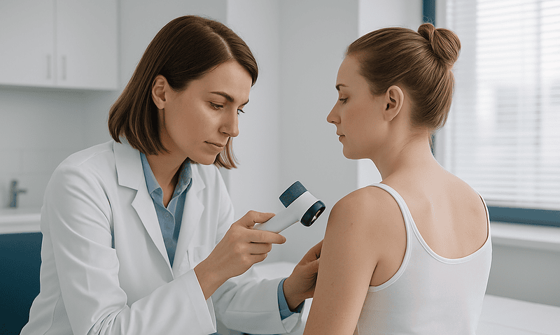
Planning Melanoma Skin Cancer Treatment by Stage: Surgery, Drug Therapy or Both?
Melanoma treatment is planned by stage. Stage 0, 1 and most stage 2 melanomas are usually treated with surgery alone, removing the tumor with…