Amelanotic Melanoma: Early Signs, Risk Factors, and How It Is Treated
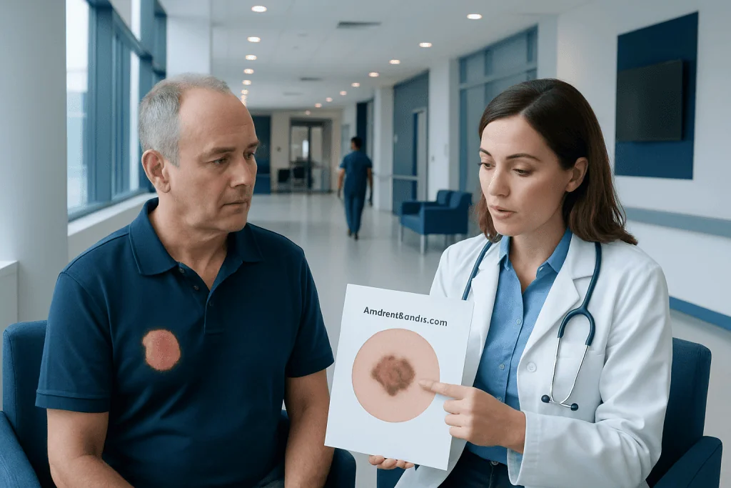
Amelanotic melanoma is a melanoma that lacks the usual dark pigment, so it can look pink, red, or flesh-colored and be mistaken for a pimple, scar, or sore. See a dermatologist promptly if a spot is new, growing, bleeding, or not healing within a few weeks. Diagnosis usually requires a skin biopsy.
Key Takeaways
- Amelanotic melanoma is a melanoma that lacks the typical dark color many people associate with skin cancer.
- It may look pink, red, skin-colored, or slightly tan, which can delay recognition.
- Warning signs include a new or changing lesion, persistent bleeding, crusting, itching, or a spot that does not heal.
- Diagnosis usually involves a skin examination, dermoscopy, and biopsy.
- Treatment depends on stage and may include surgery, lymph node evaluation, immunotherapy, targeted therapy, or radiation.
- Sun protection and regular skin checks can help support earlier detection.
Most of us are told to watch for a dark, irregular mole. But what if the spot that matters is pink?
Amelanotic melanoma is a type of melanoma with little or no dark pigment, so it may appear pink, red, flesh-colored, or lightly brown rather than black or dark brown. Because it can resemble harmless skin changes, early evaluation of a new, changing, or unusual spot is important.
Overview: what amelanotic melanoma is
Amelanotic melanoma is a form of melanoma, a skin cancer that begins in pigment-producing cells called melanocytes. Unlike more typical melanomas, it contains little or no melanin, the pigment that usually gives melanoma a dark brown, black, or blue-black color. As a result, it may appear pink, red, pale, flesh-colored, or only lightly pigmented.
This lack of obvious color is one reason amelanotic melanoma can be harder to recognize. Some lesions resemble a pimple, scar, patch of eczema, inflamed mole, or a small nonhealing sore. Others may be flat at first and then become raised or irritated over time.
Amelanotic melanoma can develop anywhere on the body. It may arise on sun-exposed skin, such as the face, neck, arms, and legs, but it can also appear in less expected areas. Like other melanomas, early diagnosis matters because treatment is generally simpler and outcomes are better when the cancer is found before it has spread.
How it can look and feel: early signs and symptoms
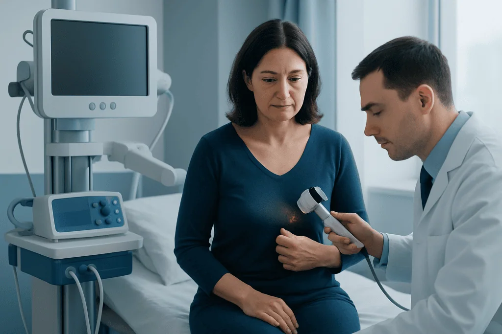
The appearance of amelanotic melanoma varies, but a common clue is a spot that looks different from surrounding skin and continues to change. Instead of relying only on the classic “dark mole” image, it helps to watch for a lesion that is new, growing, asymmetrical, irregular at the edges, or uneven in surface texture.
People may notice a pink or red bump, a shiny patch, a flat pale lesion, or a sore that crusts and then returns. Some lesions itch, sting, bleed with minor contact, or fail to heal over several weeks. In others, there may be no discomfort at all, which is why visual change is such an important sign.
In practice, clinicians often consider the “ugly duckling” idea: a spot that stands out from a person’s other moles or skin marks. Helpful warning signs include:
- A new pink, red, or skin-colored spot that persists
- A lesion that grows quickly or changes shape
- Bleeding, crusting, or repeated irritation
- A patch or bump that does not heal as expected
- A mole or mark that looks different from nearby lesions
Not every unusual skin spot is melanoma. Rosacea, psoriasis, eczema, basal cell carcinoma and plenty of harmless growths can look much the same. Even so, if a lesion sticks around or keeps changing, have it looked at — particularly when you cannot explain where it came from.
Causes and risk factors

Amelanotic melanoma develops when melanocytes begin to grow in an uncontrolled way. The exact trigger is not always clear, but it likely involves a combination of genetic changes within skin cells and environmental exposure, especially ultraviolet radiation from sunlight or tanning devices.
Risk factors overlap with those for other forms of melanoma. People with fair skin, light eyes, a history of sunburns, heavy cumulative sun exposure, or indoor tanning use may have a higher risk. A personal or family history of melanoma also increases concern, as do having many moles, atypical moles, or a weakened immune system.
Age can play a role, but melanoma can occur in younger adults as well. Amelanotic melanoma may be especially difficult to notice in people who do not expect melanoma because the lesion lacks dark pigment. For that reason, awareness of texture change, growth, and nonhealing skin lesions is as important as watching for color changes.
Quite a few of these lesions are first treated as something else entirely. That is why dermatologists tend to follow suspicious spots closely and biopsy readily when one behaves oddly — even if it looks pale and unremarkable.
How amelanotic melanoma is diagnosed
Diagnosis begins with a careful skin examination and a discussion of when the lesion appeared, how it has changed, and whether there has been bleeding, itching, or failure to heal. A dermatologist may use dermoscopy, a handheld tool that magnifies the skin and helps reveal vascular and structural patterns not visible to the naked eye.
If a lesion looks suspicious, the key next step is a biopsy. This means removing all or part of the lesion so a pathologist can examine it under a microscope. A biopsy is the only way to confirm whether a lesion is amelanotic melanoma or another skin condition. If melanoma is confirmed, the pathology report helps guide treatment by describing features such as depth and ulceration.
Further testing depends on the stage and clinical findings. Some patients may need imaging studies or evaluation of nearby lymph nodes, especially if the melanoma is thicker or there are signs it may have spread. In selected cases, doctors may discuss sentinel lymph node biopsy as part of staging.
Because it mimics so many things, amelanotic melanoma is sometimes mistaken at first for another skin cancer or an inflammatory rash. Seeing a specialist sorts this out and keeps treatment from being delayed.
Treatment options and what they depend on
Treatment for amelanotic melanoma depends mainly on the stage of disease, the thickness and location of the lesion, and whether it has spread to lymph nodes or other organs. For localized melanoma, surgery is usually the main treatment. The goal is to remove the melanoma completely with a margin of normal-looking skin around it.
If the melanoma has certain higher-risk features, doctors may recommend additional staging or treatment. This can include sentinel lymph node biopsy to check whether cancer cells have reached the nearest lymph node. The results help estimate risk and guide further decisions.
For more advanced melanoma, treatment may involve systemic therapies such as immunotherapy or targeted therapy, depending on the tumor’s molecular profile. Some patients may also receive radiation therapy in specific situations, such as treatment of lymph node areas or symptom control. Surgical management may still be part of care when feasible, including melanoma surgery.
Care is often coordinated by specialists in dermatology, surgical oncology, pathology, radiology, and medical oncology. In some cases, the broader treatment pathway overlaps with strategies used for melanoma in general, while still accounting for the unique diagnostic challenges of the amelanotic form. Near the end of the care pathway, some international patients choose centers such as Acibadem International, where multidisciplinary specialists in JCI-accredited hospitals evaluate and treat melanoma-related conditions.
Prevention and self-checks
Amelanotic melanoma cannot always be prevented, but reducing ultraviolet exposure is an important practical step. Using broad-spectrum sunscreen, seeking shade, wearing protective clothing, and avoiding tanning beds may help lower the risk of skin damage associated with melanoma. Children and adolescents especially benefit from consistent sun protection habits that continue into adulthood.
Regular skin self-checks can support earlier detection. Since amelanotic melanoma may not be dark, self-checks should focus on the full picture: a new spot, a spot that grows, a lesion that bleeds, or an area that simply does not look or behave normally. Examining hard-to-see areas with a mirror or asking a partner for help can be useful.
People at higher risk may benefit from routine dermatologist visits, particularly if they have a history of melanoma, many atypical moles, or significant sun exposure. Clinical photography or mole mapping may sometimes help monitor changes over time. If a spot changes between scheduled visits, it should not wait for the next routine appointment.
Everyday skin habits count as well. Do not pick at a lesion, keep track of scabbing that keeps coming back, and use photos to record change — not to replace a doctor’s opinion. If a spot worries you, get it checked without waiting.
When to seek medical care
Medical attention is advisable if a skin spot is new, changing, unusual for that person, or not healing after several weeks. This is especially true for pink, red, or skin-colored lesions that bleed easily, crust repeatedly, or grow over time. A lesion does not need to be dark to require evaluation.
Prompt review is also important if there is a personal or family history of melanoma, a weakened immune system, or many atypical moles. Patients should seek care sooner rather than later if a lesion becomes painful, ulcerated, or associated with swollen lymph nodes nearby. Early examination may allow simpler treatment if melanoma is confirmed.
Most suspicious lesions turn out to be nothing, so getting one checked is not a reason to panic. But amelanotic melanoma is quiet by nature — do not brush off a change that keeps going, and do not decide on your own that it is harmless.
Frequently asked questions
Is amelanotic melanoma more dangerous than other melanomas?
It is not necessarily biologically more dangerous in every case, but it can be harder to recognize because it lacks the usual dark pigment. That means diagnosis may sometimes happen later than with more typical melanomas. The stage at diagnosis is one of the most important factors affecting treatment and outlook.
What color is amelanotic melanoma?
Amelanotic melanoma is often pink, red, flesh-colored, or lightly tan rather than black or dark brown. Some lesions may also look shiny, inflamed, or like a nonhealing sore. Because color can be misleading, change over time is an important warning sign.
Can amelanotic melanoma look like a pimple or rash?
Yes. It may resemble a pimple, eczema patch, scar, irritated mole, or small sore, which is one reason it may be overlooked. A spot that persists, grows, bleeds, or does not heal should be checked by a qualified clinician.
How is amelanotic melanoma confirmed?
It is confirmed with a biopsy, which allows a pathologist to examine the tissue under a microscope. A skin exam and dermoscopy help identify suspicious lesions, but biopsy is needed for a definite diagnosis. If melanoma is found, additional staging tests may be recommended depending on the pathology results.
Can amelanotic melanoma be cured?
Many cases can be treated effectively, especially when found early and removed completely with surgery. More advanced cases may still be treatable with modern therapies such as immunotherapy or targeted treatment. Prognosis varies from person to person, so a specialist should explain the expected outlook based on stage and pathology.
Should every pink spot on the skin be evaluated?
Not every pink spot is cancer, and many are harmless or related to common skin conditions. However, a pink or skin-colored lesion should be evaluated if it is new, changing, bleeding, crusting, or not healing. If there is uncertainty, a dermatologist can decide whether monitoring or biopsy is appropriate.
This article is for general information only and is not a substitute for professional medical advice. Please consult a qualified doctor about your individual situation.
Melanoma in Turkey — costs, top hospitals & a free quote
7 JCI-accredited hospitals · board-certified surgeons · reply within 24h
Add us as a Preferred Source to see more of our trusted health content across Google Search, AI Overviews and Discover.

Medically reviewed by the Acıbadem International Medical Board — August 24, 2026
See our medical review board →
Update history
- PublishedJuly 29, 2026
- Medical review approvedAugust 24, 2026
- Last content updateAugust 24, 2026
References3
- Melanoma Treatment (PDQ®) – Patient Version — cancer.gov
- Melanoma – MedlinePlus — medlineplus.gov
- Melanoma skin cancer – NHS — nhs.uk
Medical Oncology Department
Chemotherapy, immunotherapy and targeted therapy planned by a multidisciplinary tumor board — coordinated cancer care across Acibadem's JCI-accredited hospitals.
61 specialists in this unitDermatology Specialists at Acibadem





Related Treatments
Related Conditions
More from the Health Library

Eczema Treatment: Symptoms, Causes, and Treatment Options

Alopecia Universalis Causes Total Hair Loss but Follicles Survive

Cracked Skin Reflects a Dry, Damaged Barrier, Not Poor Hygiene

Hair on the Penile Shaft Is Usually a Normal Variation
Bed Sores Start as Skin Redness That Does Not Fade