Cryotherapy
Cryotherapy uses controlled extreme cold to destroy abnormal tissue, such as skin lesions or selected tumors. It is a minimally invasive option planned according to lesion type, size, and location.
Quick answer
Cryotherapy destroys abnormal tissue by freezing it with controlled extreme cold, usually liquid nitrogen or argon gas. Dermatologists use it for warts, actinic keratoses and selected superficial skin lesions. Image-guided cryoablation treats certain small tumours in organs such as the kidney, lung, liver, bone and prostate. The frozen cells die, and the body clears or heals the treated area over the following weeks.
Cryotherapy: Treating Abnormal Tissue with Controlled Extreme Cold
Cryotherapy is a medical treatment that destroys abnormal tissue by freezing it. Controlled extreme cold is applied to a precise target: a wart, a sun-damaged patch of skin, a precancerous area of the cervix, or in selected cases a small tumour inside the body, and the frozen cells die. It is used for people whose lesion has been properly diagnosed and is small and well-defined enough for a focused local treatment to make sense.
Being told you have an abnormal skin lesion, a precancerous change or a small tumour raises immediate questions. Is it dangerous? Does it need to come off? Will treatment hurt? Will there be a scar? If the abnormal area sits inside the body, the questions multiply. Most patients want a treatment that is as focused as possible, disrupts daily life as little as possible, and comes with a recovery plan they can understand before committing.
Cryotherapy answers some of those wishes, but not all of them, and not for everyone. The principle is simple; the planning is not. The medical team must understand the lesion type, its size, its depth, its exact location and your overall health before recommending freezing over the alternatives. A wart on a finger, a suspicious pigmented spot and a small kidney tumour all sit under the same word, yet they call for completely different assessments and completely different levels of caution.
At Acibadem, cryotherapy sits inside a diagnostic and treatment pathway rather than standing alone as a technical act. Depending on the case, dermatologists, gynaecologists, interventional radiologists, oncologists, surgeons, pathologists and radiologists weigh in on whether freezing is appropriate, whether another treatment would be safer or more effective, and what follow-up the treated area will need. The aim throughout is the same: destroy the abnormal tissue while preserving as much healthy tissue as possible. For many patients that means treatment without a large incision, outpatient care or a short hospital stay, and a recovery that is manageable with clear instructions. It remains a medical procedure, and it has to be matched carefully to the right diagnosis and the right patient.
What Is Cryotherapy?
Cryotherapy, sometimes called cryoablation when it is used for tumours or deeper tissues, is the deliberate application of very low temperatures to abnormal cells. The cold causes ice crystals to form inside and around the targeted cells, disrupts the blood supply to the treated tissue and leads to cell death. Over the following days and weeks, the body clears or heals the treated area, depending on where it sits and what was treated.
It is not a single uniform treatment. The dose of cold, the number of freeze–thaw cycles, the way the cold is applied and the recovery plan all vary. A small wart is not treated the way a suspicious skin lesion is, and neither resembles the treatment of a cervical precancerous area or a selected kidney, lung, liver, bone or prostate lesion. That is why careful diagnosis comes first. Some lesions need a biopsy before anyone freezes them; others need imaging to establish whether cryotherapy can reach them safely, or whether surgery, radiotherapy, medication, observation or a different minimally invasive method would serve you better.
How does cryotherapy work?
Cryotherapy works by freezing tissue fast enough, and deeply enough, to kill the targeted cells. Ice crystals rupture cell structures from within. Freezing also damages the tiny blood vessels feeding the treated zone, so cells that survive the initial freeze are starved of blood supply afterwards. Many protocols freeze the tissue, allow it to thaw, then freeze it again: the thaw phase makes the second freeze more destructive. In dermatology the cold reaches the lesion through a spray, a probe or a small applicator held against the skin. For internal treatment, thin needle-like probes carry the cold directly into the target, and imaging shows the physician exactly where the ice is forming so that the freeze covers the lesion and spares what surrounds it.
How cold is cryotherapy?
Cryotherapy uses temperatures far below anything encountered in daily life. Liquid nitrogen, the agent most often used on the skin, boils at around −196°C, and the spray or applicator delivers a freeze cold enough to turn the treated spot white within seconds. Internal cryoablation probes typically use pressurised argon gas, which cools the probe tip to temperatures well below the point at which tissue can survive, forming a visible ball of ice around the target. The physician controls how long the cold is applied and how far the ice is allowed to spread, because the depth and duration of the freeze, not just the temperature, determine both how effective the treatment is and how much surrounding tissue is affected.
Is cryotherapy the same as cryosurgery?
Cryosurgery is essentially another name for the same treatment, used when freezing takes the place of a surgical procedure, destroying a lesion that might otherwise have been cut out. Dermatologists, gynaecologists and interventional radiologists may use the terms cryotherapy, cryosurgery and cryoablation somewhat interchangeably. The label matters less than the plan behind it: what is being frozen, why, to what depth, and what happens afterwards. One practical difference from conventional surgery is worth understanding early. Freezing destroys tissue in place rather than removing it intact, so there is usually no complete specimen for the pathologist to examine afterwards. When cancer is possible, the diagnosis has to be secured before the freeze, not after it.
What is a cryo chamber?
A cryo chamber is a whole-body cold-exposure unit: a cabin or room chilled to very low temperatures in which a person stands for a few minutes, and it is a different thing from the medical cryotherapy described on this page. Whole-body cold exposure, often shortened to cryo, is used in sports recovery and wellness settings; you can read about that approach on our CRYO page. It does not destroy tissue and it does not treat lesions, precancerous changes or tumours. Medical cryotherapy is targeted, lesion-directed and performed by a physician for a diagnosed condition. If you have searched for one and found the other, the distinction is worth keeping in mind while you read.
What does cryotherapy do for your body?
That depends entirely on which cryotherapy you mean. Medical cryotherapy does one thing: it destroys a defined area of abnormal tissue so the body can clear it and heal, which is why its value rests on choosing the right target. Whole-body cold exposure in a cryo chamber works differently, brief cold narrows blood vessels in the skin and may temporarily ease muscle soreness, but it is a general exposure, not a treatment aimed at a lesion, and the evidence base for its broader wellness claims is still developing. This page deals with the medical use: focused freezing of diagnosed abnormal tissue.
What Is Cryotherapy Used For?
Cryotherapy is used across several specialties, from common skin conditions to selected tumours treated by image-guided ablation. In dermatology, it treats warts, seborrheic keratoses, skin tags in selected situations, actinic keratoses caused by sun damage, and other benign or precancerous lesions. Certain superficial forms of skin cancer may be considered for freezing in carefully chosen patients, particularly when surgery is not preferred and the lesion’s characteristics make cryotherapy reasonable. In gynaecology, it treats selected precancerous changes of the cervix once strict criteria are met. In interventional radiology and oncology, cryoablation is used for selected small tumours in organs such as the kidney, lung, liver, bone, soft tissue and prostate, and sometimes to reduce pain from bone or soft-tissue lesions as part of palliative care. In some settings it is also applied to nerves to interrupt pain signals, an application that demands careful selection to avoid nerve damage or functional problems.
One caution belongs in this list rather than in a footnote. Pigmented lesions should not be frozen on appearance alone. A spot that could be melanoma needs biopsy and specialist assessment first, because destroying it without a diagnosis removes the evidence, delays proper cancer care and may allow a dangerous tumour to progress unrecognised. The same logic applies to any suspicious or high-risk lesion: diagnosis first, destruction second.
Does cryotherapy work?
For the right lesion, yes, cryotherapy is a long-established, well-studied method of destroying abnormal tissue, and for many benign and precancerous skin lesions it is a standard first-line option. How well it works in an individual case depends on the diagnosis, the lesion’s size and depth, the skill of the application and the follow-up plan. Some lesions clear after a single session; others, such as stubborn warts, need repeat treatments. Some are simply the wrong target for freezing, too large, too deep, too aggressive, or sitting where the freeze would threaten an important structure, and for those, a different treatment works better. Honest answer: cryotherapy works when the selection is right, and selection is where the real medical skill lies.
Cryogenic skin tag removal
Cryogenic skin tag removal freezes the narrow base of a skin tag so that the tag loses its blood supply, darkens and falls away as the area heals. Skin tags are small, soft, usually painless growths that are harmless in themselves, but they can catch on clothing or jewellery, become irritated, or simply bother you cosmetically. Freezing is one of several ways to deal with them, alongside snip excision and cautery, and the choice depends on the tag’s size, location and your skin type. A word of caution here too: not everything that looks like a skin tag is one. A growth that has changed, bleeds, or looks different from your other tags deserves a proper look before anyone freezes it off.
Who May Be Considered for Cryotherapy
Patients usually arrive at a cryotherapy discussion by one of three routes: a visible lesion on the skin, an abnormal screening result, or an imaging study that has detected a small mass or a localised area of disease. Which route you took shapes the questions the team needs to answer before treating anything.
For skin conditions, the trigger is often something you noticed yourself: a rough patch, a wart-like growth, a small raised lesion, a scaling area, or a spot that has changed. Some of these are harmless but bothersome. Others, such as actinic keratoses on sun-exposed skin, are precancerous and may be treated to reduce the risk of progression. A few will turn out to be superficial skin cancers for which cryotherapy is one of several options, depending on type, depth, location, recurrence risk and whether a tissue diagnosis has been obtained.
For gynaecological indications, the route usually runs through screening: Pap testing, HPV testing, colposcopy and biopsy. Cervical precancerous changes generally cause no symptoms at all, which is precisely why screening finds them. Not every abnormal result is suitable for freezing: the clinician must be able to see and assess the entire abnormal area and must be confident there is no invasive disease that would demand a different treatment.
For internal tumours, the conversation typically starts after ultrasound, computed tomography, magnetic resonance imaging or another study identifies a localised lesion, sometimes discovered while investigating something else entirely. Cryoablation enters the discussion when the tumour is small, positioned where probes can reach it safely, and located where freezing will not put critical structures at unacceptable risk. It is also considered for patients who are not ideal candidates for major surgery because of age, other medical conditions, reduced organ reserve or previous treatments.
Symptoms, when they exist, vary enormously: a skin lesion may itch, bleed or catch on clothing; an internal lesion may cause pain, bleeding, urinary symptoms or cough, or nothing at all. None of these symptoms is specific to conditions treatable by freezing, which is why the diagnosis, not the symptom, drives the decision. Assessment usually combines clinical examination, medical history, imaging, laboratory tests and sometimes biopsy, all aimed at two questions: has this lesion been adequately characterised, and can cryotherapy reach it safely? Having previous pathology reports, imaging files, operative notes and treatment summaries gathered in one place makes it far easier for a specialist to judge whether an evaluation is likely to end in cryotherapy or in a different recommendation.
Conditions and Indications: Where Freezing Fits, and Where It Doesn’t
Suitability for cryotherapy rests on diagnosis, lesion behaviour, anatomical location and the goal of treatment, and it is worth understanding the boundaries as clearly as the indications.
In dermatology, freezing suits well-defined, superficial lesions with a confirmed or confidently assessed diagnosis. For suspicious or high-risk lesions, biopsy comes first, because a lesion destroyed without diagnosis can hide a cancer that needed wider treatment. Some skin cancers require surgical excision with a margin of healthy tissue, and larger excisions on the face or limbs sometimes need reconstruction after skin disease or skin grafting to restore the area, considerations that belong in the treatment decision from the beginning, not after a freeze has failed.
In gynaecology, cryotherapy treats selected precancerous cervical changes. It is not a treatment for invasive cervical cancer, and it is not appropriate when the abnormal area cannot be fully visualised or when the evaluation suggests a more advanced lesion. Follow-up screening after treatment is part of the plan, not an optional extra.
In interventional oncology, cryoablation may serve as a primary local treatment for small tumours, as a pain-relieving measure for bone or soft-tissue lesions, or as local control of limited metastatic disease in selected patients. These decisions usually pass through a multidisciplinary board, because the best answer may be surgery, radiotherapy, systemic medication, active surveillance or a combination, and because cryoablation treats only the frozen zone. If disease is systemic, or carries a high risk of microscopic spread, a purely local treatment cannot address it alone.
The recurring theme is the pathology question. Because freezing destroys tissue in place, it rarely yields a complete specimen afterwards. In some tumours a biopsy is taken before or during the same session; in others, surgical removal is preferred precisely because the team needs full information about margins, depth or lymphatic spread. A well-run cryotherapy programme is defined as much by the cases it declines as by the cases it treats.
How Cryotherapy Is Performed
Every cryotherapy pathway begins with planning. Your physician reviews your history, medications, allergies, previous treatments and the reason freezing is on the table. Reviewing existing reports and images early in the process clarifies what additional tests are still needed and whether cryotherapy looks appropriate at all.
What happens during skin cryotherapy?
Skin cryotherapy is usually brief and follows a predictable sequence:
- Assessment. The doctor examines the lesion, confirms the diagnosis or decides whether dermoscopy or biopsy is needed first, and explains the expected healing and cosmetic changes.
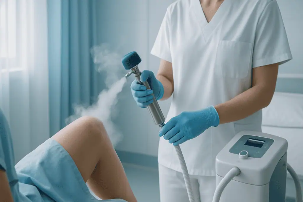
- Application. The cold agent, most often liquid nitrogen, is applied directly to the lesion by spray, probe or applicator for a controlled period. The spot turns white briefly, then reddens and may swell. You may feel stinging, burning or aching during the application and for a short time afterwards.
- Aftercare instructions. You are told how to keep the area clean, what the blister or crust will look like as it forms, and what changes to report while it heals.
- Review. Small lesions may need only minutes of actual treatment; larger or multiple lesions may need staged sessions, and some lesions require more than one visit before they clear.
Cervical cryotherapy follows a similar logic in a gynaecological setting: the clinician visualises the cervix, places the cryotherapy probe against the treatment area and applies freezing according to the planned protocol. Cramping or pressure is common during the procedure, which is typically brief, though assessment and post-procedure observation extend the visit. A watery discharge lasting several weeks is common afterwards as the treated tissue sloughs and the cervix heals, and your clinician will advise how long to avoid tampons and intercourse while that happens. Follow-up matters here, both to confirm healing and to continue cervical screening on the schedule your clinician sets.
How is image-guided cryoablation performed?
Internal cryoablation is a more involved procedure, and its preparation reflects that:
- Fitness and planning. Blood tests assess clotting, kidney function and general fitness for anaesthesia or sedation. Decisions about blood-thinning medication rest with the treating doctor, who weighs the bleeding risk of the procedure against the reason the medication was prescribed. Imaging is reviewed to plan the safest path to the lesion, taking account of nearby organs, vessels, nerves, bowel, airways and anything else that must be protected.
- Anaesthesia. On the day, you may receive local anaesthesia, conscious sedation, regional anaesthesia or general anaesthesia, depending on the treatment site, the expected duration and your condition.
- Probe placement. Under ultrasound, CT, MRI or other imaging guidance, the physician inserts one or more thin cryoprobes into the target. Imaging confirms position before any freezing begins.
- Protective measures. Where a sensitive structure sits too close, the team may create distance, for example, by introducing fluid or gas into a space to push or insulate nearby tissue away from the developing ice.
- Freeze–thaw cycles. The tissue is frozen, thawed and usually frozen again. The visible ice zone is monitored on imaging so that it covers the lesion, with a planned margin where appropriate, while sparing healthy tissue.
- Recovery and monitoring. Afterwards you are observed in a recovery area. Some patients go home the same day; others stay overnight or longer depending on the organ treated, the anaesthesia used and their overall condition.
One practical advantage of cryoablation over heat-based ablation methods is that the growing ice zone is directly visible on CT and MRI, which lets the team confirm in real time that the freeze covers the target before the procedure ends. Total procedure times vary widely. A simple skin treatment takes minutes; image-guided cryoablation may take one to several hours once probe placement, imaging, freeze cycles and recovery monitoring are counted. Modern practice leans heavily on precision: planning software, temperature-aware techniques and carefully designed cryoprobes for internal work, and controlled application tools for the skin, all aimed at hitting the target and nothing else.
Is cryotherapy very painful?
Skin cryotherapy stings and burns during the freeze and often aches for a short while afterwards, uncomfortable rather than unbearable for most people, and brief. As the area heals, blistering and tenderness can add a few days of soreness. Cervical cryotherapy commonly causes cramping similar to period pain. Internal cryoablation is performed under anaesthesia or sedation, so the procedure itself is managed for comfort; afterwards, mild pain, bruising and fatigue at or around the treatment site are common and are usually controllable with the pain-relief plan your team prescribes. Nobody should promise you a comfortable week, but for most indications the discomfort is short-lived and predictable, and your team will tell you honestly what to expect for your specific treatment.
Why Acting Early Matters
Not all abnormal tissue behaves the same way, which is why early evaluation matters more than early treatment. A small lesion may be harmless, precancerous or malignant. Some can be observed safely; others should be treated before they grow, spread, bleed, become painful or come to require more extensive therapy. Cryotherapy is at its most useful when the target is well defined and still small enough for focused treatment.
For sun-damaged skin and precancerous lesions, early treatment may reduce the chance that abnormal cells progress to something more serious, and it can stop lesions becoming larger, thicker or harder to treat cosmetically. For warts and benign growths, early care can relieve discomfort, reduce irritation and, in appropriate cases, limit spread. For cervical precancerous changes, timely evaluation and treatment can be important in preventing progression to invasive disease, and because early cervical abnormalities usually cause no symptoms, delaying follow-up after an abnormal screening result allows changes to advance unnoticed.
For internal tumours, timing shapes the menu of options. A small tumour suitable for cryoablation today may become harder to treat if it enlarges or grows towards critical structures, and in cancer care delay can allow disease to move beyond a purely local strategy. That does not mean every lesion needs immediate intervention, some small tumours are monitored deliberately, and active surveillance is a legitimate plan. But observation should be a decision based on evidence, not a drift caused by uncertainty or poor coordination. The realistic costs of postponement are growth of the abnormal tissue, loss of eligibility for minimally invasive treatment, more complex procedures later, worsening symptoms and the ongoing anxiety of an unresolved diagnosis.
Cryotherapy Benefits: What a Focused Freeze Can Offer
The cryotherapy benefits that matter most to patients follow directly from the method: the treatment is local, controlled and comparatively gentle on everything outside the target. When the indication is right, that translates into practical advantages.
| Benefit | What It Means for You |
|---|---|
| Focused treatment | The cold is directed at the abnormal tissue, with the goal of limiting damage to surrounding healthy structures. |
| Minimally invasive approach | Many cryotherapy procedures avoid a large incision, which may reduce wound-related concerns and support a shorter recovery. |
| Outpatient or short-stay care in many cases | Some treatments are completed during a clinic visit, while selected internal procedures may require only brief hospital monitoring. |
| Repeatable in selected situations | If medically appropriate, some lesions can be treated again, or monitored with additional therapy planned if needed. |
| An option when major surgery carries higher risk | For certain internal tumours, cryoablation may be considered when a patient is not an ideal surgical candidate or when preserving organ function is especially important. |
| Integration with broader cancer care | In oncology cases, cryotherapy can be planned alongside imaging, biopsy, systemic therapy, radiotherapy or surveillance when a combined strategy is needed. |
What Are the Negatives of Cryotherapy?
The main negatives of cryotherapy are local: pain during and after treatment, blistering, crusting, swelling, and changes in skin colour that can be temporary or lasting, lightening of the treated area is a particular consideration for people with darker skin. Loss of pigment can be permanent, because the pigment-producing cells in the skin are more sensitive to cold than the surrounding tissue, which is one reason cautious dosing matters on the face and other visible areas. Scarring is possible, especially with deeper freezes or repeated treatment of the same spot, and hair may not regrow in a treated area. Some lesions do not clear with a single session and need retreatment, and some recur, which is one reason follow-up belongs to the treatment rather than sitting outside it.
Two structural limitations deserve equal weight. First, freezing usually leaves no intact specimen for the pathologist, so a lesion frozen without prior diagnosis can conceal a cancer that needed different handling. Second, cryotherapy treats only the frozen zone; it cannot address disease that has spread beyond it, and it cannot substitute for treatments that assess margins or lymph nodes when those matter.
Internal cryoablation adds organ-specific risks: bleeding, infection, injury to structures near the treatment zone, and, depending on the organ, problems such as air leak around a treated lung lesion or injury to the urinary tract near a kidney treatment. Nerve freezing carries the risk of numbness or weakness if a nerve is affected unintentionally. These risks are why probe paths are planned on imaging, why protective techniques are used near sensitive structures, and why the decision to freeze is weighed against surgery, radiotherapy and surveillance rather than made in isolation. A candid consultation should walk you through the risks that apply to your lesion and your anatomy specifically, not a generic list.
Recovery Timeline After Cryotherapy
Recovery varies by treatment site, lesion size and whether the freeze was applied to the skin, the cervix or an internal organ. The table below sets out the broad pattern; your own instructions take precedence over any general timeline.
| Time Period | What Patients Can Expect |
|---|---|
| Day 1 | Skin treatments may cause stinging, redness, swelling or blistering. Internal cryoablation patients are monitored for pain, bleeding, anaesthesia effects and early complications. |
| First Week | Skin lesions may crust or peel. Mild soreness or bruising may follow image-guided procedures. Patients receive specific instructions about wound care, bathing, activity and medications. |
| First Month | Surface areas usually continue healing, although colour changes can persist. Internal treatments may require a gradual return to normal activity and scheduled clinical review. |
| Longer Term | Follow-up may include repeat skin examination, cervical screening, pathology review or imaging to assess the treated zone and check for recurrence or residual disease. |
A few practical notes flesh out the table. Treated skin typically blisters, forms a scab and heals over days to weeks; the crust should be left to separate on its own, and the area kept clean while it does. Colour change, mild swelling and tenderness are part of normal healing, and treated skin benefits from sun protection while it settles. After internal cryoablation, mild pain, bruising, fatigue or a low-grade fever can occur in the early days, and follow-up imaging is often scheduled to assess the treated zone over time. One point spares patients unnecessary alarm: a treated internal lesion may shrink gradually or simply change its appearance on scans rather than vanishing, so post-ablation imaging should be interpreted by clinicians familiar with what a treated zone is supposed to look like.
What Influences Outcomes and a Good Result
The outcome of cryotherapy depends first on correct diagnosis. Treating a lesion before understanding what it is invites incomplete care, especially if cancer is present. A good result starts with proper examination, imaging where needed, pathology review where indicated, and a clear-eyed decision that freezing genuinely is the best available option.
Lesion size comes next. Smaller, well-defined lesions suit focused freezing far better than large, irregular or deeply invasive abnormalities. Depth matters in the same way: a superficial skin lesion needs a different plan from one extending into deeper layers, and an internal tumour must be reachable by a safe probe path with the entire target coverable by the intended ice zone.
Location shapes both effectiveness and risk. A lesion near the eye, a major nerve, bowel, a bile duct, an airway, a blood vessel or a ureter demands special caution. In some locations cryotherapy is simply not recommended because the risk to neighbouring structures is too high; in others, protective measures and imaging guidance make treatment feasible. The biology of the condition matters too. Slow-growing, localised disease behaves differently from aggressive disease likely to spread, and since cryotherapy treats only a local zone, anything systemic needs systemic thinking, which is why tumour cases pass through multidisciplinary boards where local and systemic options are compared side by side.
Your own health influences safety and recovery. Diabetes, circulation problems, immune suppression, bleeding disorders, infection risk, smoking, kidney function, heart and lung disease, and anticoagulant use all feed into the planning, and your team may order additional testing or steer the plan towards a different method because of them. Technique then has to deliver on the plan: the right amount of cold for the right duration, appropriate freeze–thaw cycles, accurate probe placement for internal work, and depth control on the skin: the last of which determines both lesion clearance and the cosmetic result.
Finally, follow-up is part of the treatment. Skin checks, cervical screening or imaging surveillance establish whether the abnormal tissue was adequately treated and whether anything new has appeared; if residual or recurrent disease turns up, further treatment can be planned early rather than late. Patients hold part of this outcome in their own hands, following wound-care instructions, protecting treated skin from the sun, avoiding trauma to the area, keeping follow-up appointments, and reporting the changes your team has asked you to watch for during healing.
Is cryotherapy expensive?
The honest answer is that cost varies so much with the indication that a single figure would mislead. A brief clinic visit to freeze a wart sits at one end of the spectrum; an image-guided cryoablation involving anaesthesia, advanced imaging, hospital monitoring and follow-up scans sits at the other. The main cost drivers are the type and number of lesions, the setting (clinic versus operating or interventional suite), the anaesthesia required, the imaging used for guidance and follow-up, the length of any hospital stay, and whether repeat sessions are needed. Whoever quotes you a price should be able to itemise it against those drivers and tell you what is and is not included: that transparency is a better test of a provider than the number itself.
How Acibadem Approaches Cryotherapy
At Acibadem, cryotherapy is treated as one option within a complete diagnostic and therapeutic pathway rather than a stand-alone technique. The team first clarifies the diagnosis, then evaluates whether the lesion is suitable for freezing, and only then weighs cryotherapy against the alternatives, including the possibility that another treatment, or watchful surveillance, would serve you better. A recommendation against freezing is treated as a legitimate outcome of the assessment, not a failure of it.
Multidisciplinary review carries particular weight when cryotherapy is being considered for cancer or precancerous disease. Dermatological lesions may draw on dermatology and pathology; cervical abnormalities on gynaecology, pathology and screening expertise; internal tumours on interventional radiology, medical oncology, radiation oncology, surgery, radiology, nuclear medicine and organ-specific specialists. In board reviews, physicians examine imaging, pathology, staging, the patient’s overall health and international treatment guidelines before recommending a plan for the individual case. Technology supports rather than replaces that judgement: dermoscopic assessment for surface lesions, modern imaging for identifying targets and guiding probes, anaesthesia and monitoring systems for the more complex procedures, and laboratory and pathology services to confirm what is actually being treated.
Planning also looks past discharge. Patients leave with clear wound-care instructions, activity guidance and a follow-up schedule, and the treating team remains available to review results, skin checks, screening tests or imaging of the treated zone, as they come in. The measure of a well-run pathway is simple: you leave knowing what was treated, what to expect during recovery, which changes matter, and when follow-up is due.
What a Specialist Review Looks Like
Cryotherapy can be a valuable treatment for selected abnormal tissue, from common skin lesions to carefully chosen tumours. Its appeal lies in precision and a minimally invasive character; its safety and effectiveness rest on accurate diagnosis, careful selection, skilled technique and appropriate follow-up. If freezing has been suggested to you, or if you want a second opinion about a lesion, a precancerous condition or a localised tumour: it helps to know what a thorough review actually involves.
A specialist review typically weighs your previous pathology reports, imaging files, photographs of skin lesions where relevant, screening results, medication list and treatment summaries, alongside a fresh examination and any tests still missing from the picture. Good questions to have answered during that process: why is cryotherapy recommended over the alternatives in my specific case; what risks apply to my lesion and my anatomy; how will success be assessed and on what schedule; and what is the plan if residual or recurrent disease is found. A team confident in its recommendation will answer all four plainly, and a recommendation that survives those questions is one you can act on with clarity, whether it points to cryotherapy or to something better suited to your condition.
Preparation
- Before cryotherapy, the doctor reviews your diagnosis, medical history, medications, and any imaging or biopsy results. Blood thinners may need adjustment if an invasive cryoablation is planned. The treatment area is examined and the expected benefits, risks, and aftercare are explained.
Aftercare
- After treatment, mild pain, swelling, blistering, or crusting may occur depending on the treated area. Keep the area clean and follow dressing or medication instructions. Attend follow-up visits so the doctor can check healing and assess whether additional treatment is needed.
Frequently Asked Questions
What affects the cost of cryotherapy?
Cost depends on the treated area, lesion type, lesion size, depth, location, imaging needs, anesthesia, pathology, specialist fees, hospital setting, and follow-up requirements. A personalised quote is only possible after medical review.
How can I get a personalised quote for cryotherapy in Turkey?
You can request a free consultation by sharing medical reports, photos when relevant, biopsy results, imaging, and a short history of previous treatments. The care team can then advise whether cryotherapy is suitable and what the planned package may include.
Is cryotherapy always performed as an outpatient procedure?
Many skin treatments are performed in an outpatient setting, but deeper or image-guided procedures may require hospital resources, anesthesia assessment, and observation. The setting is determined by the specialist based on safety and treatment goals.
What is usually included in an international patient package?
A package may include specialist consultation, treatment planning, the procedure, hospital services, interpreter support, and follow-up guidance. Items such as additional imaging, biopsy, pathology, medication, travel, and accommodation may vary by case and should be confirmed in writing.
Why do cryotherapy quotes differ between countries?
Quotes differ because hospital billing systems, facility fees, specialist fees, anesthesia, imaging, pathology, insurance rules, waiting pathways, and travel support vary by country and provider. The most accurate comparison is based on the same diagnosis and treatment plan.
Is cryotherapy the right option for every lesion or tumor?
No. Suitability depends on diagnosis, size, depth, location, proximity to important structures, cosmetic expectations, and overall health. This information is general and not medical or financial advice; a specialist consultation is needed for an individual recommendation.
Medically reviewed by the Acıbadem International Medical Board August 30, 2026
See our medical review board →
Update history
- PublishedJune 6, 2026
- Medical review approvedAugust 30, 2026
- Last content updateAugust 31, 2026
References2
- Cryotherapy my.clevelandclinic.org
- Cryotherapy for prostate cancer medlineplus.gov
Trusted care for international patients
From the Acibadem Blog
Cryotherapy Side Effects: Blisters and Pale Marks vs Infection Signs to Report
After skin cryotherapy with liquid nitrogen, stinging, redness, swelling, a clear or blood-tinged blister, and later a crust are expected side effects, and a…