

Physical Medicine & Rehabilitation
Robot-assisted rehabilitation with the Lokomat, Armeo and Erigo systems, intensive residential programmes after stroke and spinal cord injury, spasticity management — and the everyday work on back pain, joints and sports injuries, with free remote review of the records you already have.
What each machine is actually for
Robot-assisted therapy earns its place by letting you practise far more repetitions, safely and measurably, than a therapist could deliver alone. It is an adjunct to skilled hands-on therapy, not a replacement for it — and no device restores function the injury has made unrecoverable.
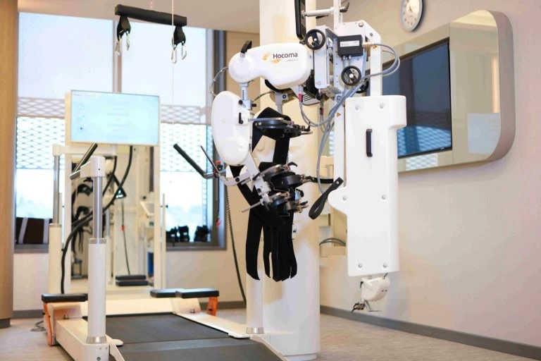 Robot-assisted gait training Lokomat Pro
 Robotic leg orthoses move the legs through a walking pattern on a treadmill while a harness carries part of your weight. Guidance and support are reduced as you take over more of the work.
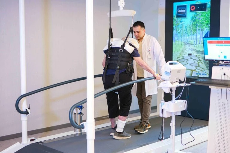 Gait and balance with virtual tasks C-Mill VR+
 An instrumented treadmill that projects targets and obstacles onto the belt. You generate the movement yourself, which is why it suits a later stage than the Lokomat.
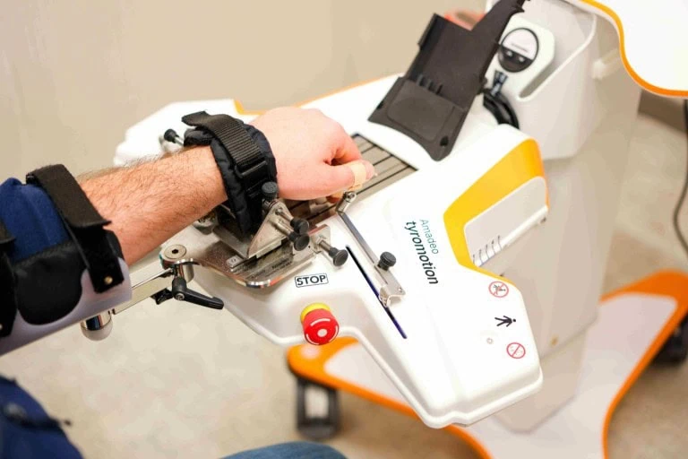 Arm, hand and finger training Armeo Spring & Amadeo
 Armeo cancels gravity so a weak arm can practise reaching. Amadeo trains the fingers individually, including where there is almost no active movement left.
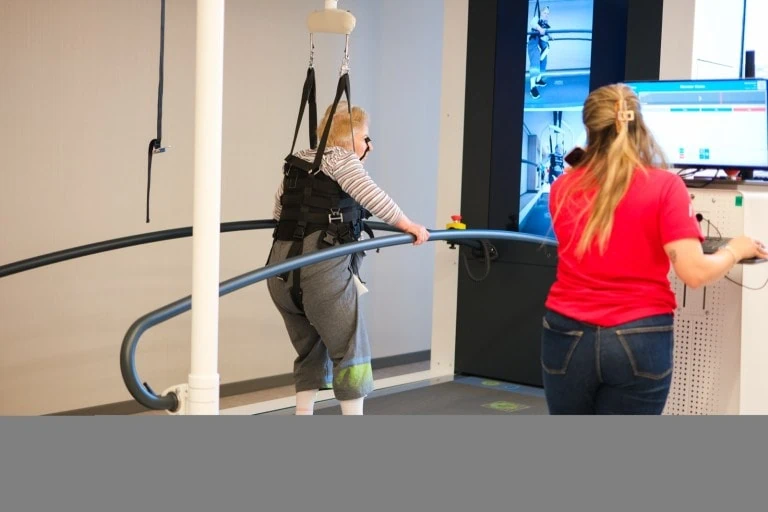 Getting upright, early Erigo Pro
 A robotic tilt table that adds stepping while it raises you. The leg movement supports blood pressure, so patients tolerate being upright sooner than on a plain tilt table.
Two very different reasons people come here
One is a family researching intensive rehabilitation after a stroke or a spinal injury. The other is someone with months of back, neck or joint pain who has run out of answers. The unit is built for both.
Neurological rehabilitation
Stroke, brain and spinal cord injury, and the conditions where regaining movement is the whole point of the programme.
Nerve and muscle disorders
Progressive and inherited conditions where rehabilitation maintains function, prevents complications and adapts as things change.
Pain, joints and everyday function
The musculoskeletal work: back and neck pain investigated properly, arthritis, chronic pain, and getting back to what you did before.
Goals first, then the machine
Rehabilitation is the one speciality where the wrong answer is easy to sell. A family that has just been told a relative may not walk again will buy almost anything, and a room full of robots photographs well. So the sequence here is fixed: assess, agree specific functional goals, then choose the tools that serve them — which is sometimes a robot and sometimes an hour of skilled hands and a set of parallel bars.
Progress is measured with the same instruments at the start and at the end, so that what changed is a number rather than an impression. If the numbers stop moving, we say so.
What we will not do
- Promise that someone will walk again.
- Sell a robotic programme to someone whose goals it cannot serve.
- Quote a programme length before the assessment has happened.
- Treat spasticity aggressively without a functional goal for doing it.
- Keep a programme running after the measurements have stopped moving.
Who would run your programme
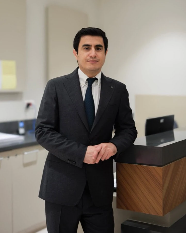
Assoc. Prof. Dr. Mustafa Çorum
Physical medicine and rehabilitation, with his clinical work centred on robot-assisted rehabilitation, stroke and brain-injury care, and spasticity management including botulinum toxin. He also performs ultrasound-guided injections and nerve blocks, PRP and dry needling for musculoskeletal pain.
Read the full profileHow a residential programme actually runs
Send records and a short video
The discharge summary, imaging on disc, the medication list — and a phone video of walking or of the affected arm, which tells us more than any written description.
Candidacy review
Whether an intensive programme is the right instrument at all, and which parts of it would apply. We say no when the answer is no.
Assessment and goal setting on arrival
Formal measurement, then goals agreed with you and your family rather than set for you. The programme length follows the assessment.
The programme
Therapy blocks alternating robotic sessions with hands-on physiotherapy, occupational and speech therapy, under daily medical review, with rest built in.
Reassessment and handover
The same measures repeated, a written home programme, the equipment list, and follow-up arranged before you travel back.
Six things worth knowing first
A robot is not a therapist
Robotic systems let you do far more repetitions safely and measure them. They are an adjunct to skilled hands-on therapy, never a replacement for it.
No device undoes an injury
If the neural pathways for a movement are gone, nothing that supports or guides the limb will bring them back. Robotics work with the capacity that remains.
Recovery does not stop at six months
It slows. That is not the same thing, and the belief that the door closes at six months costs people function they could still have gained.
Some spasticity is useful
Tone can be what allows a weak leg to bear weight. The goal is not always to abolish it, and treating it without a functional goal can make walking harder.
A disc bulge on MRI is common
Disc findings appear on scans of people with no pain at all. The report is one input; what you can and cannot do matters more.
Ask what happens if progress stops
Any centre should tell you in advance how progress is measured, who decides the programme has run its course, and what is offered instead.
Jump to what you came for
Quick answer
Physical Medicine and Rehabilitation is the medical unit that helps restore movement, function, and independence for people affected by injury, illness, pain, or disability. At Acibadem in Turkey, this unit evaluates each patient’s physical limitations and supports recovery with coordinated non-surgical treatments such as exercise therapy, pain management, assistive approaches, and personalized rehabilitation programs.
What this unit covers
Two very different people tend to arrive at a physical medicine and rehabilitation department, and they arrive in very different states of mind.
The first is usually a family rather than a patient. Someone has had a stroke, a spinal cord injury or a serious head injury. The acute hospital has done its work, the immediate danger has passed, and now a daughter or a husband or a brother is reading late at night trying to work out what happens next — whether intensive rehabilitation is worth arranging, what a robotic device actually does, whether the person they love will walk, hold a cup, speak. Nobody has given them a straight answer about the outcome, because at that stage an honest straight answer does not exist. What does exist is a way of working: measured, repeated, supervised practice, reassessed as the person changes, with the plan rewritten when the evidence in front of the team changes.
The second reader is one person, usually alone, who has had back pain or neck pain or a shoulder that will not settle for months. They have seen more than one doctor. They have had a scan and been told it looks more or less normal, or been told it looks bad and offered surgery they are not sure they want. They are not in danger. They are tired of being told what the pain is called and would like somebody to work out where it is actually coming from.
Both belong in the same department, and that is not an accident of hospital administration. Physical medicine and rehabilitation — physiatry — is the medical speciality organised around function rather than around a single organ. It asks what you can do, what you cannot do, how much pain you are in while you try, and what can be changed. That question is the same whether the person asking cannot yet sit upright unsupported or is a runner whose heel hurts on the first step of the morning. The tools differ enormously. The reasoning does not.
The work divides into strands that overlap constantly in practice. A stroke patient with a painful, stiff shoulder is receiving neurological rehabilitation, spasticity management and musculoskeletal pain treatment at the same time, from the same team, in the same week.
- Neurological rehabilitation — retraining movement, balance, swallowing, speech and daily independence after damage to the brain, spinal cord or nerves. This is the largest part of the intensive work and covers rehabilitation after stroke, spinal cord injury and traumatic and acquired brain injury, alongside conditions such as multiple sclerosis, Parkinson’s disease and Guillain-Barré syndrome. The nervous system’s capacity to reorganise is real, it is not unlimited, and nobody can tell you in advance how much of it a particular person has.
- Robot-assisted therapy — devices that support, guide or resist movement while measuring every repetition. The unit’s robotic rehabilitation programme is built around Lokomat Pro for gait, C-Mill VR+ for walking and balance under real-world demands, Erigo Pro for patients who cannot yet tolerate being upright, and Armeo Spring and Amadeo for the arm and hand. These machines are used because they let a patient practise more, more safely, with numbers attached. They are not a shortcut past the work.
- Inpatient intensive programmes — residential rehabilitation for people who need several therapy disciplines every day and medical and nursing supervision between sessions. Inpatient rehabilitation exists because an hour of outpatient therapy twice a week cannot deliver the volume of practice a recovering nervous system needs, and because problems like pressure areas, bladder management, swallowing safety and mood do not wait for the next appointment. A day in the programme sets out what that actually looks like from waking to evening.
- Spasticity management — the tight, involuntary muscle overactivity that follows many brain and spinal injuries, and that can undo good movement, cause pain, deform joints and make washing and dressing difficult. Treatment combines stretching and positioning, splinting and orthoses, oral and injected medicines including botulinum toxin, targeted nerve blocks, and in selected cases an implanted pump. Treating spasticity is not the same as removing it — some tone is what a person is standing on.
- Musculoskeletal and pain medicine — the high-volume everyday work: back pain and sciatica, neck and shoulder pain, tendon problems, plantar fasciitis, arthritis, sports and overuse injuries, and recovery after orthopaedic surgery. Diagnosis comes first, with ultrasound and clinical examination doing more of the work than most people expect, and treatment ranges from graded exercise to dry needling and ultrasound-guided injections. Where pain has become persistent and its own problem, care is shared with pain management.
- Paediatric and specialist rehabilitation — children with cerebral palsy, spina bifida, neuromuscular disease or injury, who need a different approach because they are still growing and because the family is part of the treatment; lymphoedema after cancer surgery or radiotherapy; vestibular rehabilitation for dizziness and balance disorders; and hydrotherapy where weight-bearing on land is not yet possible.
The sections that follow are written to be read out of order. If you are looking for one thing, take that section and ignore the rest. If you are trying to decide whether to bring a relative here for intensive rehabilitation, the honest sequence is what robotic therapy does and does not do, then who benefits and who does not, then what a residential programme involves, then planning your care.
Rehabilitation patients live with symptoms that would alarm anyone else, which makes it harder, not easier, to notice the ones that are genuinely dangerous.
No numbers for duration, frequency, staffing or cost appear here as promises, because they are set by an individual assessment and not by a web page.
Emergency red flags
Rehabilitation is slow, patient work, and almost nothing in it is urgent. The exceptions are absolute. People recovering from neurological injury already have altered sensation, altered blood pressure control, weakness, stiffness and pain, so the ordinary warning signals the body uses are muffled or missing. Families learn to normalise symptoms, which is usually correct and occasionally dangerous. The list below is the set of things that must never be normalised.
- New stroke symptoms — think FAST. Face: one side of the face has dropped, the smile is uneven. Arms: one arm cannot be raised or drifts down. Speech: words are slurred, jumbled or absent, or the person cannot understand you. Time: time is the treatment. Sudden loss of vision in one or both eyes, sudden severe unexplained headache, sudden loss of balance or coordination, and sudden numbness down one side belong in the same group. This applies with full force to someone who has already had a stroke — a second stroke is not less likely because there has been a first, and it is easy to mistake for a bad rehabilitation day.
- Autonomic dysreflexia in someone with a spinal cord injury at or above the T6 level. This is the emergency most families have never been warned about. A pounding headache comes on suddenly, the face and chest above the level of injury flush and sweat, the skin below may be cold and pale, the person may feel anxious, have blurred vision or a blocked nose, and the heart rate may slow. The blood pressure is surging, and it can rise high enough to cause a bleed into the brain, a seizure or death. It is triggered by something the body cannot feel — most often a blocked or kinked urinary catheter or a full bladder, sometimes constipation, a pressure sore, an ingrown toenail, tight clothing or a fracture.
- Suspected deep vein thrombosis or pulmonary embolism. A calf or thigh that becomes newly swollen, warm, red, tight or painful — often on one side only — may be a clot, and immobility after a stroke, spinal injury, fracture or major surgery is exactly the circumstance that produces one. In a limb with reduced sensation there may be no pain at all, only swelling and warmth. If the clot travels to the lungs, the signs are sudden breathlessness, sharp chest pain that is worse on breathing in, a racing heart, coughing blood, light-headedness or collapse.
- New or worsening leg weakness with a change in bladder or bowel control. Difficulty starting to pass urine, losing control of urine or stool, or numbness in the saddle area — the inner thighs, buttocks and around the genitals and anus — together with weakness or severe back pain, can mean the nerve bundle at the base of the spine is being compressed. This is cauda equina syndrome, and it is time-critical: delay is what turns a treatable compression into permanent loss of bladder, bowel and sexual function.
- A fall with a head injury, particularly in anyone taking anticoagulants. Falls are common during rehabilitation. Most are harmless. A head injury in someone taking a blood thinner is not, because bleeding inside the skull can build slowly and the person can seem entirely normal for hours before deteriorating. Drowsiness, confusion, repeated vomiting, worsening headache, weakness, unequal pupils, a seizure, or clear fluid from the nose or ear after a knock to the head all demand immediate assessment — as does any head injury on anticoagulants even when the person feels fine.
- A suspected fracture in a limb with reduced sensation. Where sensation is impaired by spinal cord injury, stroke, diabetes or nerve damage, the pain that normally announces a broken bone is absent. Instead you may see swelling, unusual warmth, bruising, a limb lying at an odd angle, a new grinding or clunk when the limb is moved, or — in someone with a spinal injury above T6 — an episode of autonomic dysreflexia with no other explanation. Bones weakened by disuse after paralysis can break during ordinary transfers and handling, with no dramatic accident to point to.
- Intrathecal baclofen pump failure or withdrawal. Anyone with an implanted pump delivering baclofen into the spinal fluid must know this. If the supply is interrupted — an empty reservoir, a missed refill, a catheter that has kinked, migrated or broken, a pump alarm or a programming fault — withdrawal can begin within hours. The first sign is usually spasticity that rebounds worse than it ever was before the pump, together with itching without a rash, fever, agitation, confusion and a rising heart rate. Left untreated it can progress to rhabdomyolysis, organ failure and death, and it is repeatedly mistaken for sepsis or a drug reaction. Never let anyone start, stop or alter baclofen, or any other medicine here, outside the care of the doctor responsible for it.
What is a physiatrist, and what does a physiatrist do?
Most people meet the word for the first time when someone hands them a referral. A physiatrist is a medical doctor — a specialist in physical medicine and rehabilitation, the speciality also called physiatry, and in much of Europe rehabilitation medicine. The distinguishing feature is not a body part. It is a question: what is limiting this person’s function, and what can be done about it?
Every other speciality is organised around an organ or a technique. A cardiologist owns the heart, a neurologist owns the nervous system, a surgeon owns the operation. A physiatrist owns what happens to a life afterwards, and, in the musculoskeletal clinic, what happens instead of an operation. That is why the same doctor can be found directing an intensive stroke programme in the morning and putting an ultrasound probe on a painful shoulder in the afternoon.
The training is a full medical degree followed by a speciality residency in physical medicine and rehabilitation, covering neurology, orthopaedics, rheumatology, musculoskeletal ultrasound, electrodiagnosis, prosthetics and orthotics, injection technique and the management of rehabilitation teams. Many then subspecialise — in neurorehabilitation, spinal cord injury, paediatrics, sports medicine, interventional pain or spasticity management.
What a physiatrist actually does in clinic
Stripped of the job description, the work is this.
- Diagnosing the cause of pain, weakness or loss of function. This is the part patients underestimate. A physiatrist examines how you move, not only where it hurts, and reads imaging against that examination rather than in place of it. A written report is a summary of what one person saw on one day; the patient in front of the doctor is the evidence. Diagnostic tools include musculoskeletal ultrasound, nerve conduction studies and electromyography, and formal gait analysis, with imaging arranged only where it will change the plan.
- Prescribing and directing therapy. A physiatrist does not usually deliver the hands-on treatment. They decide what the treatment should be, how intensive, in what sequence, with what precautions, and they change it when the reassessment says it is not working. “Do physiotherapy” is not a prescription. “Six weeks of eccentric loading with this progression and these limits” is.
- Performing injections and nerve blocks. Ultrasound-guided joint, tendon sheath, bursa and nerve injections, diagnostic blocks that answer a question about where pain is coming from, and regenerative approaches such as PRP where they are indicated.
- Managing spasticity and tone. Deciding which muscles are the problem, choosing between stretching, splinting, oral medicines, botulinum toxin injection, nerve blocks and pump therapy, and — as often — deciding to leave tone alone because the patient is using it.
- Prescribing equipment. Wheelchairs, orthoses, splints, prostheses, walking aids, seating and pressure management. Badly chosen equipment causes new problems; well chosen equipment is sometimes the single largest change in someone’s independence.
- Leading the rehabilitation team and holding the medical picture. Bladder and bowel management, skin integrity, pain, nutrition, mood, sleep, spasticity, blood pressure control and medication burden all sit with one doctor while several therapists work on separate goals.
Physiatrist or orthopaedic surgeon: who to see
The question is usually typed into a search box as physiatrist vs orthopedist, and the honest answer is that for most non-emergency musculoskeletal pain either door leads to the same place, but they start from opposite ends. A surgeon’s core question is whether an operation is indicated. A physiatrist’s core question is whether function can be restored without one, and how to prepare for or recover from surgery if it is needed. Neither is a gatekeeper for the other, and in this hospital group they run parallel clinics with shared patients through orthopaedics and traumatology.
| Professional | Trains in | What they decide | When to see them |
|---|---|---|---|
| Physiatrist (physical medicine and rehabilitation doctor) | Medicine, then a residency in function, disability and rehabilitation across neurology, orthopaedics and rheumatology; ultrasound, electrodiagnosis, injections, orthotics | What is causing the loss of function, whether surgery is needed at all, what therapy is prescribed and at what intensity, which injections or tone treatments are appropriate, what equipment is required | Pain or weakness with no clear diagnosis; after stroke, spinal or brain injury; before or after orthopaedic surgery; when therapy has stalled; when several problems interact |
| Orthopaedic surgeon | Medicine, then a surgical residency in bones, joints, ligaments and tendons | Whether an operation will help, which operation, and the surgical plan and its risks | Fracture, mechanical instability, a torn structure that will not heal, advanced joint destruction, or non-surgical treatment that has genuinely been exhausted |
| Physiotherapist | A university degree in physiotherapy; hands-on assessment, exercise prescription, manual therapy and movement retraining | How the prescribed treatment is delivered session by session — technique, load, progression, and when to hold back | Directly for straightforward musculoskeletal problems where local rules allow it, and always as the person who does the actual work of any rehabilitation plan |
| Neurologist | Medicine, then a residency in diseases of the brain, spinal cord, nerves and muscles | What the neurological diagnosis is and what medical treatment the disease itself requires | When the diagnosis is unclear, or the underlying disease is active or progressing; care is then usually shared with rehabilitation |
The practical rule: if you know what is wrong and it needs fixing mechanically, start with the surgeon. If you do not know what is wrong, or you know and want to avoid an operation, or the problem is what you can no longer do rather than what a scan shows, start with the physiatrist.
Who else is in the room
Rehabilitation is the least solo speciality in medicine. The physiatrist sets direction; the results are produced by a team, and knowing who does what makes the programme far easier to follow.
- Physiotherapists — movement, strength, balance, transfers, standing and walking, and the operation of the gait and balance technology.
- Occupational therapists — the tasks that make up a day: dressing, washing, eating, cooking, writing, handling a phone, returning to work, and adapting the home.
- Speech and language therapists — communication after brain injury or stroke, and swallowing, which is a safety issue as much as a comfort one.
- Rehabilitation nurses — skin and pressure care, bladder and bowel programmes, medication, and the twenty-two hours a day that are not therapy sessions.
- Clinical psychologists and neuropsychologists — mood, adjustment, motivation, and the memory, attention and planning problems that quietly decide how much of the physical work transfers into real life.
- Orthotists and prosthetists — splints, braces, footwear and artificial limbs, fitted and adjusted as the body changes.
- Dietitians and social workers — nutrition for tissue healing and energy, and the practical arrangements for going home.
Robotic rehabilitation: what it is, and what it is not
Robot-assisted rehabilitation is the reason many international families find this unit, so it deserves the plainest description available rather than the most impressive one.
A rehabilitation robot is a machine that supports, guides or resists a movement while measuring it. It does not think and it does not heal. It holds a limb against gravity, or moves it through a pattern, or lets the patient move it themselves and only intervenes when they cannot complete the range. Every device in this unit is built around that idea, applied to a different part of the body and a different stage of recovery.
Why it helps: repetition, intensity, consistency
The nervous system relearns movement through practice. Not gentle practice — a large amount of correct, focused, difficult practice, repeated until the pattern becomes available again. That is the whole mechanism, and it is unglamorous.
The problem in conventional therapy is arithmetic. One therapist manually supporting a hemiplegic leg through a gait cycle, or lifting a paralysed arm through a reach, is doing hard physical labour. They tire, the number of repetitions in a session is limited by what a human body can lift, and the quality of the twentieth repetition is not the quality of the first. For a patient who cannot yet bear weight, more than one member of staff may be needed for a short period of assisted standing.
A robot changes that arithmetic.
- Repetition. The number of guided movements in a session rises substantially, because the machine does not fatigue.
- Intensity and consistency. Every repetition is delivered with the same trajectory, speed and support, so the nervous system receives a clean, unvarying signal rather than a pattern that degrades as the therapist tires.
- Earlier and safer starting. Body-weight support, harnesses and tilt mechanisms allow upright, task-specific practice with patients who could not otherwise be safely stood or walked, and long before independent standing is possible.
- Objective measurement. The device records how much force the patient contributed, how symmetrical the movement was, how much assistance was needed and how that changed across sessions. This replaces impressions with data — and data cuts both ways, because it also shows plainly when a programme is not producing change.
There is a further benefit that matters more than clinicians once assumed: engagement. Robotic sessions are usually delivered with screen-based tasks and feedback, so the patient is playing a game with their affected limb rather than counting repetitions. Attention and motivation are part of motor learning, not decoration around it.
What the evidence supports, and what it does not
This part is the reason the section exists, and every other robotics section here refers back to it rather than repeating it.
Robot-assisted therapy is an adjunct. The evidence supports it as a way of delivering high-repetition, measurable practice under safe conditions — a delivery method, not a treatment in its own right. Used alongside skilled hands-on therapy, it lets a patient do more of the work that drives recovery. Used instead of skilled hands-on therapy, it removes the clinical judgement that decides what should be practised, how a compensation is being built in, and when the plan is wrong. A therapist adapts within a repetition. A robot repeats what it was set to repeat.
Second, and harder: no device restores function that the underlying injury has made unrecoverable. If the neural pathways required for a movement are gone, nothing that supports, guides or measures the limb will bring them back. Robotic therapy works with the capacity that remains — it can help a person exploit that capacity far more fully than they would otherwise, and that difference can be the difference between dependence and independence. It cannot create capacity that is not there. How much remains is not knowable in advance from a scan, a diagnosis or a video call, which is precisely why no honest clinician will tell you before the assessment what a programme will achieve.
Third: a centre that presents robots as a cure is selling you something other than rehabilitation. Watch for outcome percentages, before-and-after videos offered as evidence, guaranteed programmes, and any suggestion that a device is unique or that this technology is unavailable elsewhere. The machines named here are commercially manufactured systems in use in rehabilitation centres worldwide. What varies between centres is the assessment, the therapy hours around the machine, and whether anyone is willing to tell you when the answer is no.
Robotic therapy suits patients who have enough medical stability, cognitive ability to follow instruction, skin integrity and joint range to use a device safely, and a plausible target to work towards. It does not suit everyone, and being turned down for it is a clinical judgement rather than a verdict on the person — who benefits and who does not sets out the reasoning in detail. Where the underlying neurological disease is active or progressing, the rehabilitation plan is made jointly with neurology, because treating the disease and training the function are different jobs done at the same time. The unit’s robotic programme is directed by Assoc. Prof. Dr. Mustafa Çorum, whose work covers robotic rehabilitation, stroke and brain-injury care, and spasticity management.
The devices in use
- Lokomat Pro — a treadmill with body-weight support and robotic leg orthoses that move the legs through a physiological walking pattern.
- C-Mill VR+ — an instrumented treadmill that projects targets and obstacles onto the walking surface, training the adaptive, reactive part of walking that a fixed pattern cannot.
- Erigo Pro — a robotic tilt table with stepping and cyclic leg movement, used to bring very early or unstable patients towards upright safely.
- Armeo Spring — a spring-loaded exoskeleton arm support that cancels gravity so a weak arm can practise reaching.
- Amadeo — finger-by-finger robotic training for the hand, from fully passive movement to active resisted work.
Robotic work never runs on its own. It sits inside a programme with conventional therapy, functional electrical stimulation, gait analysis, tone management and — for a smaller group of patients — wearable exoskeletons. The device is the tool. The programme is the treatment.
Lokomat Pro: robotic gait training
The Lokomat is the device most people mean when they ask about robotic rehabilitation, and it is the one that most needs describing accurately, because what it does is narrower and more useful than the marketing around it suggests.
A Lokomat Pro is a treadmill with two additions. The first is an overhead body-weight support system: a harness worn around the trunk and thighs, attached to a frame that takes a set proportion of the patient’s weight, so that a person whose legs cannot yet hold them can still be upright and loaded through the feet. The second is a pair of robotic leg orthoses — powered exoskeleton legs strapped to the patient’s own — that drive the hips and knees through a physiological gait pattern in time with the belt. A screen in front of the patient displays feedback from the sensors in the orthoses.
Put together, this allows something that is otherwise very hard to obtain: a large number of anatomically correct steps, taken upright, with the trunk loaded and the feet in contact with a moving surface, by someone who cannot walk.
It is used in the rehabilitation of stroke, incomplete spinal cord injury, traumatic brain injury, multiple sclerosis, Parkinson’s disease and cerebral palsy, and after some spinal and neurosurgical procedures, where the plan is made together with the operating surgeon. The common feature is not the diagnosis. It is a gait problem in someone who has some potential for walking, or for whom safe upright loading itself is the goal.
What a Lokomat session involves
Patients and families are almost always surprised by the same thing: how much of the session is not walking.
Setup takes roughly fifteen to twenty minutes at the start, and less as the team learns your body. You transfer onto the platform, usually from a wheelchair, sometimes with a hoist. The harness is fitted around the trunk and thighs and checked for pressure points. The robotic orthoses are then sized to your legs — thigh and shank lengths adjusted, hip width set, cuffs fastened at thigh, knee and shin, foot straps attached — and this fitting is the part that determines whether the session is comfortable or not. A poorly aligned knee joint on the orthosis will rub within minutes. Say so immediately; it is adjusted, not endured.
You are then raised until your weight is partly supported and the belt starts slowly. The first minutes are spent finding the right combination of speed, body-weight support and guidance force while the therapist watches your hips, knees and trunk. Blood pressure is monitored in patients who have been immobile, because being upright is itself a physiological demand.
Actual walking time is typically thirty to forty-five minutes once you are tolerating the device, built up gradually from much shorter periods. It is genuinely tiring, more so than it looks, and the fatigue is often the limiting factor early on rather than the legs. Getting out and back into the chair takes another few minutes. How many sessions you have, and how they sit alongside the rest of your therapy, is set by your programme rather than by the device — see inpatient rehabilitation and a day in the programme.
How the difficulty increases
The point of the Lokomat is not to be walked by the machine. It is to hand the work back to the patient as fast as they can take it, and the settings that do this are guidance force and body-weight support.
Guidance force is how strictly the orthoses hold the legs to the programmed pattern. At the highest setting, the legs are moved for you. As guidance is reduced, the robot allows more deviation and only assists where you cannot complete the movement yourself, until it is doing little more than catching errors.
Body-weight support is how much of your weight the harness carries. Reducing it progressively loads the legs, the trunk and the balance system, which is what the nervous system needs in order to treat the movement as real walking rather than as an assisted exercise.
Alongside these, treadmill speed rises and the augmented-feedback tasks get harder. Those tasks are the games on the screen — stepping over projected obstacles, hitting targets with a specific leg, matching a symmetry bar between left and right. They exist because they convert a repetitive exercise into a goal with a score, and because directed attention improves motor learning. A patient who is bored is practising less than a patient who is competing.
The sensors also generate objective output: how much force each leg contributed, how symmetrical the step lengths were, how much assistance the robot supplied, how the range of motion at hip and knee changed. This is the record used to decide whether the programme continues unchanged, is progressed or is stopped. It is also, bluntly, the record that shows when a device is not the right tool for a particular patient.
Who the Lokomat is not suitable for
The screening is real, and a good unit refuses patients. The following are the reasons a session may be ruled out, delayed or modified.
- Fixed joint contractures. The orthoses move through a set arc. A hip, knee or ankle that cannot reach the required range will be forced or will fight the machine. Contractures are treated first — stretching, splinting, tone management — and the device revisited.
- Unstable or unhealed fractures, and recent lower-limb or spinal surgery that has not been cleared for weight-bearing by the surgeon.
- Severe osteoporosis or fragile bone from prolonged disuse. Loading and driven movement carry a fracture risk in bone that has demineralised after paralysis, which is why bone density is assessed before gait robotics in long-standing spinal cord injury rather than afterwards.
- Open wounds, pressure sores or fragile skin at harness and cuff contact points — trunk, groin, thighs, knees and shins. Skin that cannot tolerate the interface is a hard stop until it heals, because a pressure injury costs far more recovery time than the sessions gain.
- Orthostatic intolerance. Patients whose blood pressure falls when they are brought upright cannot start here. They usually start on the Erigo Pro, which is designed exactly for that problem.
- Weight and height limits. The harness, frame and orthoses have manufacturer-set limits for body weight and for leg segment length, and a person outside them cannot be fitted safely. Whether the device fits a particular body is checked at assessment.
- Uncontrolled spasticity or severe involuntary movement that resists the programmed pattern. Working against the orthoses is uncomfortable, unproductive and can injure soft tissue. Tone treatment often comes before gait robotics rather than alongside it.
- Severe cognitive or behavioural difficulty that prevents cooperation, or an inability to tolerate the harness. Agitation in a body-weight support harness is unsafe.
- Medical instability — uncontrolled cardiac disease, active infection, unstable blood pressure, deep vein thrombosis under investigation, uncontrolled seizures.
One last point, and it is the honest limit of the machine. The Lokomat trains a pattern. Real-world walking is not a pattern; it is a continuous negotiation with uneven ground, obstacles, turns, doorways, other people and your own attention divided between walking and everything else. Improvement measured on the device does not automatically become improvement on the ward corridor or the street. Carry-over requires floor practice — over-ground walking with a therapist, stairs, turning, obstacle work, walking while doing something else — which is why gait robotics is scheduled alongside conventional gait training and C-Mill VR+ rather than instead of it, and why the assessment that matters is how you walk off the machine. Any unit that reports your progress only in device numbers is measuring the wrong thing.
Armeo Spring and Amadeo: arm and hand rehabilitation
Walking recovers more readily than the arm does. That is an uncomfortable fact, and families deserve to hear it before they are shown the equipment.
There are structural reasons. The leg performs one dominant task in a repeating cycle, largely in one plane, with both sides cooperating in a fixed rhythm. The arm has far more degrees of freedom at the shoulder, elbow, forearm, wrist and each finger, and it performs an enormous variety of tasks that share almost no pattern. Reaching for a glass, buttoning a shirt and turning a key are different problems for the nervous system. Recovery in the upper limb tends to be slower, more partial and less predictable than in gait, and after stroke it commonly lags well behind walking. Meanwhile, hand function is what determines independence: a person who can walk but cannot dress, eat or wash without help is not independent.
The arm also gets less practice. A leg is used every time the person stands. A weak arm is quietly abandoned, the good arm takes over, and the affected side is used less and less — learned non-use, which makes the underlying weakness worse than the injury alone requires. Much of upper-limb rehabilitation is a fight against exactly that.
The devices in this unit address different ends of the problem, and both sit inside the framing set out in robotic rehabilitation: they deliver practice, they do not deliver recovery.
Armeo Spring: reaching against gravity
Armeo Spring is an exoskeleton arm support — a mechanical arm on a frame, with spring-loaded segments that carry the weight of the patient’s own limb. Gravity is the main obstacle for a weak arm: a person may have enough neural drive to move the arm horizontally on a table but nothing like enough to lift it. Cancel the weight and movement that was invisible becomes possible, and once movement is possible it can be practised.
The patient’s forearm and upper arm sit in adjustable cuffs, the amount of weight relief is set to the individual, and a grip sensor may be added so that grasp and release become part of the task. The workspace is large and three-dimensional: reaching forward, out to the side, up towards a shelf, back towards the mouth. Screen tasks provide the goal — moving an object across a display, catching, sorting, cleaning a surface, pouring — and the system records the reachable workspace, movement smoothness and how it changes.
Armeo Spring suits patients with some active movement in the shoulder and elbow but not enough to work against gravity for any useful length of time. It is used from the early subacute phase onwards in stroke, brain injury, incomplete spinal cord injury, multiple sclerosis and after brachial plexus or peripheral nerve injury. It does not suit an arm with no active movement at all, because there is nothing for the weight relief to release — that arm is handled differently, with passive range work, positioning, electrical stimulation and tone management, until active movement appears, if it appears.
Amadeo: finger-by-finger hand training
Amadeo works where the arm ends and the real difficulty starts. Each fingertip, and the thumb, is attached by a small magnetic slider to an individual actuator, so each digit can be moved, assisted or resisted on its own. The modes available cover the whole range of ability.
- Passive mode — the device opens and closes the fingers with no contribution from the patient. This is used where there is no active movement at all: it maintains joint range, keeps soft tissue from shortening, provides sensory input to a hand the brain is ignoring, and gives the patient something to attempt against.
- Assisted mode — the patient initiates the movement and the device completes what they cannot. This is where minimal active movement is amplified into a full, task-shaped repetition, which is the point at which the practice becomes motor learning rather than stretching.
- Active and resisted modes — the patient moves each finger themselves, against measured resistance, with force feedback on screen. Individual finger control, not gross grip, is what returns fine function.
Amadeo also measures force per finger, which is diagnostically useful in its own right: it shows which digits are contributing, whether the thumb is participating, and whether an apparent grip improvement is real or is the whole hand flexing together.
How this fits with hands-on therapy, and what limits it
Neither device replaces the occupational therapist. Robotic upper-limb work is scheduled around conventional treatment, most usefully alongside two well-established approaches. Constraint-induced movement therapy restricts the unaffected hand for set periods so that the affected one has to be used, directly attacking learned non-use; it demands a minimum of active wrist and finger movement to be appropriate. Mirror therapy uses a mirror placed so that the reflection of the good hand appears where the affected hand is, and the visual illusion of normal movement can improve motor output in a hand with very little of its own. Task practice with real objects — a cup, a fork, a shirt, a key — remains the destination for everything the robots train, and the transfer to real objects has to be deliberately taught, not assumed.
The limits on upper-limb robotics are mostly about the shoulder, and they are firm.
- Pain changes everything. A painful shoulder is treated before it is trained. Post-stroke shoulder pain is common and has several causes — subluxation, rotator cuff injury, capsulitis, spasticity, complex regional pain syndrome — and training through it makes each of them worse and teaches the patient to avoid the limb. Diagnosis first, often with ultrasound and where appropriate a guided injection, then training. See neck and shoulder pain.
- Shoulder subluxation. When the shoulder muscles are flaccid, the weight of the arm pulls the humeral head partly out of the socket. The arm must be supported and handled correctly, and the amount and direction of movement permitted is a clinical decision, not a device setting.
- Severe spasticity and fixed contracture. A hand that cannot be opened cannot be placed in the sliders, and a flexed elbow or internally rotated shoulder that will not release restricts the workspace to the point where practice is meaningless. Botulinum toxin injection and tone management frequently come first, precisely so that robotic and manual therapy become possible afterwards.
- Sensory loss and neglect. An arm the patient cannot feel, or does not attend to, needs the therapy aimed at sensation and attention as much as at strength, and screen-based tasks must be positioned with that in mind.
- Skin fragility and oedema at cuff and slider contact points, which need checking at every session.
What none of this changes is the honest position stated in the robotics section: these devices increase the amount and quality of practice a weak arm can do. They do not decide how much recovery is available. Two patients with apparently similar strokes can end in very different places, and the difference is largely in the injury rather than in the equipment. What is within your control is the amount of correct practice, which is exactly what the devices are there to raise.
Erigo Pro and early mobilisation: getting a patient upright safely
Lying still is not neutral. After a severe stroke, a brain injury or a spinal cord injury, the body starts losing ground within days, and the losses have nothing to do with the injured brain or cord. Muscle is lost fastest in the legs and trunk. The circulation stops rehearsing the reflexes that hold blood pressure steady when a person sits or stands, so the first attempt to sit upright brings dizziness, a racing pulse, sweating and nausea — orthostatic intolerance. Joints that are never taken through their full range begin to shorten, and a shortened ankle, knee or hip is far harder to recover than it was to prevent. Skin over the sacrum and the heels carries load for hours at a time that it was never built to carry. Bone demineralises. The bases of the lungs do not fully inflate, which is one reason chest infections are so common in this group.
None of this is inevitable, and almost all of it is easier to prevent than to reverse. That is the entire argument for early mobilisation: the window in which these secondary losses can be avoided is early, and it closes quietly while everyone is understandably focused on the primary injury.
When early mobilisation starts
Early mobilisation begins as soon as the treating team judges the medical picture stable enough to allow it — often within the first days, sometimes while the patient is still in intensive care and still ventilated. It is a daily judgement, not a fixed date on a calendar. Blood pressure control, intracranial pressure, spinal stability, fracture fixation, respiratory status and the presence of an untreated clot all shift the answer, and the answer can change between morning and afternoon.
Earlier is not automatically better in an unqualified sense, and it is worth saying so plainly, because families sometimes arrive convinced that a hospital which did not stand their relative up on day one has failed them. In the very acute phase after stroke, the pattern that has held up best is short, frequent, carefully graded sessions rather than long or forced ones. What matters is that something purposeful happens early and often — passive range of movement, positioning changes, supported sitting, upright time — rather than that a particular threshold is crossed on a particular day.
What the Erigo Pro is
The Erigo Pro is a robotic tilt table. A conventional tilt table simply raises a strapped patient from lying towards standing so the cardiovascular system relearns how to cope with being upright. The problem is that a motionless person tilted upright pools blood in the legs, and many patients in the early phase after a severe injury cannot tolerate more than a shallow angle before their blood pressure drops.
The Erigo adds two things to that plain tilt. The first is cyclic leg stepping: motorised footplates move the legs through a walking-like pattern while the table is tilted. The second is optional functional electrical stimulation to the leg muscles, timed to the step cycle. Both drive the calf and thigh muscles to contract rhythmically, and rhythmic contraction is what pushes venous blood back towards the heart. That muscle pump is why patients on an Erigo commonly tolerate being upright sooner, and at a steeper angle, than they would on a static table. The stepping also delivers repetitive, patterned sensory input to a nervous system that is otherwise receiving almost nothing from the legs.
Who it is used for
It is used in the phase where a patient cannot yet stand, transfer or sit unsupported: the early weeks after severe stroke, traumatic brain injury or spinal cord injury, and after long intensive care stays where critical illness weakness has taken hold. It is also used with patients who have a prolonged disorder of consciousness. In that group the aim is not gait training in any meaningful sense. It is verticalisation, cardiovascular and postural loading, and a standardised, repeatable setting in which the team can observe whether responses change over weeks — which is more informative than an impression formed at the bedside on one afternoon.
What a session involves
The patient is transferred onto the table lying flat, secured with chest, pelvis and knee straps, and the feet are fixed to the footplates. Blood pressure and heart rate are recorded before starting and monitored throughout, usually continuously. The table is raised to a shallow angle first and the stepping begins at a slow cadence. The angle is then increased in small increments, with the team watching the numbers and the patient between each step up — a drop in blood pressure, a rising pulse, pallor, sweating, yawning, nausea or a change in responsiveness means the table comes back down rather than the session being pushed through. Sessions in the early phase are typically short, in the region of twenty to thirty minutes, and are repeated on most days rather than being made long and rare. Progress is measured by the angle tolerated, the time at that angle and the stability of the blood pressure, and those numbers are written down so the next session starts from evidence rather than memory.
Limits, monitoring and what it does not do
The Erigo is not appropriate for everyone, and the checks are specific:
- Spinal stability and fractures. An unstable spinal column, an unfixed fracture, or a fracture in a limb that would be loaded rules the session out until the surgical team clears it.
- Cardiovascular and respiratory status. Unstable blood pressure, uncontrolled arrhythmia, active sepsis or a respiratory picture that is deteriorating means waiting.
- Clots. A suspected or newly diagnosed deep vein thrombosis stops mobilisation until it has been assessed and treated.
- Skin. The strap and footplate sites are checked before and after every session. Broken or fragile skin at those points, or an existing pressure injury in the loaded area, changes the plan.
- Bone density. Marked osteoporosis or established disuse bone loss raises the risk of a fracture during loading, and is weighed before stepping is added.
- Contracture and joint range. A hip or knee that cannot be brought near extension may not fit the device safely or comfortably.
- Device limits. The equipment has weight and height limits, and a patient outside them cannot be positioned safely.
- Autonomic instability. In spinal cord injury, poorly controlled autonomic dysreflexia or severe orthostatic hypotension is managed before verticalisation is pushed.
It is also worth being clear about what this machine is. It is a way of delivering upright posture and repetitive leg movement safely and measurably at a stage when a patient cannot produce either alone. It does not replace the unglamorous work that prevents most of the damage of bed rest — turning, positioning, passive range of movement, sitting out of bed, chest care and getting nutrition right — and it does not alter the underlying injury. Its value is that it makes early upright practice possible sooner, and repeatable, for people who would otherwise spend those weeks flat.
C-Mill VR+: gait, balance and virtual reality rehabilitation
Once a patient can take steps, the problem changes. Walking in a straight, empty, well-lit corridor beside a therapist is not the skill that is missing. What is missing is walking on a pavement with a kerb, turning while someone speaks to you, stepping over a dog, recovering from a stumble, and doing all of it without the constant fear that the next step is the one that puts you on the floor. That is what the C-Mill VR+ is built to train.
What the machine actually is
The C-Mill is an instrumented treadmill and walkway with a projector above it and force sensors underneath. The projector puts targets, stepping stones, obstacles, lines and paths directly onto the belt in front of the patient. The sensors record where each foot actually lands, how long it stays there, how the load is distributed and how symmetrical the two sides are. The patient walks in a safety harness that will hold them if the leg gives way. Some tasks are projected onto the belt; others are displayed on a screen as a virtual environment the patient walks through.
The important design point is that the belt can also change speed suddenly or shift under a foot, delivering a controlled perturbation — a small, deliberate near-stumble that the patient has to recover from.
Why this is not a Lokomat
It is worth being precise about the difference, because the two devices are often described in the same breath and they belong to different stages. With robot-assisted treadmill gait training, the exoskeleton guides the legs through a walking pattern; the machine supplies part of the movement, which is what makes it usable by someone who cannot yet step at all. On the C-Mill, the machine supplies nothing. The patient generates every step. The device measures, challenges and provides feedback.
That makes it a later-stage tool. Broadly, guided robotic gait training belongs to the phase where stepping has to be made possible at all, and instrumented, task-based treadmill work belongs to the phase where stepping exists but is unsafe, asymmetrical, slow or fragile. Many patients use both across a programme, in that order.
What it trains that a corridor cannot
- Stepping accuracy. Projected targets force the patient to place the foot in a specific spot, which is exactly the demand that kerbs, stairs and uneven ground make, and the sensors show whether they hit it.
- Obstacle avoidance. Objects appear on the belt with little warning and have to be stepped over or around, training the visual and motor timing that a straight corridor never asks for.
- Reactive balance. A sudden belt perturbation trains the recovery step. This is one of the few things in rehabilitation that targets the actual mechanics of a fall rather than the general fitness around it.
- Dual-tasking. Walking while counting, answering, or watching for a signal. Real walking is almost never the only thing a person is doing, and gait that looks fine in silence often deteriorates the moment attention is divided — particularly after stroke or brain injury.
- Speed and adaptability. Changing pace on demand, starting, stopping and turning.
- Confidence through safe failure. In the harness, a patient can get it wrong, lose balance and not fall. People who have fallen once, or who have never trusted the leg since the injury, need somewhere to find out what their limit is without paying for the discovery. That is not a soft benefit; fear of falling drives people to walk less, and walking less makes them weaker and more likely to fall.
Because everything is measured, progress is described in step length, symmetry, variability and how reliably the task is completed, rather than in someone’s impression that things look better than last week.
Who it suits
Patients who can bear weight and take steps with or without an aid, and who have enough attention and comprehension to follow a task. In practice that means people recovering from stroke, people with incomplete spinal cord injury, brain injury patients past the early confused phase, people with Parkinson’s disease and other neurological gait disorders, older patients with a history of falls or near-falls, and orthopaedic patients — after a hip fracture, a joint replacement, a complex leg fracture or a ligament reconstruction — whose mechanics and confidence both need rebuilding.
It is not suitable for someone who cannot yet sustain standing, who cannot follow the task, or who is outside the harness limits. Marked visual field loss or visual neglect needs the tasks adapted rather than abandoned. A small number of people feel motion-sick or visually disoriented with projected and virtual environments, and anyone with a seizure disorder triggered by visual stimuli should say so before starting.
The honest note on virtual reality in rehabilitation
Virtual reality is sold hard in this field, so here is the plain version. VR does two useful things. It makes repetitive practice tolerable, because a person will do more repetitions of a game than of the same movement counted out loud, and repetitions are the active ingredient. And it lets difficulty be graded precisely and increased in small steps, which is hard to do by eye.
What the evidence supports is virtual reality as a method of delivering therapy, not as a distinct treatment with powers of its own. Comparing it against the same amount of well-designed conventional practice, the advantage narrows considerably; comparing it against less practice, it looks impressive. So the question to ask about any VR-based programme is not how advanced the system is but how much task-specific practice the patient is actually getting, and whether the tasks resemble what they need to do at home. There is also a transfer problem worth naming: skill trained on a treadmill in a harness has to be taken outdoors, onto real pavements, in real weather, and that step belongs in the programme rather than after it.
Falls prevention is the outcome worth training for here, and it is a legitimate one — a fall in an older or neurologically impaired person is frequently the event that ends independent living. Training reactive balance, obstacle negotiation and dual-tasking addresses the actual circumstances in which falls happen, which is more than can be said for strengthening alone.
Stroke rehabilitation and the recovery timeline
Stroke rehabilitation is the largest part of what this unit does, and it is where families arrive with the most urgent questions and the least reliable information. The most useful thing to establish first is what the shape of recovery actually is, because almost every decision — how hard to push, when to travel, whether to keep going — depends on it.
What the timeline really looks like
Recovery after a stroke is not linear, and it is not the same process throughout. In the first days and weeks there is biological repair happening independently of anything anyone does: swelling settles, tissue around the damaged core recovers function, and the brain begins reorganising. This is the period of fastest change, and it is also the period in which the nervous system is most responsive to training — which is why rehabilitation starts in hospital rather than after discharge.
Change continues through the first three months, still relatively quickly, then through the following months at a slower rate. By around six months to a year, most people find that the pace of spontaneous change has dropped a long way, and progress from that point comes mainly from training, adaptation and equipment rather than from further biological repair.
Then there is the belief that has to be corrected, because it does real damage: that recovery stops at six months. It does not. What happens at around six months is that the rate of improvement slows — there is no cliff edge, no door that closes, and no evidence that a nervous system becomes untrainable on a particular date. People who resume intensive, task-specific practice years after a stroke can and do improve, particularly in walking capacity, endurance, arm use in daily tasks and independence. What is true is that gains later on tend to be smaller for the same effort and to come more from relearning how to do a task than from restoring the original movement pattern. Both things are worth having. Someone who is told at six months that this is as good as it gets is often being told something that suits a service’s capacity rather than something that is true of them.
It is also worth separating two questions that get merged. Recovery of the impairment — the arm moving the way it used to — follows a different course from recovery of function, which is the ability to get the task done, with adaptation, aids or the other hand if necessary. Function keeps improving for far longer than impairment does. Families who measure only the first are often watching the wrong number.
What the team does, and when
In the acute phase the work is protective and preparatory: positioning to protect the shoulder and prevent contracture, early mobilisation where the medical picture allows, swallowing assessment before anything is given by mouth, and prevention of clots, chest infection and pressure injury. In the subacute phase — the weeks and first months when change is fastest — the programme becomes intensive and specific: repeated practice of the movements and tasks the person needs, gait work, arm and hand work, speech and language therapy, cognitive rehabilitation, and training the family in what to do. In the later phase the focus shifts towards endurance, community walking, return to work or study, driving assessment where relevant, equipment and home adaptation, and periodic bursts of intensive therapy rather than a continuous programme.
The techniques, and what each is actually for
- Task-specific repetitive practice. The core of everything else. The brain reorganises around what is practised, in the amounts it is practised. This is why rehabilitation intensity matters more than any individual technique.
- Constraint-induced movement therapy. The stronger arm is restrained for part of the day while the affected arm is drilled through graded tasks. It works for a defined group — people who already have some active wrist and finger extension — and it is demanding, both in hours and in frustration tolerance. It is not for a hand with no movement, and it is not something to attempt without supervision.
- Mirror therapy. A mirror is placed so that the moving unaffected limb appears to be the affected one. It is used mainly where movement is very limited or absent, and sometimes for pain, as a way of engaging the motor system when there is not yet enough movement to practise with.
- Functional electrical stimulation. Used to assist a specific movement, most often ankle dorsiflexion in a dropped foot or wrist and finger extension, and used during practice so that the stimulation and the intention happen together.
- Robot-assisted and treadmill gait training. A way of getting a large number of stepping repetitions with support, most useful when independent stepping is not yet possible.
- Speech and language therapy. For aphasia, for speech clarity, and for swallowing, which is a safety issue rather than a comfort one.
- Strength, fitness and dose. Cardiovascular fitness after stroke is usually poor, and it limits everything else. It is trained deliberately.
The complications rehabilitation exists to prevent
A large part of the value of a proper rehabilitation programme is not in what it adds but in what it stops from happening: hemiplegic shoulder pain and subluxation, which is largely a handling problem and is why nobody should ever pull a weak arm to move the patient; contracture in the ankle, elbow, wrist and fingers; falls; aspiration and the pneumonia that follows it; pressure injury; and constipation and bladder problems that quietly wreck a rehabilitation programme.
Two complications deserve particular attention because they are the most under-treated in stroke care. Depression after stroke is common, and it is treatable, and it is frequently dismissed as an understandable reaction rather than assessed as a condition that blocks recovery. Post-stroke fatigue is not ordinary tiredness, does not resolve with rest in the way people expect, and is often the single biggest limiting factor on how much therapy a person can absorb.
Preventing the next stroke
Secondary prevention — blood pressure, atrial fibrillation, cholesterol, diabetes, smoking, and the medication that goes with them — belongs to the stroke physicians and the referring team, not to the rehabilitation programme, and no medication should be started, stopped or adjusted without them. Rehabilitation supports it by rebuilding the fitness and activity that reduce the risk, and by making sure nothing falls between services.
Spinal cord injury rehabilitation
Spinal cord injury rehabilitation is one of the most technically demanding pathways in this specialty, because it has to do two different jobs at once: teach a completely new set of physical skills, and manage a set of medical problems that will be part of daily life from now on. Getting the second part wrong undermines everything achieved in the first.
Complete or incomplete: the distinction that governs everything
The first thing any rehabilitation team establishes is the level of the injury and whether it is complete or incomplete. This is done with a formal, standardised neurological examination — sensation tested at defined points, muscle groups graded, and crucially the sacral segments checked, because preserved sensation or voluntary contraction there changes the classification. It is repeated over time, because the picture in the first days is distorted by swelling, spinal shock, sedation and pain.
An injury is complete when there is no sensory or motor function preserved in the lowest sacral segments. It is incomplete when something is preserved below the level, however small — and that “however small” matters, because incomplete injuries carry a genuinely different range of possibilities. This is why the same-sounding injury in two people leads to two entirely different programmes, and why a rehabilitation plan copied from someone else’s case is worthless.
Level, and what it generally means
Described in general terms, and with the constant caveat that individuals vary enormously and incomplete injuries break every rule:
- High cervical injuries affect breathing, and respiratory management — ventilation, weaning where possible, secretion clearance and cough assistance — dominates the early programme. Independence is achieved largely through powered mobility with alternative controls, environmental control systems and directed assistance.
- Mid and lower cervical injuries preserve increasing shoulder, elbow and then wrist function, and rehabilitation concentrates on transfers, pressure relief, manual or powered wheelchair skills and self-care with adapted equipment. Where hand function is partly preserved, the gain in independence is disproportionate to how small the movement looks.
- Thoracic injuries leave full arm function, so the work is trunk control, independent transfers, advanced wheelchair skills including kerbs and slopes, and standing programmes for the secondary benefits.
- Lumbar and sacral injuries may leave enough leg function for walking with orthoses and aids, and the programme then weighs walking against the energy cost of it — many people use both walking and a wheelchair, choosing by distance and terrain, and that is a sensible outcome rather than a failure.
What rehabilitation targets
Transfers in every direction, from bed, chair, car and floor. Wheelchair skills as a genuine athletic skill set. Standing and gait training where feasible. Bladder and bowel management as a reliable daily routine. Skin protection and pressure relief. Respiratory function. Upper limb preservation, because shoulders that will be doing the work of legs for decades need protecting from the start. Equipment, seating and cushion prescription. Home adaptation planning. Sexual function and fertility, which are frequently left unmentioned by clinicians and are among the questions people most want answered. Driving, work and study.
The medical problems that come with the injury
- Autonomic dysreflexia. In injuries at the upper thoracic level and above, a painful or irritating stimulus below the injury — most often a full bladder or blocked catheter, a loaded bowel, a pressure sore, an ingrown toenail or tight clothing — can trigger a sudden, dangerous rise in blood pressure with a pounding headache, flushing and sweating above the injury, and a slow pulse. It is a medical emergency. The full description sits with the other emergency red flags.
- Neurogenic bladder. Managed with a catheter programme chosen for the individual, with the long-term aim of protecting the kidneys as much as achieving continence. Recurrent infection is common and needs a considered approach rather than repeated antibiotic courses.
- Neurogenic bowel. A structured, predictable routine matters more here than almost anything else for quality of life and for confidence about leaving the house.
- Pressure injury. The dominant preventable complication. Skin that cannot feel pressure will not report it, so it has to be inspected and offloaded on schedule, and the right cushion and mattress are medical equipment rather than comfort items.
- Spasticity. Almost universal after the initial phase, and treated when it interferes rather than simply because it is present.
- Neuropathic pain. Burning, electrical or squeezing pain at or below the injury level, which is generated by the injured nervous system and does not mean the injury is worsening. It is managed with a combination of medication, physical and psychological approaches, in coordination with pain management.
- Heterotopic ossification. Bone forming in the soft tissue around a joint, usually the hip, presenting as swelling, warmth and a loss of range. It is looked for actively, because it is easy to mistake for infection or a clot.
- Other consequences that are watched for routinely: deep vein thrombosis, orthostatic hypotension, impaired temperature regulation, bone loss below the level of injury with the fracture risk that follows, and respiratory decline.
Where robot-assisted gait training fits, honestly
Robotic gait training has a real place here. It allows a person who cannot step to be brought upright and to perform a large number of stepping repetitions safely, with body weight supported, in a way that no number of therapists could deliver manually. The secondary benefits of regular standing and stepping — on bowel function, spasticity, circulation, posture and mood — are worth having in their own right, and patients frequently value the experience of being upright at eye level with other people more than the therapy value.
What it is not is a treatment that reverses the injury. Guided stepping does not regenerate the cord, and in a complete injury it does not create voluntary movement where the connection is gone. In incomplete injury it is one way — not the only way, and not clearly a superior way — of delivering the intensive walking practice that may help preserved pathways do more. Any programme that presents a robot as the thing that will make a spinal cord injury walk again is misrepresenting it; the broader position is set out under robot-assisted rehabilitation.
“Will they walk again?”
Every family asks this, usually within the first week, and it deserves a straight answer about why a straight answer is not available yet. The likelihood of walking depends principally on whether the injury is complete or incomplete, on the level, on how much is preserved at the initial examination, and on how the picture changes over the first weeks as spinal shock resolves. Those change the odds enormously, and they are assessed formally and repeatedly rather than guessed at. No honest clinician gives a definitive answer in the first week, and anyone who does — in either direction — is telling you something they cannot know.
What can be said is this. False hope and false despair do equal damage: one delays the practical skills that produce independence, the other abandons possibilities that were real. Good rehabilitation runs both tracks at once. It trains standing and stepping where there is a basis for it, and at the same time it builds the transfers, wheelchair skills and self-management that make a life work regardless of the answer. Independence is the goal. Walking is one of several routes to it.
Traumatic brain injury rehabilitation
Brain injury rehabilitation looks, from the outside, like the physical work of getting someone moving again. That is the smaller half of it. The limp is visible and the loss of attention is not, and it is the invisible half that determines whether a person returns to work, keeps relationships and lives independently.
The two kinds of consequence
The physical consequences — weakness, poor balance, coordination problems, spasticity, double vision, swallowing difficulty, fatigue — respond to the same rehabilitation methods used elsewhere in this specialty and tend to improve visibly, which makes them satisfying to work on. The cognitive and behavioural consequences are different: reduced attention and processing speed, memory problems, difficulty planning and organising, impaired judgement, poor insight, irritability, emotional flatness or emotional volatility, and loss of the social filter.
These cause more long-term difficulty than the physical problems and receive less attention, partly because they are hard to see and partly because a person who walks into a room unaided is assumed to have recovered. Families describe it as the person being physically back but not quite the same, and they are usually right and usually not believed.
Disorders of consciousness
Where the injury is severe, consciousness may return slowly and in stages: unresponsive wakefulness, in which the eyes open but there is no sign of awareness; a minimally conscious state, in which responses are inconsistent but real; and emergence, marked by reliable communication or object use.
The critical point is that these states are assessed over time with structured, repeated examinations, not judged from a single visit. Responses fluctuate through the day. Sedation, infection, seizures, hydrocephalus, pain and metabolic problems all suppress them and are all treatable, which is why reversible causes are hunted for before any conclusion is drawn. Families are entitled to be told plainly what is known, what is not known, and what would change the assessment — and to be told that a period of watching is itself the correct action rather than a delay. Neurology input is routine here for seizure and imaging questions.
The agitation phase
As a person emerges, many pass through a period of confusion, disorientation and agitation, often with no continuous memory being laid down — post-traumatic amnesia. They may be restless, shout, swear, try repeatedly to get out of bed, pull at lines, fail to recognise family, or behave in ways entirely unlike themselves.
Families need warning that this phase exists, because it is frightening and it is routinely misread as the person’s new personality or as psychiatric illness. In most cases it is a stage rather than an endpoint. It is managed mainly by shaping the environment — a quiet room, consistent staff, reduced stimulation, familiar objects, a predictable routine, and safety measures instead of confrontation — while treatable contributors such as pain, infection, constipation, urinary retention and sedating medication are identified. Behaviour in this phase is a symptom of the injury, not a choice, and it is not something to argue with.
Cognitive rehabilitation
Cognitive rehabilitation works on attention and processing speed, memory, and executive function — planning, sequencing, initiating, monitoring and correcting your own performance. It uses two approaches together: retraining the underlying capacity through graded practice, and building compensatory systems, which in practice means external aids — diaries, alarms, phone reminders, checklists, a fixed place for everything — and the training to use them habitually.
Occupational therapy takes this into real tasks: preparing a meal, managing money, using transport, planning a day, returning to work in graded steps. Speech and language therapy addresses communication, which after a brain injury is often not about words but about pacing, turn-taking, staying on topic and reading other people, as well as swallowing safety. Neuropsychology contributes formal assessment, which is what makes a return-to-work or driving decision defensible rather than a matter of opinion, and support for mood, insight and adjustment.
Medical consequences that are actively looked for
- Post-traumatic epilepsy. Seizures can appear months or years after the injury, and any new episode of staring, jerking, confusion or unexplained collapse is investigated rather than attributed to the injury.
- Hydrocephalus. A build-up of cerebrospinal fluid presenting as a stall or decline in progress, worsening walking, incontinence or increased confusion. It is treatable and it is a specific reason a rehabilitation team refers back to neurosurgery, along with shunt problems and planned cranioplasty.
- Paroxysmal sympathetic hyperactivity. Episodes of high heart rate, high blood pressure, fever, sweating and posturing in the early phase after severe injury, which need recognising and managing rather than treating as agitation.
- Endocrine consequences. Pituitary function can be disturbed by head injury, producing fatigue, low mood, poor recovery and hormonal disturbance that is easily mistaken for depression or laziness. It is worth testing for rather than assuming.
- Spasticity, contracture, heterotopic ossification, sleep disturbance and persistent headache, all of which are common and all of which are treatable to some degree.
The family’s role, and the honest position on time
In no other rehabilitation pathway does the family carry as much. They provide the continuity that a person with no reliable memory cannot provide for themselves; they enforce the routines and the use of aids; they notice the change in behaviour that nobody else has the baseline to notice; and they absorb the cost of insight that has not returned. They also need protecting. Caregiver exhaustion in brain injury is severe and predictable, and a rehabilitation programme that does not plan for it — with training, respite, written information and someone to ask — has planned badly.
On timescale: brain injury recovery is measured in years rather than months, it is uneven, and periods that look like a plateau are frequently followed by further change, particularly in cognition and behaviour. Nobody can tell you at the outset where it ends. What a programme can commit to is regular reassessment, treating what is treatable, and being straight with you about which problems are being worked on and which are being adapted around. Where the injury followed a road accident there are usually other injuries in play — fractures, chest and abdominal trauma — and the rehabilitation has to be sequenced around what the surgeons will and will not allow to be loaded.
Spasticity treatment: botulinum toxin and baclofen pumps
Spasticity is one of the most common consequences of injury to the brain or spinal cord, and one of the most frequently mismanaged — usually by being treated too aggressively, occasionally by being ignored until a joint has fixed.
What spasticity is, and the point patients rarely hear
Spasticity is an increase in muscle tone that depends on speed: move the limb slowly and it may go; move it quickly and the muscle catches and resists. It comes from the loss of the brain’s normal damping control over spinal reflexes, and it usually appears weeks after the original injury rather than immediately. It is not the same thing as weakness, and it is not the same thing as contracture — a contracture is a physically shortened muscle or joint capsule that will not lengthen even under anaesthetic, and it is what untreated spasticity and poor positioning eventually produce. It also travels with spasms, clonus, stiffness and pain.
Here is what clinicians know and patients are rarely told: some tone is useful. A leg that is too weak to hold a person’s weight may hold it precisely because the extensor tone is there. Abolishing that tone can turn a person who transfers and stands into a person who does neither. Tone also supports circulation and helps maintain muscle bulk. The goal is therefore not to eliminate spasticity but to reduce it where it interferes and leave it where it works — which is why the first conversation is about goals rather than about drugs.
When it is worth treating, and how goals are set
Spasticity is treated when it causes a problem: pain, spasms disturbing sleep, difficulty with washing and hygiene in the palm, armpit or groin, difficulty with dressing or catheterisation, problems positioning in a wheelchair, skin breakdown where a limb presses on itself, an emerging contracture, or direct interference with walking, hand use or care.
Assessment involves examining the limb at different speeds, grading the resistance with a standard scale, watching the person walk or use the arm, and identifying exactly which muscles are producing the problem — a stiff hand and a stiff foot involve different muscles in different people. Goals are then agreed and written down in concrete terms: get the palm open enough to clean it, get the heel to the floor, reduce night spasms, make dressing possible with one carer instead of two. Written goals are what make it possible to say afterwards whether the treatment worked.
The treatment ladder
Treatment is layered, and the earlier layers are never dropped when a later one is added.
- Look for the trigger first. Tone that suddenly worsens usually has a cause below the surface: a urinary infection, a full bladder or bowel, a pressure sore, an ingrown toenail, a fracture, tight clothing or a badly fitting splint. Treating the cause often removes the problem without touching the spasticity itself.
- Physical management. Stretching, positioning programmes, seating and wheelchair set-up, splints and orthoses, serial casting for a joint that is losing range, and standing programmes. This layer does the work of preventing contracture, and no injection substitutes for it.
- Oral medication. Several classes of drug reduce tone generally — they are prescribed and monitored by a physician, and they are always a trade-off, because a dose high enough to reduce tone across the whole body frequently causes sedation, weakness or low mood. Because they act everywhere, they are best suited to widespread spasticity rather than a single problem muscle. Do not start, stop or change the dose of any of them yourself.
- Focal botulinum toxin injection, for spasticity concentrated in specific muscles.
- Intrathecal baclofen, for severe generalised spasticity that has defeated the layers above.
- Surgery, considered in selected cases — tendon lengthening or transfer, orthopaedic correction of a fixed deformity, and selective dorsal rhizotomy chiefly in children — and always after the reversible options have been tested.
Botulinum toxin
Botulinum toxin is injected directly into the overactive muscles, where it blocks the chemical signal from nerve to muscle and reduces the contraction in that muscle only. Its advantage over tablets is precision: the hand flexors can be relaxed without weakening the whole body.
Accuracy of placement is what determines whether it works, so the injection is guided — with ultrasound, electrical stimulation or electromyography — rather than placed by feel. In this unit that work is done by physiatrists trained in the technique; Assoc. Prof. Dr. Mustafa Çorum’s listed areas include spasticity management, botulinum toxin and ultrasound-guided injection.
What to expect: the effect begins over the following days, builds over a couple of weeks, holds, and then fades, with most people returning for repeat injection at intervals of around three to four months. It is not a one-off procedure and it is not permanent.
The part that gets skipped is the part that matters most. Botulinum toxin creates a window of reduced tone; it does not by itself create movement, strength or function. What converts that window into a lasting gain is the therapy done inside it — stretching, casting, splinting, strengthening of the opposing muscles and task practice, scheduled deliberately for the weeks when the tone is lowest. An injection given without a therapy plan attached is largely a wasted opportunity.
Real risks and limits: unwanted weakness in the injected muscle, which is the same mechanism as the intended effect and is the reason dose and muscle selection are conservative; spread to nearby muscles, causing weakness that was not wanted; pain, bruising or infection at the injection site; flu-like symptoms for a few days; and rarely, spread beyond the injected muscle causing difficulty swallowing or difficulty breathing, which can appear days or even weeks after the injection. Some people gradually lose response over years. It is avoided or reconsidered in neuromuscular junction disorders, in the presence of infection at the injection site, and in pregnancy and breastfeeding, where the decision is individual. It also cannot help a fixed contracture — once the muscle has physically shortened, relaxing it changes nothing, which is the argument for treating early rather than waiting.
Intrathecal baclofen and the baclofen pump
For severe, widespread spasticity — most often in spinal cord injury, multiple sclerosis, cerebral palsy and severe brain injury — where oral medication either fails or requires doses that leave the person sedated, baclofen can be delivered directly into the fluid around the spinal cord. Because it reaches its target directly, a very small dose achieves what a large oral dose could not, with far less sedation.
The pathway is deliberate. First a trial dose is given by lumbar puncture and the response is observed over the following hours, which answers the question of whether this will help this person before anything is implanted. If it does, a pump about the size of a hockey puck is implanted under the skin of the abdomen, with a fine catheter tunnelled to the intrathecal space. The dose is programmed externally and adjusted in clinic over the following weeks until the balance between tone reduction and useful strength is right. The reservoir is refilled through the skin at scheduled clinic appointments, and the pump has a finite battery life and is replaced when it reaches it.
Surgical and device risks are real and should be discussed: infection, catheter kinking, disconnection or migration, cerebrospinal fluid leak and headache, and pump or programming failure.
The safety point that everyone with a pump and everyone caring for them must know: abrupt interruption of intrathecal baclofen is a medical emergency. A missed refill appointment, an empty reservoir, a catheter that has failed or a pump that has stopped can cause withdrawal, which appears as a sudden and severe rebound in spasticity beyond the person’s normal baseline, high fever, intense itching, agitation and confusion, and which can progress to organ failure and death. Overdose, at the other end, causes excessive drowsiness, profound weakness, slowed breathing and loss of consciousness.
Inpatient rehabilitation: the residential intensive programme
Inpatient rehabilitation means living in the hospital while a rehabilitation programme runs, with therapy treated as the main business of the day and medical care organised around it. That is the whole distinction. It is not a hospital stay with some physiotherapy attached to the end of it. The admission exists because of the therapy, and the nursing, the medication rounds, the meals and the rest periods are arranged to protect the therapy rather than to interrupt it.
Most people arrive at this question from one of a few directions. Someone has had a stroke and the acute hospital is ready to discharge, but going home would mean two or three outpatient appointments a week and long empty days in between. Someone has a spinal cord injury and needs bladder care, skin checks, blood pressure monitoring and pain control alongside the therapy, so an outpatient timetable is not safe. Or someone is travelling from another country, cannot make repeated trips, and needs the whole programme delivered in one continuous block. The common thread is that the need exceeds what scattered outpatient sessions can carry.
Who residential rehabilitation is for
Residential rehabilitation suits someone who has a defined injury or condition, identifiable functional goals, enough medical stability to tolerate daily effort, and enough alertness and stamina to participate. Participation is the operative word. Therapy is not something done to you while you lie still. If a person cannot engage at all yet, admission may still be right for a different reason — positioning, prevention of contracture, respiratory care, early mobilisation on a device such as the Erigo Pro described in the early mobilisation section — but the goals of that admission are different and should be stated as such rather than dressed up as intensive rehabilitation.
It suits people less well when the medical picture is still unstable, when a surgical decision is pending that will change everything anyway, or when the realistic goal is comfort and maintenance rather than functional gain. The candidacy section deals with that honestly and at length, because it is the part families most need and least often get.
The admission assessment, and setting goals with you
Admission begins with an assessment rather than a booking. A physical medicine and rehabilitation physician takes the history, examines strength, tone, sensation, balance, joint range and skin, reviews imaging and operative notes, and looks at the things that quietly derail programmes: swallowing, continence, nutrition, sleep, mood, pain, and any pressure area that has started to form. Physiotherapy, occupational therapy and, where relevant, speech and language therapy and neuropsychology each carry out their own baseline assessment. Objective measures are recorded at this point, including instrumented walking measurement where it applies, so that later change can be compared against something rather than remembered.
Goals are then set with the patient and the family, not for them. This matters more than it sounds. A therapist’s default goal might be independent transfers; the patient’s actual goal might be getting to the bathroom at night without waking anyone, or holding a grandchild, or returning to a job that requires standing. Those are different training targets. A goal that belongs to the patient survives the hard weeks; a goal imposed by a timetable does not. Good goals are specific, observable and dated, so that everyone can tell whether they were met. Vague goals — “improve mobility”, “get stronger” — cannot be measured and cannot be honestly reported on.
What intensity actually means
The unit’s residential programme is built around four to six hours of therapy a day under daily medical supervision. That is the figure to hold in mind when comparing options, and it is the practical difference between an intensive programme and a hospital bed. Those hours are divided into separate blocks with rest between them, and they include robot-assisted sessions, hands-on physiotherapy, occupational therapy and other disciplines as the plan requires. The day-by-day walk-through sets out how the blocks fit together.
Daily medical supervision is the other half of it. Blood pressure, spasticity, pain, bowel and bladder function, skin, mood and medication tolerance change constantly during intensive rehabilitation, and each of them can stop therapy dead if it is missed. A physician reviewing the patient every day is what allows the therapy load to be pushed to a useful level safely. The unit’s robotic rehabilitation work sits under Assoc. Prof. Dr. Mustafa Çorum, whose practice covers robot-assisted therapy, stroke and brain-injury care, and spasticity management, which is the combination residential neurological programmes tend to need.
The team around the bed
An intensive programme is a team product. Physical medicine and rehabilitation physicians lead it and hold the medical picture. Physiotherapists do the movement, strength, balance and gait work. Occupational therapists deal with the arm and hand, with dressing, washing, eating, cooking and writing, and with the adaptations and equipment that make those possible. Speech and language therapists handle communication and swallowing. Neuropsychologists work on attention, memory, planning and the mood consequences that follow brain injury. Rehabilitation nursing runs continence and skin programmes and reinforces therapy techniques through the rest of the day. Dietitians, orthotists, prosthetists and social workers join where they are needed, and other specialities — neurology, urology, psychiatry, cardiology — are consulted rather than duplicated.
Reviewing progress, and what happens if goals are not met
Progress is reviewed formally, with the whole team present, against the goals that were written at admission. Three outcomes are possible and all of them are normal. Goals may be met on schedule, in which case new goals are set at the next level or discharge planning begins. Goals may be met early, which usually means either that recovery is moving faster than expected or that the goals were set too conservatively; either way the plan is rewritten upward. Or goals may not be met, and this is where a rehabilitation service shows what it is worth. The team should be able to say why: whether the goal was unrealistic for this injury, whether an obstacle such as pain, spasticity, low mood or poor sleep is blocking the work, whether the method should change, or whether the plateau is genuine. A plateau is not a failure of effort. It is information, and it should change the plan rather than trigger more of the same.
Discharge, equipment and the handover home
Discharge planning begins early, not at the end. It covers the equipment that will be needed and who will supply it — wheelchair, walking aid, ankle-foot orthosis, bathroom adaptations, hoist, pressure-relieving mattress — and any changes to the home that have to be made before arrival rather than after. It covers the home programme: a written and demonstrated set of exercises with a clear statement of how often to do them and which ones matter most, taught to the family as well as the patient. It covers medication and how it will be reviewed. And it covers the follow-up plan, including who to contact and when, and what warning signs mean an urgent local review rather than waiting for a scheduled appointment.
If you are travelling for the programme
Programme length is decided after assessment, not quoted in advance, because the honest answer depends on the injury, the goals and how the first weeks go. Implanted device details — a pacemaker, a defibrillator, an intrathecal pump, a spinal stimulator — change what therapy can be used.
A companion is strongly advisable for neurological programmes, and not only for company. The person who will be helping at home has to learn the transfers, the exercises and the safety points while there is still a therapist standing there to correct them, and they need to be present at the team reviews where the plan is discussed. Plan for the companion’s accommodation, for interpretation if English is not comfortable for either of you, and for the fact that the patient will be tired in the evenings. Visits and outings are best scheduled around the therapy blocks rather than through them.
A day in the programme
Families ask what the days are actually like, and vague answers are unhelpful when someone is deciding whether to travel. What follows is the shape of an ordinary day in a residential neurological programme. It is a pattern, not a timetable: your own schedule is written by your team after assessment, and it changes as you progress.
| Part of the day | What happens |
|---|---|
| Early morning | Nursing care, washing and dressing — treated as therapy rather than as something done for you, with the occupational therapist’s techniques used by the nursing staff. Medication. Breakfast, with swallowing precautions observed where they apply. |
| Morning medical review | The rehabilitation physician reviews the night: pain, sleep, blood pressure, bowel and bladder, spasticity, skin, mood, any new symptom. Anything that would make the day’s therapy unsafe or unproductive is dealt with before therapy starts. |
| First therapy block | Usually the heaviest physical work, because this is when energy is highest. Robot-assisted gait training or hands-on physiotherapy for standing, balance, transfers and walking. |
| Rest | Scheduled, in bed or in a chair, quiet. Part of the plan. |
| Second therapy block | A contrasting demand — arm and hand work, occupational therapy on daily tasks, or speech and language therapy. Alternating the type of load lets you do more total work than repeating the same one. |
| Lunch and the long rest | The longest break of the day. In brain injury this period is protected deliberately, and visitors are better placed elsewhere in the day. |
| Afternoon therapy block | Often the task-specific and functional work: kitchen practice, dressing, writing, the specific movement that matters to your goal. Balance and virtual-reality treadmill work sits well here. |
| Late afternoon | Group work where appropriate, hydrotherapy, or a session with a family member present so they learn the handling and the exercises. |
| Evening | Independent practice set by the therapists — a short, specific list, not a free-for-all. Then rest. Sleep is a therapy variable, and poor nights show up in the next day’s sessions. |
Why the blocks alternate
Sessions are deliberately unlike each other. A robot-assisted walking session and a hand therapy session tax different systems, so putting them back to back allows more useful practice than two identical blocks would. Robot-assisted sessions have a particular role in the day: they let you complete far more repetitions of a movement, at a controlled speed and posture, than a therapist can safely deliver by hand. What they do not do is replace the therapist. The hands-on blocks are where the quality of the movement is corrected, where the therapist feels what the machine cannot measure, and where the transfer from device to real ground is made. The combination is the point, and the robotics section sets out exactly what robot-assisted therapy adds and where the evidence stops.
Rest is part of the plan, not a gap in it
The rest periods in the table are prescribed. They are there because the adaptation you are training for happens between sessions, not during them, and because a fatigued nervous system practises badly. Practising a movement badly is not neutral — repetition writes the pattern you rehearse, and a tired, compensating pattern is exactly the one you do not want reinforced. This is why a therapist will end a session that still has time left on it. It is a clinical decision, not a shortcut.
Fatigue, and why pushing through it backfires
Intensive rehabilitation is exhausting in a way that surprises almost everyone. The tiredness after brain injury or stroke is not ordinary tiredness. It arrives abruptly, it is disproportionate to the effort, it hits attention and word-finding before it hits the legs, and it can take far longer to clear than a night’s sleep. Cognitive load counts as load: a long conversation with visitors, a video call with family in another time zone, a noisy dining room and a difficult phone call all draw on the same reserve as a therapy session.
The instinct of determined patients and determined families is to push through. In brain injury particularly, that instinct is wrong. Pushing through fatigue produces worse-quality practice on the day, more irritability and low mood, worse sleep, and a slower week overall. The rehabilitation approach is the opposite: work hard in the block, stop before the wall rather than after it, and pace across the day and the week so that the total useful work goes up. Therapists watch for the early signs — the movement quality deteriorating, the pauses lengthening, the person going quiet — and they act on them. If you are the family member, the most useful thing you can do is defend the rest periods rather than fill them.
The weekly team meeting
Once a week the whole team meets about each patient: physician, physiotherapist, occupational therapist, speech and language therapist, neuropsychologist, nursing, and others as needed. Each discipline reports measured progress against the written goals rather than impressions. The plan is then revised — a block dropped, another added, an orthosis ordered, a spasticity problem referred on, a discharge date brought forward or pushed back. Patients and families should be told the outcome of that meeting, in plain terms, every week. If you are never told what changed and why, ask. A programme that cannot explain its own weekly decisions is not really running one.
Gait analysis: measuring how you actually walk
Gait analysis is the instrumented measurement of walking. Cameras track reflective markers on the body, force plates in the floor record the loads passing through each leg, and the software reconstructs the movement of every joint through the walking cycle. Where it is available, surface electromyography records which muscles fire and when, and pressure mats or in-shoe sensors show how load is distributed under the foot. The output is numbers and graphs rather than an opinion, and that is the entire value of it.
What the laboratory measures
Spatial and temporal parameters are the simplest layer: walking speed, cadence, step and stride length, step width, how long each leg spends bearing weight, and how symmetrical the two sides are. These alone often reveal things that are invisible to the eye — a leg that looks fine but spends noticeably less time loaded, for example.
Kinematics is the geometry: the angle of the hip, knee, ankle and pelvis at every point in the cycle, in all planes. This is where the specific problem shows itself. A knee that fails to bend enough in swing, a hip that hitches to clear the foot, a pelvis that drops on one side, an ankle that never reaches a neutral position.
Kinetics is the force behind the geometry: the moments and powers at each joint, and how the ground reaction force passes through the limb. Kinetics answers the question kinematics cannot, which is why a movement looks the way it does. A knee that hyperextends because the calf is over-active is a different problem from a knee that hyperextends because the quadriceps is weak, and they need opposite treatments. Watching the person walk will not reliably tell you which one you are looking at.
Dynamic electromyography adds the timing of muscle activity, which matters when a muscle is firing at the wrong moment rather than firing too weakly. Pressure distribution shows what the foot is doing inside the shoe, which is where orthotic and footwear decisions are made.
Why measurement matters when progress is slow
Rehabilitation is a field in which subjective impressions mislead in both directions, and they mislead systematically. Families who see someone every day miss gradual improvement entirely, then are told at review that the numbers have moved and cannot quite believe it. Equally, hope makes people see gains that are not there — a good day interpreted as a trend, a compensation mistaken for recovery. Clinicians are not immune either; a therapist who has invested effort in a technique is not the most neutral observer of whether it worked.
A baseline recorded at admission and a repeat measurement later removes the argument. It also protects against the more damaging error, which is continuing a treatment that is not working because everyone involved wants it to be. If walking speed and step length have not changed after a block of training, that is a fact, and it should redirect the plan. Objective measurement is what makes it possible to stop something honestly.
What the report contains, and what it changes
A gait report describes the deviations from typical walking, ranks them by how much they matter functionally, and separates primary problems from the compensations built on top of them. That separation is the clinically useful part: treating a compensation while leaving its cause in place makes walking worse, not better.
In practice the report changes decisions in several concrete ways. Orthotic prescription becomes specific — which joint needs controlling, in which direction, and how rigidly, rather than a generic ankle-foot orthosis issued because the foot drops. Botulinum toxin targeting becomes far more precise, because the analysis identifies which muscle is actually producing the unwanted movement at which point in the cycle; the spasticity section covers that treatment itself. In cerebral palsy, instrumented analysis is a standard input to surgical decision-making, because multi-level surgery involves choosing between operations on muscles, tendons and bone, and the wrong choice is difficult to undo. Prosthetic alignment can be checked objectively rather than adjusted by feel. And robot-assisted training can be aimed at the specific deficit rather than at walking in general — Lokomat Pro gait training is far more useful when the parameters are set against measured data.
Where a surgical question is involved, the analysis usually sits alongside imaging and an orthopaedic opinion; orthopaedics and traumatology and rehabilitation make these decisions together rather than sequentially.
The honest limit
A gait laboratory captures a short walk, on a flat, level, well-lit floor, in a quiet room, while the person is rested and being watched. Real walking is none of those things. It happens on uneven pavements, in the dark, while carrying something, while tired at the end of the day, while distracted by traffic or a conversation. Fatigue changes gait substantially in neurological conditions, and the laboratory rarely sees the fatigued version. Markers can shift slightly on the skin, and a person who knows they are being recorded walks a little differently from someone who does not.
So the report is not a description of your life. It is a precise measurement of a controlled sample, and it should be read alongside what actually happens at home — how many times you stopped, whether you fell, what you avoid doing. Treadmill work with projected tasks, described in the C-Mill section, exists partly to test walking under more realistic demands. The measurement is a tool for making better decisions, not a verdict, and its main use is comparison with itself over time.
Electrical stimulation and functional electrical stimulation
Electrical stimulation covers a wide range of treatments that share only a delivery method, and the differences between them matter far more than the similarity. Understanding which kind is being offered is the difference between a reasonable adjunct and a waste of programme time.
Stimulation for comfort, stimulation for function
At one end are the stimulation modalities used for symptom relief and tissue effects: transcutaneous electrical nerve stimulation applied for pain, and neuromuscular stimulation used to maintain muscle bulk, reduce disuse atrophy and pump fluid out of a swollen limb. These have their place. A muscle that cannot be contracted voluntarily will waste, and stimulation can slow that. But the muscle contracts because a machine told it to, at a moment chosen by a timer, doing nothing in particular. Nothing functional is being practised.
Functional electrical stimulation is a different proposition. The stimulus is timed to the movement so that the contraction produces something useful at the moment it is needed — the ankle lifting exactly as the foot leaves the ground, the fingers opening exactly as the hand reaches for a cup. Timing is the whole idea. Because the movement is functional and the person is intending it at the same moment the stimulus arrives, the practice looks much more like ordinary motor learning: intention, movement, and the sensory feedback of a task completed, repeated many times. That combination is what distinguishes it from stimulation delivered to a passive limb.
Foot-drop stimulators and the ankle-foot orthosis
Foot drop — the inability to lift the front of the foot, so that the toes catch and the person trips, hitches the hip or swings the leg outward to clear the ground — is one of the commonest consequences of stroke, multiple sclerosis, incomplete spinal cord injury and peroneal nerve injury. The traditional solution is an ankle-foot orthosis, a brace that holds the foot up mechanically. It is reliable, inexpensive, needs no charging and works from the first day.
A foot-drop stimulator takes a different approach: a small device worn below the knee stimulates the peroneal nerve at the right instant in the walking cycle, and the person’s own muscle lifts their own foot. Some people much prefer it. The foot is lifted actively rather than propped, the ankle is free to move rather than splinted, shoe choice is easier, and there is a psychological difference between a brace and a device that makes your own leg work that patients report as significant.
The honest note is that it does not suit everyone. Some people find the sensation unpleasant and cannot habituate to it. Some cannot get consistent placement of the electrodes day after day, particularly with one usable hand. Some do not respond well enough for the lift to be reliable, and an unreliable foot lift is more dangerous than a brace, because you cannot trust it on a kerb. Sensation, skin tolerance, cognition and manual dexterity all bear on whether a stimulator is practical at home rather than only in a clinic. A trial period is the only sensible way to decide, and a brace remains a perfectly good answer if the trial does not go well. Neither choice is a defeat.
FES cycling
FES cycling uses sequenced stimulation of the thigh and buttock muscles to turn the pedals of a stationary cycle in a person who cannot pedal voluntarily, most often after spinal cord injury. The stimulation fires each muscle group in rotation, and the legs cycle.
What it is reasonably described as doing: providing exercise to muscle that would otherwise do none, maintaining muscle bulk in the stimulated groups, giving a cardiovascular training stimulus that is otherwise hard to obtain in someone with extensive paralysis, and moving the legs through repeated range, which is helpful for joint and circulatory reasons. Many users report reduced spasticity for a period afterwards, and value it as a form of exercise they can actually do.
What it should not be described as doing is restoring voluntary control of the legs. It is exercise delivered by stimulation, not a treatment that reverses paralysis, and any account that blurs that line is misleading you. In incomplete injuries where some connection remains, stimulation combined with active effort is part of a training programme with a different rationale, but the distinction between complete and incomplete injury is fundamental and is covered in the spinal cord injury section.
Stimulation combined with robotic training
Stimulation is increasingly paired with robot-assisted training, and the logic is straightforward: the robot provides the posture, the body-weight support and the repetition, while the stimulation makes the person’s own muscles contribute to the movement rather than being carried through it passively. Active participation is what distinguishes practice from being moved, and stimulation is one way of obtaining some when voluntary effort alone cannot produce a visible contraction. The same caution applies as to robotics generally: this is a way of getting more and better-quality practice, not a separate mechanism of recovery, and it does not restore function that the underlying injury has made unrecoverable.
Who should not have electrical stimulation
Stimulation is not harmless and the limits are specific.
- Implanted cardiac devices. Pacemakers and implantable defibrillators are a firm reason for caution, and stimulation near the chest is avoided. Any use requires cardiology input first. Tell the therapist about the device before the first session, every time, even if it is in your notes.
- Active malignancy in the treatment area, and stimulation over a known or suspected tumour.
- Pregnancy, particularly stimulation over the abdomen, pelvis or lower back.
- Insensate skin. Reduced or absent sensation is common in the people most likely to be offered stimulation, and it removes the warning system that would normally report a burn or an irritation under an electrode. Skin under and around the electrodes has to be inspected before and after every session by someone who can see it properly.
- Epilepsy, for certain placements, particularly around the head and neck. This needs a neurological opinion rather than a therapist’s judgement.
- Broken, infected or recently irradiated skin, active thrombosis in the limb, and stimulation over the front of the neck, over the eyes, or across the chest in a way that would send current through the heart.
- Unstable fractures or recent surgery in the stimulated segment, until the surgeon confirms that muscle contraction is safe.
Metal implants such as joint replacements and fixation plates are generally not a barrier in themselves, but they should be declared so that placement can be planned. If you have any implanted device at all — a pump, a spinal cord stimulator, a deep brain stimulator — say so before treatment is planned rather than after it starts.
Exoskeletons: what they are and what they are not
A wearable powered exoskeleton is a rigid frame strapped to the legs and trunk, with motors at the hips and knees, that moves the legs through a walking pattern while the wearer stands upright and uses crutches or a walking frame for balance. Sensors detect weight shift and intention, and the device steps in response. Interest in these systems is enormous, coverage of them is frequently overstated, and the questions families ask about them deserve a straight answer.
How an exoskeleton differs from a treadmill robot
The robot-assisted gait training described in the Lokomat section takes place on a treadmill, with the body weight supported from above by a harness and the legs guided by a fixed frame. The ground moves and the person does not. An exoskeleton walks over ground: the person carries the device, controls their own balance with crutches or a frame, negotiates real floor, and travels from one place to another.
That difference is both physical and psychological. Physically, over-ground walking demands balance, weight transfer and the ability to arrest a fall in a way that a supported treadmill does not, so the training loads more of the real task. Psychologically, standing at full height in front of other people and moving across a room is not the same experience as walking on the spot in a harness, and patients consistently say so. Neither approach is simply better. The treadmill system delivers far more repetitions per session under tightly controlled conditions; the exoskeleton delivers fewer repetitions of something closer to real walking. They answer different questions at different stages.
Therapy tool or personal mobility device
These are two entirely different products, and conflating them is the single biggest source of confusion in this area.
As a therapy tool, an exoskeleton is used inside a rehabilitation programme, in a clinic, supervised by therapists, for a block of sessions with a training goal. The device is the centre’s, it is set up and adjusted by trained staff, several patients use it, and the point is the training effect that remains when the device is taken off.
As a personal mobility device, an exoskeleton is bought or funded for one person to use at home and in daily life. That is a different proposition with different requirements: purchase and insurance, a home environment that suits it, someone available to help with donning and doffing, regular servicing, battery management, and formal training and certification for the user and their helper before independent use. Reimbursement rules vary by country and by device. Anyone comparing options should ask which of the two is being discussed, because a promising clinical experience says relatively little about whether personal ownership is workable.
The named systems
Several systems appear in the literature and in the news. ReWalk is a personal and rehabilitation exoskeleton associated primarily with spinal cord injury, using tilt-based intention detection and crutches for balance. Ekso Bionics produces rehabilitation-focused systems widely used in stroke and spinal cord injury programmes, with modes that allow the device to provide only as much assistance as the patient’s own effort fails to supply. HAL is a Japanese system that reads surface bioelectrical signals from the wearer’s own muscle activity to trigger assistance, which makes it a distinctly different concept and one that requires some residual voluntary signal. There are others, and the field moves.
The unit’s own robotic equipment is treadmill and table based — Lokomat Pro, C-Mill VR+ and Erigo Pro for the legs and early mobilisation, Armeo Spring and Amadeo for the arm and hand. “Robotic rehabilitation” does not mean the same technology everywhere.
What the device requires of your body
Exoskeletons have hard candidacy requirements, and they are mechanical rather than negotiable. Each system states a height range and a weight limit, and a person outside them cannot be fitted safely. Hip, knee and ankle joints must have enough passive range to be moved through a walking pattern, so fixed contracture excludes people until it is treated. Bone density matters, because osteoporosis after prolonged immobility raises the risk of fracture when load is reapplied to bones that have not carried any for a long time, and a bone density assessment is often required first. Skin must be intact where the device bears, and pressure areas rule it out until healed. The wearer generally needs functioning arms and hands to manage crutches or a frame, which excludes many people with higher-level or upper-limb-affecting injuries. Cardiovascular fitness and orthostatic tolerance have to be sufficient for sustained upright activity. Spasticity must be controlled well enough that the limb does not fight the device. Unstable fractures and unhealed spinal fixation are absolute barriers until cleared by the surgeon. Cognition and the ability to follow safety instructions matter, because the wearer is responsible for their own balance.
What standing and stepping are reported to offer
Beyond walking practice itself, users and clinicians report secondary benefits from regular supported standing and stepping: better bowel function, easier bladder management, reduced spasticity for a period afterwards, less pain in some people, better sleep, and the cardiovascular and psychological effects of upright exercise. Being at eye level with other people is repeatedly described by wheelchair users as mattering in ways that are hard to quantify. These reports are consistent enough to be worth taking seriously and soft enough that they should not be presented as guaranteed outcomes.
The plain statement
Walking in an exoskeleton is not walking. It is slower, it requires crutches or a frame, it needs help to put on and take off, it takes concentration, and it does not handle stairs, crowds, uneven ground, rain or long distances the way legs do. Most people who own a personal exoskeleton continue to use a wheelchair for most of their daily mobility, and they are entirely clear-eyed about this — the device is used for specific purposes, for periods of exercise and standing, and not as a replacement for the chair. An exoskeleton does not repair a spinal cord and it does not restore voluntary control. It is a way of standing and stepping when the body cannot do it unaided, and it is worth what it is worth on those terms rather than on the terms used in promotional videos.
Who benefits from intensive rehabilitation, and who does not
Every rehabilitation centre in the world can list who it treats. Rather fewer will tell you who it cannot help, and that omission is how families end up spending money, time and hope on a programme that was never going to deliver what they were hoping for. This section is written to be useful before you commit, which means it has to be blunt in places.
Who benefits most
The person most likely to gain from an intensive, robot-assisted programme has a defined neurological injury with an identifiable functional goal. A stroke, a traumatic brain injury, a spinal cord injury, a nerve injury, a condition that has caused damage at a known point in time and is not currently getting worse. The distinction that matters is between an injury the nervous system is recovering from and a disease that is still advancing, because the training is aimed at the recovery process.
They are medically stable enough to work hard: blood pressure, heart, breathing, blood sugar, nutrition, sleep and pain all sufficiently under control that four or more hours of exertion is safe and repeatable. They can engage — well enough to follow instruction, remember something from one day to the next, and intend the movement they are practising. Intention matters mechanically here, not just motivationally: the training effect depends on the person trying, and a limb moved through a pattern by a machine while the owner is absent is being exercised rather than retrained.
They have goals that are specific and reachable from where they are now. Not “walk again”, but “transfer from bed to chair alone”, “climb the stairs at home with one rail”, “use the affected hand to steady a plate”, “return to a desk job”. And they have support for what comes afterwards, because the programme is a concentrated push, not the whole of recovery. What happens in the months after discharge — whether the home programme is actually done, whether someone helps, whether the environment allows practice — determines how much of the gain is kept.
Who benefits less, or not at all
Progressive conditions in their later phase. In advanced neurodegenerative disease, the honest goal is maintaining comfort, preserving what function remains for as long as possible, preventing contracture and pressure damage, and supporting the family. That is real and valuable rehabilitation work. It is not what an intensive gain-oriented programme is designed for, and selling one as the other is exploitation of the least defensible kind. Earlier in the course of many progressive conditions, intensive blocks can be genuinely worthwhile — the phase matters more than the diagnosis.
Severe uncontrolled spasticity or fixed contracture. A joint that will not move cannot be trained, and a limb that fights every movement cannot be positioned in a device. These are treatable problems and they come first, through the approaches in the spasticity section and sometimes surgery. Admitting someone to an intensive gait programme with a fixed equinus foot wastes the admission.
Unstable medical or psychiatric illness. Uncontrolled cardiac disease, active infection, unhealed fractures, poorly controlled epilepsy, severe untreated depression or active psychosis. Some of these are temporary obstacles that make the programme unsafe now and reasonable in a few months; some need treating in parallel. Depression in particular is worth naming, because it is extremely common after stroke and brain injury, it is routinely mistaken for lack of motivation, and it responds to treatment — an unaddressed mood disorder will flatten the results of an otherwise well-designed programme.
Profound cognitive impairment or disorders of consciousness. Where a person cannot follow instruction, retain learning or intend a movement, active retraining is not available to them. Careful positioning, contracture prevention, respiratory care, sensory programmes and family support remain entirely appropriate, and there is skilled work to be done. It should simply be described accurately rather than billed as intensive rehabilitation.
The person whose expectation is restoration of what the injury has taken permanently. This is the hardest one, and it is usually the family rather than the patient who is holding the expectation. A complete spinal cord injury at a high level, an extensive stroke with no motor return after a long interval, a nerve injury with no reinnervation on repeated testing — these have limits that no amount of technology alters. Robot-assisted therapy is a way of delivering more repetitions of practice under safe, measurable conditions. It is an adjunct to skilled hands-on therapy, not a substitute for it and not a mechanism that recovers function the injury has made unrecoverable. Any programme, anywhere, that suggests otherwise is not being straight with you.
What is offered instead
Being told that an intensive programme is not the right answer should never be the end of the conversation, and if it is, that is a failure of the service rather than a statement about you.
Where the goal is maintenance rather than gain, a maintenance programme is a real programme: positioning and seating, contracture prevention, respiratory physiotherapy, spasticity management, skin and pressure care, and a home exercise plan that a carer can sustain. Where an obstacle is blocking training, treating the obstacle becomes the plan — a spasticity intervention, a pain intervention through pain management, a mood assessment, a sleep or nutrition problem, a surgical opinion — with a rehabilitation programme reconsidered afterwards. Where the person is not ready yet, a shorter preparatory admission or an outpatient block can build the tolerance an intensive programme would require.
Where the realistic ceiling is lower than hoped, the work shifts from restoring the movement to achieving the goal a different way: compensation, adaptive equipment, environmental changes, one-handed technique, wheelchair skills, driving adaptations, and the occupational therapy that makes a life work as it is. This is not a consolation prize. For many people it delivers more actual independence than another block of gait training would.
Questions to ask any rehabilitation centre, including this one
Ask these of every service you are considering, and compare the answers rather than the brochures.
- What specifically are the goals? Written down, observable, and belonging to the patient. If the answer is “improve function”, ask again.
- How will progress be measured? Which measures, taken at admission, repeated when. If nothing is measured at baseline, nothing can be demonstrated later.
- What happens if progress stops? A service should be able to describe what triggers a change of plan and what the alternatives are, without treating a plateau as the patient’s fault.
- Who decides when the programme ends, and on what basis? Goals met, plateau reached, or a date fixed in advance.
- Who is medically responsible day to day, and how often are they seen?
- How much of the therapy is hands-on and how much is device-based? A programme that is mostly machine time is not an intensive rehabilitation programme.
- What is the discharge plan, and what equipment and follow-up come with it?
- What would make you say this programme is not appropriate for us? A service that cannot answer this question has not thought about it, or does not intend to.
Back pain and sciatica
If you have arrived here after weeks of back pain and an MRI report you did not fully understand, begin with the fact that matters most: the great majority of back pain, including most pain caused by a disc, settles without surgery. That is not a line offered to move you along. It is the most dependable single statement in this whole area, and it changes what a sensible plan should look like.
The second fact is harder to accept, because it seems to contradict the scan. Imaging findings correlate poorly with symptoms. Disc bulges, disc degeneration, annular tears and worn facet joints are found in very large numbers of people who have never had a painful day in their lives, and they become more common with each decade in much the same way grey hair does. A report describes a spine. It does not describe your pain. Two people can have nearly identical images and completely different lives — one of them immobilised, the other running. So a finding on the film only matters when it explains your particular pattern of symptoms and your particular examination. The question a physiatrist asks when reading your images is narrow and useful: does this picture account for what I can see and test in front of me? If it does not, the picture is not the target, and treating it will not treat you.
Sciatica is a specific thing, and the word is used far too loosely. True sciatica is pain generated by irritation or compression of a lumbar nerve root, most often in the lower lumbar spine, which then travels along the territory that nerve supplies. It is felt in the buttock and down the leg, characteristically below the knee, and it usually follows a band rather than spreading over the whole limb. It often burns, shoots or feels electric rather than aching. It commonly comes with numbness, pins and needles, or weakness in a pattern that matches one nerve root — a weak big toe, a foot that slaps, a lost ankle reflex. Coughing, sneezing or sitting may make it worse.
Referred back pain is different. A facet joint, a sacroiliac joint or the deep muscles of the back can all send pain into the buttock and the back of the thigh, and it is often described as heavy, dull and diffuse. It rarely travels below the knee in a nerve pattern, and it does not usually come with numbness or true weakness. This distinction is not academic. Referred pain and radicular pain respond to different things, and a treatment aimed at the wrong one wastes months.
Herniated disc: treatment without surgery, and when surgery is considered
Disc-related sciatica also tends to improve. Even a frank disc herniation pressing on a nerve root often shrinks over time, and pain that is severe at the outset is not a prediction of a permanent problem.
The features that change the picture
A small number of findings take back pain out of the ordinary.
Cauda equina syndrome is the one that cannot wait. It presents with difficulty passing urine or a loss of the sensation of a full bladder, new incontinence of urine or faeces, numbness in the saddle area — the inner thighs, genitals and around the back passage — or weakness in both legs that is getting worse, particularly alongside severe sciatica. Nerve function that is decompressed early has a far better chance than nerve function that has been compressed for days.
Other features mean back pain should be assessed properly rather than managed with patience alone:
- Fever, night sweats or feeling generally unwell alongside back pain.
- A history of cancer, at any point in the past.
- Unexplained weight loss.
- Back pain that began after significant trauma, such as a fall or a road accident.
- Back pain in someone with osteoporosis, or on long-term steroid medication.
- Leg weakness that is progressing rather than fluctuating.
- Back pain in someone with a suppressed immune system, or who injects drugs.
They change which investigations are appropriate and how quickly they are needed.
What actually helps first
The evidence-based first line for ordinary back pain and for most sciatica is unglamorous and it works: stay active, keep moving within what your pain allows, and give the problem structured exercise and time. Bed rest was standard advice for generations and it has been abandoned for good reason. Lying still deconditions muscle quickly, stiffens joints, worsens mood and sleep, and reliably makes people slower to recover than those who kept walking. This does not mean ignoring pain or pushing through a flare with heavy lifting. It means that the default position is movement, modified in intensity, not immobility.
Structured exercise means a programme built for you rather than a printed sheet: graded loading, work on the muscles that control the trunk and hip, restoration of normal movement patterns, and a gradual return to the activities you have been avoiding. Progress is usually measured in what you can do, not in how the images look. Sleep, general activity levels, workplace ergonomics and — where pain has become long-standing — the fear of movement that grows around it all belong in the same plan, because they all affect the outcome. Long-standing pain is not imaginary and saying that psychological factors matter is not the same as saying the pain is in your head. It means the nervous system’s sensitivity is part of the problem and part of the solution.
Finding the actual pain generator
The most valuable thing a physiatrist adds to back pain is diagnostic precision. Low back pain is a symptom with many possible sources: a disc, a nerve root, one or more facet joints, the sacroiliac joint, muscle and fascia, the hip joint masquerading as the back, and in some cases a problem outside the spine altogether. Examination separates most of these — how the pain behaves with position and load, what the neurological testing shows, how the hip moves, what happens with specific provocation tests. Where uncertainty remains, a carefully targeted diagnostic injection under imaging guidance can answer the question directly, because a structure that is anaesthetised and stops hurting has just identified itself. That reasoning is described under injections and PRP, along with what injections can and cannot do therapeutically. In short: an injection is most useful when it is either answering a specific question or buying a window of reduced pain in which rehabilitation can finally progress. It is least useful as a repeated substitute for that rehabilitation.
A surgical opinion becomes appropriate in defined situations rather than by default: cauda equina syndrome, a progressive neurological deficit, and radicular pain that has not settled despite a genuine and properly delivered course of conservative care, where the imaging clearly corresponds to the symptoms and the examination. Our colleagues in neurosurgery assess those cases. Surgery for the right indication can be transformative, and surgery for pain that does not match the scan frequently disappoints.
Neck and shoulder pain
Neck pain deserves the same caution about imaging as the lower back. Degenerative changes in the cervical spine are close to universal from middle age onwards, and a report listing disc dessication, osteophytes and reduced disc height in a person with neck pain has usually described their age as much as their complaint. The examination decides what is relevant. Most mechanical neck pain — stiffness, an ache across the top of the shoulders, pain with sustained postures, a limited turn to one side — improves with movement, targeted exercise for the deep neck and shoulder-blade muscles, and attention to the way you spend your working day.
Cervical radiculopathy is the neck’s equivalent of sciatica: irritation of a nerve root in the neck producing pain that travels into the shoulder blade and down the arm, often with numbness or tingling in specific fingers and sometimes weakness of a particular movement. It can be severe, and it frequently improves over time as the irritation settles. The pattern of the arm symptoms tells an examiner which root is involved more reliably than the scan does. Progressive weakness, clumsiness of the hands, changes in walking or balance, or problems with bladder control point to compression of the spinal cord itself rather than a single root, and need prompt specialist assessment rather than a rehabilitation programme.
Whiplash — a neck injury from rapid acceleration and deceleration, typically in a vehicle — is where early movement matters most. Prolonged use of a soft collar was once standard and is now discouraged for ordinary whiplash, because immobilising a neck weakens it, stiffens it and prolongs recovery. Reassurance about the natural course, early gentle range of movement, a return to normal activity and graded exercise produce better results. Serious bony injury must be excluded first where the mechanism or the examination warrants it; that exclusion is a clinical decision, not a matter of how bad the pain feels.
Frozen shoulder
Frozen shoulder, properly called adhesive capsulitis, is a distinct condition and not a general term for a stiff painful shoulder. The capsule surrounding the shoulder joint becomes inflamed and then thickened and contracted, so the joint physically loses room to move. The hallmark is loss of passive external rotation: someone else moving your relaxed arm cannot rotate it outwards either. That sign separates it from a rotator cuff problem, where the joint itself still moves when the muscles are not being asked to work.
It is classically described in phases that overlap rather than switch cleanly. A painful freezing phase, in which pain dominates, is often severe at night, and movement is progressively lost. A frozen phase, in which pain settles somewhat but stiffness is at its worst and daily tasks such as reaching a back pocket, fastening a bra or reaching a seatbelt become difficult. Then a thawing phase, in which movement gradually returns.
Frozen shoulder is described as self-limiting, and in the sense that it does not usually last forever, that is true. But telling someone in the painful phase that it will resolve on its own is an inadequate answer, and often a cruel one. The painful phase can last many months. Sleep is disrupted throughout it. Function and mood suffer. Left entirely alone, some shoulders also do not recover their full range. There is real work to be done, and what helps depends on which phase you are in.
In the painful phase, aggressive stretching makes things worse. The priorities are pain control, sleep, and gentle movement within a comfortable range. This is the phase in which an intra-articular corticosteroid injection is most useful, because reducing capsular inflammation early can shorten the miserable part and open a window in which physiotherapy is tolerable; the trade-offs of corticosteroid are set out under injections and PRP. Hydrodilatation, in which fluid is injected under guidance to stretch the capsule from the inside, is used in some cases. In the stiff phase, the emphasis shifts to sustained stretching and progressive mobilisation of the joint, done regularly and over a long period, with strengthening added as range returns. Frozen shoulder is more common in people with diabetes, and in that group it tends to be more stubborn and longer-lasting; it is also associated with thyroid disease, and it may follow a period of immobilisation after injury or surgery. Both conditions affect the expected course and the decision about injection.
Rotator cuff problems — tendinopathy, partial tears and impingement-type pain — are the other large group of shoulder complaints, and they behave differently. Pain is typically felt on the outer upper arm, is worse with overhead reaching and with lying on that side, and the joint’s passive range is preserved. Progressive loading of the cuff and the muscles that control the shoulder blade is the mainstay, and it works for a large proportion of people, including many with tears visible on imaging. Tears found on a scan are common with age and do not automatically require repair. Where a full-thickness tear follows a clear injury in a younger arm, or where a properly delivered rehabilitation programme has not changed a meaningful loss of function, a surgical opinion from orthopaedics and traumatology is the appropriate next step.
Tennis elbow
Tennis elbow, or lateral epicondylitis, is a problem of the common extensor tendon on the outside of the elbow, and the honest modern account of it differs from what most people have been told. The suffix “-itis” implies inflammation, and the tissue in a persistent case does not, on examination under a microscope, look inflamed. It looks degenerative and disorganised — a tendon that has been overloaded and has failed to repair properly. The name has stuck, but the implication is wrong, and it matters because it explains why anti-inflammatory approaches so often disappoint.
Most people who have it have never played tennis. It comes from repeated gripping and wrist extension: manual work, tools, a keyboard and mouse, lifting a child, a new hobby taken up too enthusiastically. The pain is felt over the bony point on the outside of the elbow, is provoked by gripping, by lifting a kettle or a cup, and by turning a door handle, and it may radiate down the forearm.
Loading exercise is the mainstay. A tendon adapts to graded, progressive load, and a structured programme of strengthening for the wrist extensors and the whole arm — introduced at a level the tendon tolerates and increased steadily — is the treatment with the best support. It is also the one people abandon, because it is slow and undramatic. Most passive treatments have weak evidence: braces and straps may reduce symptoms while they are worn and change nothing underneath, and manual therapy and various modalities may help comfort in the short term without altering the course. Corticosteroid injection is a specific caution here. It often produces impressive short-term relief and, in several trials, worse outcomes at a year than doing nothing much at all. That is one of the clearest examples in musculoskeletal medicine of a treatment that feels effective and is not. Shockwave therapy and platelet-rich plasma are both used in resistant cases and both are discussed with their evidence elsewhere in these sections.
The realistic message is about time. A tendon that has taken many months to become painful does not resolve in a fortnight, and recovery is measured in months of consistent loading, with adjustments to the activities that keep re-provoking it. Told that in advance, most people cope with it. Told nothing, most people conclude after three weeks that the exercises do not work.
Dry needling
Dry needling is the insertion of a thin filament needle through the skin into a myofascial trigger point — a taut, tender band within a muscle — in order to change how that muscle behaves. The word “dry” is the entire point: nothing is injected. No local anaesthetic, no steroid, no substance of any kind. The needle itself is the treatment, and it is the same type of fine solid needle used in acupuncture, not the hollow needle used to give an injection.
A trigger point is a discrete, exquisitely tender spot within a tight band of muscle that reproduces a recognisable pattern of pain when pressed, often at a distance from the spot itself. A trigger point in the upper trapezius may send pain up the side of the neck to the temple. One in the gluteal muscles may send pain down the outside of the thigh in a way that people reasonably mistake for sciatica. This referral pattern is why identifying the source by where the pain is felt so often fails, and why a careful examination of the muscles matters. When the needle enters the taut band accurately, the muscle may produce a brief involuntary contraction — the local twitch response. It feels like a deep cramp or a jolt, it lasts a moment, and it is generally taken as a sign the needle has found the right tissue. It is not painful in the way patients expect, though it is a strange sensation the first time.
Dry needling is used for myofascial pain in the neck and shoulders, the muscles around the lower back and hip, tension-type headache with cervical trigger points, jaw and chewing muscle pain, calf and foot pain, and the muscular component that accumulates around a joint or tendon problem. It is also used alongside spasticity management in neurological patients, where a shortened, overactive muscle contributes to discomfort and limits positioning. It is almost always one part of a programme rather than a treatment in its own right, because releasing a muscle without addressing what is loading it means the same muscle tightens again.
What a session feels like, and whether dry needling hurts
A session is short. The area is examined by hand, the points are located, and the needles are inserted, sometimes moved briefly within the tissue, and removed within minutes; some techniques leave them in place for a while. Most people describe a deep ache rather than a sharp pain. Afterwards, expect soreness in the treated muscle, similar to the ache after unaccustomed exercise, usually for a day or two. That soreness is normal and expected, and it is not a sign anything has gone wrong. Bruising is common in areas with superficial vessels. Applying warmth, drinking water, moving the area gently and avoiding heavy loading of that muscle for the rest of the day are the standard aftercare. As for how many sessions are typical: a short course of a few sessions spread over a small number of weeks is the usual pattern, with the effect on a given trigger point often apparent quickly. If several sessions produce nothing at all, the reasonable conclusion is that the pain is not primarily myofascial, and the diagnosis rather than the dose is what should be revisited.
What dry needling can and cannot do
The evidence position, stated honestly, is this: dry needling can give useful short-term relief of myofascial pain and can reduce tenderness and improve range of movement, which makes it valuable as a way of opening a window for exercise and manual therapy to work. Trials generally support a short-term effect. What it is not is a stand-alone cure for chronic pain. Anyone offering a course of needling as the treatment for long-standing widespread pain, without a broader programme of loading, activity, sleep and function around it, is overselling a modest tool. Used inside that broader programme, it earns its place; used as the whole programme, it does not.
Safety, side effects and who should not have it
Safety depends almost entirely on the skill and anatomical knowledge of the person holding the needle. Tell the clinician if you take anticoagulant or antiplatelet medication, or have a bleeding disorder — needling is not automatically excluded, but the choice of muscles and technique changes, and bruising is more likely. Beyond that:
- It is not performed through infected or broken skin, or over a fresh surgical wound.
- It is not performed into a limb affected by lymphoedema, or one at risk of it after lymph node surgery or radiotherapy to the armpit or groin.
- It is avoided in areas of significantly reduced sensation, where you cannot report what you feel while the needle is in.
- It is avoided in genuine needle phobia, where the physical response can be severe, and in anyone who cannot give informed consent.
- In pregnancy it is used cautiously and certain regions are avoided — discuss it rather than assuming either that it is fine or that it is forbidden.
- Immunosuppression, uncontrolled diabetes, and metal or implant in the immediate area all warrant discussion first.
The specific and serious risk is pneumothorax — a punctured lung — from needling muscles over the chest wall, the upper back and around the shoulder blade, where the chest cavity lies immediately beneath. It is rare, and it is the reason operator training is not a formality. It can declare itself in the hours or days after needling, as chest pain that is worse on breathing in, or shortness of breath.
Dry needling and acupuncture: what is actually different
This is the question almost everyone asks, and it deserves a fair answer rather than a dismissive one, in either direction.
The physical act overlaps almost completely. Both use fine solid filament needles, both insert them through the skin, and in a number of cases both end up needling the same anatomical spot — many classical acupuncture points sit exactly where common trigger points are found, which is unlikely to be a coincidence.
What differs is the reasoning and the training. Dry needling is grounded in Western anatomy and neurophysiology: the target is a palpated trigger point in a named muscle, chosen because examination suggests that muscle is generating the pain, and the intended mechanisms are local — disruption of the taut band, changes in local blood flow and biochemistry, and effects on how the nervous system processes the input. It is generally delivered by physicians and physiotherapists as one component of a musculoskeletal rehabilitation programme, within their existing scope of practice. Traditional acupuncture is grounded in a different and much older theoretical system, selects points according to that system rather than by palpating a muscle, treats a wider range of complaints than musculoskeletal pain, and is taught through its own separate training and qualifications.
So: same instrument, different map, different training, different framing of what is being treated. Neither description makes the other fraudulent. If you have had acupuncture and found it useful, dry needling will feel familiar. If you are choosing between them for a specific muscular pain problem, the more useful question is not which tradition is correct but who is examining you, what they think is generating your pain, and what the rest of the plan looks like once the needles are out.
Injections and PRP
An injection is a way of putting a specific substance in a specific place. Almost everything that determines whether it helps is contained in those two words — which substance, and how precisely it reaches the intended target.
Ultrasound-guided injection: why placement decides the result
That is why ultrasound guidance changes the picture. Injections placed by feel alone, using surface landmarks, miss their intended target far more often than most patients would guess, and the smaller and deeper the target, the worse the odds. With ultrasound, the needle is watched in real time as it travels: the clinician sees the tip enter the joint space, the tendon sheath, the bursa or the plane beside a nerve, sees the injected fluid spread where it is meant to spread, and confirms the target has been reached rather than assuming it. Just as importantly, guidance shows what to avoid — an artery, a vein, a nerve that should not be touched, the pleura beneath a shoulder — and it allows the needle to be steered around them. It also permits the diagnostic use of injections to work properly, because a structure that is anaesthetised and then stops hurting has identified itself only if the anaesthetic actually went into that structure. Ultrasound also shows the tissue itself, so the examination and the procedure happen in the same sitting. For deeper spinal targets, imaging guidance with fluoroscopy is used instead for the same reasons.
Corticosteroid injection
Corticosteroid is a powerful local anti-inflammatory and the most familiar injection. Placed accurately, it can reduce pain substantially in an inflamed joint, bursa or tendon sheath, often within days, and that relief can be genuinely valuable — not as an end in itself but as a window in which rehabilitation becomes possible for someone whose pain has made exercise unbearable. The trade-offs are real and are frequently glossed over. A flare of pain in the first day or two is common before the benefit arrives. Skin can thin and lose pigment at the injection site. Blood glucose rises for several days afterwards, which matters if you have diabetes. Repeated corticosteroid into or around a tendon is associated with weakening of the tendon and, rarely, rupture, which is why it is used with particular restraint around the Achilles and the patellar tendon and why the trajectory in tennis elbow is worse with it than without. Repeated intra-articular corticosteroid has been associated in some studies with loss of cartilage over time. The practical consequence is that injections are spaced deliberately and are not repeated indefinitely into the same structure; if a joint needs steroid again and again, that is information about the joint, not a reason for another injection.
Hyaluronic acid
Hyaluronic acid, sometimes described as viscosupplementation, is injected into an osteoarthritic joint — most commonly the knee — to supplement the joint’s own lubricating fluid. Onset is slower than steroid and the effect, where it occurs, tends to last longer. Evidence across trials is inconsistent, with some guidelines recommending it and others declining to; it is best understood as a reasonable option for some people with osteoarthritis rather than as a reliable one for everybody, and the response varies between individuals more than anyone can predict in advance.
PRP injection: what platelet-rich plasma is, and what the evidence shows
Platelet-rich plasma deserves the plainest account available, because this is exactly where private clinics overpromise. Your own blood is drawn, spun in a centrifuge to concentrate the platelets, and that concentrate is injected into the target tissue. Platelets carry growth factors, and the rationale is that delivering them in concentration into a degenerative tendon or an arthritic joint supports a repair response that is not happening on its own. It is autologous — your own tissue — which limits the type of adverse reaction possible, and the procedure itself is straightforward.
The evidence is genuinely mixed and varies by indication. It is strongest for lateral epicondylitis and for knee osteoarthritis, where a number of trials show benefit, particularly in earlier-stage disease. It is weaker or conflicting for many of the other applications it is offered for. A specific and important reason for the conflict is that PRP is not one product. Preparation protocols differ between centres in platelet concentration, in whether white cells are included or removed, in whether the platelets are activated, in spin speed and in injected volume. Two studies both labelled “PRP” may have injected substantially different preparations, which makes pooling their results problematic and makes it fair to ask any clinic what preparation it actually uses. What PRP is not is a substitute for surgery where surgery is indicated. It will not reattach a retracted tendon, it will not rebuild a bone-on-bone joint, and it will not make a structurally unstable joint stable. Offered as an alternative to an operation that is genuinely needed, it delays the operation. Offered for a degenerative tendon or an arthritic joint that is not yet at surgical stage, it is a reasonable option to consider with clear eyes.
Prolotherapy and ozone
Prolotherapy — injection of an irritant solution, usually dextrose, intended to provoke a local healing response — and ozone injection are both used in musculoskeletal practice. Described neutrally: some patients report benefit, the mechanisms proposed are plausible, and the published evidence is limited in both quantity and quality compared with the options above. Neither should be presented to you as established treatment, and you are entitled to be told where the evidence stands before agreeing to a course.
Nerve blocks and radiofrequency
Nerve blocks place local anaesthetic, sometimes with steroid, around a specific nerve or nerve root, both to identify whether that nerve is carrying the pain and to interrupt it. Where a diagnostic block has repeatedly and reliably confirmed a specific pain generator — medial branch nerves supplying a painful facet joint being the standard example — radiofrequency treatment can be used to interrupt that nerve’s conduction for a longer period using controlled heat. These procedures belong to a wider interventional field, and complex or long-standing pain is managed jointly with pain management, where the full range of interventional and multidisciplinary options sits.
Aftercare is simple and the timeline matters more than most people are told. Expect the treated area to be numb for a few hours if local anaesthetic was used; that early numbness confirms placement and is not the therapeutic effect. Avoid heavy loading of the area for the rest of the day, and follow the specific instruction you are given about how soon to resume exercise, because it differs by injection type — after PRP in particular the loading programme is deliberately staged. Corticosteroid typically declares itself within days. Hyaluronic acid and PRP work slowly, over weeks, and judging PRP at two weeks is judging it too early. Some soreness after any injection is normal. What is not normal is increasing pain, swelling, redness and heat in the days afterwards, especially with fever or feeling generally unwell, which can indicate infection of the joint. Finally, figures quoted online are no guide to what happens in an individual joint, at an individual diagnosis and stage of disease.
Shockwave therapy and plantar fasciitis
Extracorporeal shockwave therapy delivers mechanical pressure waves through the skin into a target tissue. “Extracorporeal” simply means the energy is generated outside the body and transmitted in. The waves are applied through a handpiece with gel, in the same way as an ultrasound probe, and no needle and no incision is involved.
There are two forms and the distinction is practical. Focused shockwave concentrates energy at a set depth beneath the skin, so it can reach a deeper structure such as the hip’s greater trochanteric region or a deep calcific deposit, with the energy converging at the target rather than at the surface. Radial shockwave, strictly a pressure wave rather than a true shockwave, disperses its energy outwards from the point of contact and is therefore strongest superficially, which suits structures lying just under the skin. Neither is universally superior; the choice follows the depth and nature of the target.
What it is thought to do is best stated cautiously, because the mechanism is not fully settled. The prevailing explanation is that controlled mechanical stress in a degenerative tendon provokes a biological response — local increases in blood flow and new small vessel formation, stimulation of the cells that maintain the tendon, and changes in local pain signalling, including effects on the nerve endings in the treated tissue. In calcific tendinopathy it appears also to help break up and disperse the calcium deposit itself.
Evidence is reasonable, though not uniform, in a defined group of conditions: plantar fasciitis, calcific tendinopathy of the shoulder, greater trochanteric pain syndrome at the outside of the hip, patellar tendinopathy and Achilles tendinopathy. It is also used for lateral epicondylitis and for delayed bone healing. Outside those indications it is used with much less support. Shockwave is not a cure-all, and clinics that offer it as a general remedy for pain of any origin have moved beyond what the evidence supports. It is also not a replacement for the loading programme; in the tendon conditions listed it is best understood as something that reduces pain enough for progressive exercise to be done properly, and the exercise remains the treatment that changes the tendon.
A session takes minutes. You will feel firm, rapid tapping, and it is uncomfortable — this is worth saying plainly, because people are often told it is painless and then feel misled. The intensity is adjusted to what you can tolerate and is usually increased across a course as the area becomes less sensitive. Soreness for a day or two afterwards is normal, as is some redness or bruising. The usual pattern is a short weekly course, commonly three to five sessions, with the benefit typically continuing to build for some weeks after the last one, so the result is judged well after the course finishes rather than at the end of it.
Shockwave is not applied:
- Over a malignant tumour, or over tissue being treated for cancer.
- Over the growth plates of children and adolescents, where bone is still lengthening.
- During pregnancy, and not over the abdomen or lower back in pregnancy at all.
- Over an area of active infection or broken skin.
- Directly over a major nerve or large blood vessel, over the skull, or over lung tissue.
- Into a tendon that is acutely and severely inflamed, or over a joint that has had corticosteroid injected very recently.
It is used cautiously, or avoided, in people with bleeding disorders and in those taking anticoagulant medication. Tell the clinician about anticoagulation, and about any pacemaker or other implanted device, before treatment begins rather than at the end of the first session.
Plantar fasciitis
Plantar fasciitis is the most common cause of pain under the heel, and two corrections at the outset will save you a great deal of confusion.
The first concerns the name. Despite the “-itis”, the tissue in a persistent case is not principally inflamed. What is found is degeneration and disorganisation of the plantar fascia where it attaches to the heel bone — a thick band of connective tissue that has been overloaded and has not repaired properly. Many clinicians now prefer plantar fasciopathy or plantar heel pain for that reason. This is not word-play. It explains why the condition does not behave like an inflammatory problem and why treatments aimed only at suppressing inflammation tend to give short relief and no lasting change.
The second concerns the heel spur. A great many people are shown an X-ray with a bony spur at the heel and are told, in effect, that a spike of bone is digging into their foot. Heel spurs are common findings in people with no heel pain at all, and they are frequently absent in people whose heel pain is severe. The spur forms along the line of chronic traction and is best understood as a bystander rather than the cause. It is not the thing to be treated, and removing it is not the answer to the pain.
The presentation is characteristic enough to be near-diagnostic. Sharp pain under the heel with the first steps in the morning, or after any period of sitting, which eases after a few minutes of walking as the tissue warms and loosens, then returns later in the day after prolonged standing or towards the end of a long walk. Pressing the inner front edge of the heel bone reproduces it exactly. It commonly follows a change in load: a sharp rise in walking or running, a new job on hard floors, a change of footwear, weight gain, or a long-standing tightness through the calf. Pain that is worse at night at rest, spreading numbness or tingling into the foot, or pain that began after a sudden traumatic snap during activity all suggest something other than plantar fasciitis and should be assessed rather than treated as it.
First-line measures are the ones with the best support, and they are undramatic:
- Stretching of the calf and of the plantar fascia itself, done consistently and several times a day, and specifically before the first steps of the morning rather than after them.
- Load management — not stopping activity altogether, but reducing the specific loads that provoke the pain and reintroducing them gradually, while maintaining fitness through activity the foot tolerates.
- Footwear with adequate cushioning and support, and avoiding long spells barefoot on hard floors at home, which is where many people undo the rest of their day.
- Orthoses, either an off-the-shelf insole or a custom device where the mechanics of your foot warrant it, to redistribute load away from the attachment.
- Progressive strengthening of the calf and of the small muscles of the foot, increasingly recognised as more than an afterthought.
- Symptomatic relief, such as ice or rolling the sole over a chilled bottle, used to make the rest of the programme tolerable rather than as the programme itself.
Corticosteroid injection has a specific downside in this location and it should be weighed before agreeing to it. It can give real short-term relief of severe heel pain, but it is associated with rupture of the plantar fascia and with atrophy of the fat pad that cushions the heel, and a lost heel fat pad is not recoverable. That combination is why injection here is used selectively, with guidance, and is not repeated freely. Shockwave, as described above, has reasonable evidence in this condition and is a common option when first-line measures have been given a genuine trial. Night splints, which hold the ankle in a neutral position during sleep so that the fascia is not shortened all night, help some people and are tolerated by others poorly. PRP is used for resistant cases and its evidence position sits within the wider account under injections and PRP. Surgery is a late option for a small minority.
The timeline is the part people most need to hear, and it is best delivered honestly: recovery is measured in months rather than weeks, and for many people it takes the better part of a year, with a gradual reduction in the morning pain rather than a clean end point. That does not mean nothing is working. It means the tissue changes slowly. Most people do get better. Those who do worst are usually those who tried each measure for a fortnight, concluded it had failed, and moved on to the next one.
Hydrotherapy
Hydrotherapy — also called aquatic therapy or aquatic physical therapy — is rehabilitation carried out in a warm pool by a therapist, and it is worth understanding why water is used rather than assuming it is a gentler version of the gym. Water changes the physics of movement, and that change makes possible things that are simply not possible on land.
Buoyancy unloads the body. Standing in water at chest depth removes a large part of your effective weight from your joints and your spine, so a knee that cannot tolerate standing on land can bear weight in water, and a hip that gives way under full load can be moved through range safely. For someone in the early weeks after joint surgery, or someone with severe osteoarthritis, that is the difference between walking practice and none. Buoyancy also supports a weak limb, so a movement that a muscle cannot produce against gravity may be achievable in water — and a movement that can be practised can be improved.
Hydrostatic pressure, the pressure water exerts on an immersed body, acts uniformly on the limb and helps to shift oedema and swelling. It also provides constant sensory input across the skin, which many neurological patients find helps their awareness of where the limb is in space.
Warmth matters. Therapy pools are kept considerably warmer than a public swimming pool, and that heat reduces muscle tone and eases pain, so a patient with spasticity or with a long-standing painful condition can often move more freely in water than anywhere else. It is also, for many people, the first time in months that they have not hurt while moving, and the psychological effect of that should not be underestimated.
Resistance is the last piece and the most elegant. Water resists movement in every direction, and the resistance rises with the speed of the movement. That means the dose is set by how fast the patient chooses to move, not by choosing a weight — a person can work as hard as they are able and no harder, the load adjusts itself instantly, and there is no weight to drop. It also means the same pool session can challenge a young athlete and support a frail patient.
The patients who gain most are fairly predictable. Those in the early weight-bearing phase after hip or knee replacement or after a fracture, where the surgeon’s loading restrictions can be respected while movement and gait are practised. Those with severe osteoarthritis of the hip, knee or spine who cannot exercise on land at any useful intensity. Neurological patients — after stroke, with multiple sclerosis, with incomplete spinal cord injury — whose land tolerance is short, whose balance is poor, or whose tone limits what they can do. People with chronic widespread pain or fibromyalgia, for whom warm-water exercise is one of the better-supported forms of activity. And people whose fear of falling has begun to shrink their world, because in water a loss of balance is not a fall, and practising at the edge of stability becomes possible for the first time.
A session is one-to-one or in a small group, and the therapist is in the water with you. Depth is chosen deliberately, because the proportion of body weight carried changes with immersion level, and it is often reduced through a programme as tolerance improves. The work is structured — specific exercises, gait practice along the pool, balance tasks, range of movement, strengthening against water resistance — rather than free swimming, and swimming ability is not required. Handrails, steps, and where necessary a hoist provide access. You will be asked how you feel through the session, because warm water is more demanding on the cardiovascular system than it feels and people frequently do more than they realise; tiredness afterwards is common.
The honest limit is this: hydrotherapy is a bridge, not a destination. Water makes movement possible, but daily life happens on land under full gravity, and strength gained in an unloaded environment does not transfer completely to a loaded one. Balance reactions practised in water, where a stumble is caught by the water itself, are not the same as balance reactions on a pavement. Hydrotherapy therefore works best as one component running alongside land-based work, with the pool used while it is needed and the emphasis shifted to land as tolerance allows. A programme that stays in the pool indefinitely has stopped progressing.
It is not suitable for everyone, and the exclusions are specific:
- Open wounds, ulcers and unhealed surgical incisions — entry waits until they are closed. A covered wound is not automatically acceptable.
- Faecal incontinence, which excludes pool use. Urinary incontinence needs to be managed and discussed openly rather than concealed.
- Uncontrolled epilepsy, because of what a seizure in water means.
- Severe or unstable cardiac disease, uncontrolled heart failure, and significant respiratory disease — immersion increases the volume of blood returning to the heart and warmth increases demand, so cardiology or respiratory clearance comes first.
- A tracheostomy, and any active infection, including skin, urinary and chest infections, or a fever on the day.
- Reduced or absent sensation, where water temperature cannot be judged safely, and blood pressure that drops on standing — both need care rather than automatic exclusion.
- A catheter or other indwelling device, which requires specific management, and chlorine sensitivity or skin conditions the pool chemistry may aggravate.
Some of these are absolute and some are matters for judgement and clearance, which is why the decision to use the pool is made after assessment rather than on request.
Lymphoedema (lymphedema): swelling that is managed, not cured
Lymphoedema is swelling caused by a lymphatic system that cannot move fluid as fast as the tissues produce it. The fluid that collects is protein-rich, and that detail matters. Over time it drives low-grade inflammation, the tissue thickens, fat is laid down, and the limb changes in texture as well as in size. Much of the international literature spells it lymphedema; it is the same condition and the same treatment applies.
Primary lymphoedema comes from a lymphatic system that formed abnormally. It can be present at birth, but more often it declares itself at puberty or in adult life, usually in one leg before the other. Secondary lymphoedema is damage to a system that was working normally. Cancer treatment is the commonest cause — removal of lymph nodes, radiotherapy to a nodal area, or both — which is why arm swelling after breast cancer surgery, and leg or genital swelling after gynaecological, urological and melanoma surgery, are the patterns most people have heard of. Recurrent skin infection, injury, long-standing venous disease and obesity also damage lymphatic drainage.
It can begin long after the treatment that caused it. Months is common. Years is common. Being years clear of an operation does not mean the risk has passed, and a limb that has behaved normally for a long time can start to change after an infection, a period of immobility, a long flight, an injury, or nothing identifiable at all. This is not a reason to live carefully forever. It is a reason to know what the first signs feel like, because the single thing that most changes how this condition goes is how early it is treated.
The earliest signs are felt before they are seen. A sense of heaviness in the limb by the end of the day. Tightness of the skin. A ring, a watch strap, a bra strap, a sleeve or a shoe that fits differently. Aching rather than pain. At that point the swelling may be invisible to everyone including your doctor, and measurement is often the only way to show it. A limb treated at this stage usually settles quickly and stays settled with modest maintenance. A limb treated after the tissue has become firm and fibrotic responds slowly, incompletely, and needs far more work to hold. Limb measurement recorded during follow-up after cancer treatment takes a few minutes and gives a baseline to compare against, and it belongs in a periodic check-up as readily as anywhere else.
Staging systems describe a direction of travel rather than fixed boxes. At the earliest point lymphatic transport is already impaired but the limb looks normal. Then comes swelling that pits under a finger and largely disappears overnight with elevation. Then swelling that no longer disappears, as the tissue becomes firmer and elevation stops working. At the far end the limb is grossly enlarged with thickened skin, deep folds and warty changes. Progression is not inevitable, treatment can move a limb back towards an earlier state, and a limb at a late stage is still worth treating — it is simply harder work for a smaller gain.
The standard of care is complete decongestive therapy, which is a combination rather than a single technique, and it fails when parts of it are left out.
- Manual lymphatic drainage is a light, slow, skin-stretching technique that clears the working lymphatics closer to the trunk first and then moves fluid towards them. It is not deep tissue massage, it should not hurt, and it should not leave marks. On its own it is the least powerful part of the package, which is why a course of drainage without compression rarely holds.
- Compression bandaging does most of the work in the reduction phase. Multi-layer short-stretch bandaging gives the muscle something to pump against, so every step and every hand movement becomes treatment. It is bulky, it is worn between sessions, and it is the part patients most want to skip.
- Exercise inside the compression — ordinary movement, walking, graded resistance work, breathing exercises that use the diaphragm as a pump. Strengthening a lymphoedematous limb was once discouraged; supervised, progressive loading is now part of treatment rather than a risk to it.
- Skin care, which is discussed below and is not a minor item.
Treatment runs in two phases. The intensive phase is therapist-delivered, frequent, bandaged between visits, and aimed at reducing volume and softening tissue as far as it will go. The maintenance phase begins when the limb stops reducing, and it is where results are kept or lost: a fitted garment by day, a night garment or self-bandaging where it is needed, self-drainage, exercise and skin routine. Most people who lose ground lose it in maintenance, not in the intensive phase.
Compression garments have to be measured to the limb, prescribed at the right compression class for the limb and the person, and replaced on a schedule. A garment that rolls at the top, cuts in, leaves deep marks, causes numbness or hurts is not the right garment, and wearing it that way can make swelling worse below it. Elastic fatigues with wear and washing long before a garment looks worn out, so replacement is planned rather than judged by appearance, and having more than one means the limb is never left uncompressed on washing day. Remeasure after any real change in limb volume or body weight.
Skin care prevents the complication that does the most long-term damage. Wash and dry thoroughly, especially between the toes and in skin folds; moisturise daily; treat athlete’s foot and fungal nail infection rather than ignoring them; cover cuts; take care with cuticles, insect bites, thorns, animal scratches, burns and sunburn. Cellulitis in a lymphoedematous limb needs medical attention. It looks like spreading redness, heat and tenderness, often with feeling unwell, fever or shivering, and it can develop over hours rather than days. Each episode damages the remaining lymphatics further, so the swelling that follows is usually worse than the swelling before.
Surgery has a real but narrow place. Lymphovenous anastomosis and vascularised lymph node transfer aim to create or restore drainage, and suit earlier disease where the tissue is still soft. Liposuction targeted at the fatty tissue of a long-standing limb addresses volume that fluid-based treatment cannot shift. Selection is careful, the operations are done in relatively few centres, results vary between individuals, and — the point most often missed — compression is still required afterwards. No operation currently available ends the need for a garment.
The honest summary is that lymphoedema is managed rather than cured. Volume can be reduced substantially, skin and tissue can be improved, infections can be made rarer, and most people can live and work normally. But the underlying drainage does not come back, maintenance is lifelong, and any programme that stops at the end of an intensive course has done half the job. That is a hard thing to be told early, and it is better than finding it out later.
Paediatric rehabilitation: treating a child who is still developing
Rehabilitation for a child is not rehabilitation for an adult in a smaller body. An adult who has had a stroke is working back towards a life they already had. A child is doing something harder: developing for the first time, with a condition in the way. That changes the goal. The question is not how much of a previous state can be restored, but what this child can learn to do, take part in and become independent at, given how they move. Goals are written in the language of the child’s actual life — dressing without help, keeping up in the playground, feeding themselves at the table, managing a school day, sitting comfortably enough to concentrate.
Cerebral palsy is the largest single group. It describes a permanent disorder of movement and posture caused by an injury or abnormality of the developing brain. The brain lesion itself does not progress; the body’s response to it does, which is why a child can appear to get worse during a growth spurt without anything new having happened in the brain. It is described in three ways at once, and all three matter. By movement type: spastic, dyskinetic, ataxic, or mixed. By the parts of the body affected. And by function, using classification systems that describe what a child actually does day to day — walking with or without aids or equipment, and how the hands are used to handle objects — rather than what they can produce at their best on a good day in a clinic. That functional description predicts what will help far better than the diagnostic label does, and it is the honest basis for a conversation about what to expect.
Children referred with developmental delay are a different situation: the label describes a gap between expected and actual milestones, not a cause. Some children are late and then catch up entirely. In others the delay is the first visible sign of a condition that needs identifying. Assessment is the way to tell the difference, and waiting to see is a decision with a cost attached, because the periods when children learn motor and communication skills most readily do not come back later. Related paediatric work — prematurity follow-up, acquired brain injury, spina bifida, neuromuscular conditions, torticollis and plagiocephaly, and orthopaedic problems in growing bones — is coordinated with paediatrics.
The team around a child is wider than the team around an adult and is not confined to the hospital. Alongside the physiatrist, physiotherapist, occupational therapist, speech and language therapist, orthotist and psychologist sit two members whose contribution decides most outcomes: the school and the family. A programme that a school cannot accommodate and a family cannot deliver is a programme on paper.
Managing tone in a growing child follows the same principles set out under spasticity and botulinum toxin, with additions specific to childhood. Growth pulls at muscle that is not lengthening at the same rate, so tone and range need re-checking regularly rather than once. Hip surveillance matters, because a hip that is quietly migrating out of joint is easier to treat before it hurts. Every decision about tone is made against function: tone that is doing a job — helping a child stand, transfer or bear weight — is not automatically a target. All medication decisions here belong with the treating doctor.
Orthoses and serial casting are the practical, unglamorous half of the work. Ankle-foot orthoses can change how a child stands and walks immediately; hand and wrist splints can make an arm usable for a task. Serial casting takes a joint that has lost range and regains it in stages, and it is often used to extend the benefit of tone treatment. Children outgrow all of this. A device that fitted at the last review can be causing a pressure area by the next one, so review dates are part of the treatment, not administration.
Robot-assisted gait training is used in children, and the same caution that applies to adults applies here with an extra qualification. It is a way of delivering many more repetitions of a walking pattern than a therapist can support by hand, under safe conditions, with the work measured. It is an adjunct to skilled hands-on therapy, not a replacement for it, and it does not restore function that the underlying brain injury has made unrecoverable. Practically, size is a limit: the equipment has minimum leg lengths and weight ranges, some systems have paediatric configurations and some do not, and a small child may simply not fit. Whether a particular child can use a particular device is decided at assessment.
What happens between sessions decides more than what happens during them. A child spends a few hours a week with a therapist and the rest of the week with their family, so the family-delivered home programme is the larger dose by a wide margin. The programmes that work are short, specific, built into daily routine rather than added on top of it, demonstrated until the parent is confident, and reviewed honestly — a programme nobody is doing needs changing, not repeating. Parents also need permission to be parents some of the time rather than therapists all of the time.
Finally, the part rarely discussed in advance: transition to adult services. Paediatric rehabilitation is comparatively well organised, and the move into adult care is a genuine cliff for many families — therapy that was routine becomes something to argue for, familiar clinicians are replaced, equipment funding routes change, and the young person is expected to hold their own history. Start planning it years before it happens. Keep a complete record of diagnoses, operations, tone treatments, equipment and orthoses. Involve the young person in their own appointments early, so that the handover is to someone who knows their own condition. Ask which adult service will receive them and what it does and does not provide.
Orthopaedic rehabilitation: after joint replacement, ligament surgery and fracture
Orthopaedic surgery corrects a structure. Rehabilitation is what turns the corrected structure into a working limb, and it is not an optional extra bolted on afterwards. The operation is half the treatment. Two people can have technically identical surgery from the same surgeon and end up in different places, and the difference is usually what happened in the weeks that followed. This section covers rehabilitation after the operations we are most often asked about; surgical decisions themselves sit with orthopaedics and traumatology.
After knee or hip replacement the work starts almost immediately. In the first days the priorities are standing and walking with support, breathing and circulation, controlling swelling, and beginning to move the joint — early movement is protective, not risky, within whatever limits your surgeon has set. Any restriction on how you position a new hip depends on the surgical approach used, so follow the instruction you were given rather than a general rule found online. In the following weeks the focus shifts to walking properly rather than walking at all: getting rid of the limp, restoring the muscle that switched off around a painful joint, managing stairs, and progressively reducing aids. Over the months after that it becomes strength, endurance and return to the activities you actually want. Expect swelling and stiffness to fluctuate for far longer than you expect, and expect a new knee to feel like an intruder for longer than a new hip does; that is typical rather than a sign of failure. The measure of success is not range of motion on a chart but whether you can do the things you had stopped doing.
ACL reconstruction: a criterion-based rehabilitation protocol
Anterior cruciate ligament reconstruction is the operation where rehabilitation most obviously decides the result, and it is also the one where the old model has changed most. Protocols used to be written as a calendar: at this week you do this. The approach now is criterion-based — you progress when you have met the requirements of the stage you are in, tested rather than assumed. Two people who had the same operation on the same day can be in different phases three months later, and forcing the slower one forward on a schedule is how graft failures and second injuries happen.
The calendar has not disappeared entirely, and any protocol claiming it has is overselling. A graft is biological tissue that has to revascularise and remodel, and that process takes the time it takes regardless of how strong the leg feels. So the surgeon sets the earliest date at which a stage is permitted, and the criteria decide the actual date, which is usually later. Both constraints apply.
| Phase | What the work is | What decides you move on |
|---|---|---|
| Protection and range | Settle swelling, restore full straightening as a priority over bending, wake the quadriceps up, keep the kneecap mobile, walk without a limp, respect the loading and bracing limits your surgeon set | The knee straightens as fully as the other one, swelling is minimal, the quadriceps contracts on command, and walking is normal without aids |
| Strength | Progressive loading of quadriceps, hamstrings, glutes and calf; single-leg work; control and balance; emphasis adjusted for the graft used, since a hamstring graft, a patellar tendon graft, a quadriceps tendon graft and an allograft each leave a different donor site to protect | Measured strength approaching the uninjured leg, controlled single-leg tasks, and no swelling or pain response the day after loading |
| Running | Graded return to straight-line running, then to volume, on flat ground first | Strength criteria met and measured rather than estimated, sound hopping mechanics, no reactive swelling, and the surgeon’s clearance |
| Agility and plyometrics | Landing mechanics, deceleration, change of direction, jumping and hopping progressions, then sport-specific drills | Quality of landing and cutting that holds up when you are tired, and symmetrical hop performance |
| Return to sport | Full sport-specific exposure, contact where relevant, complete training sessions before any competition | A battery of tests rather than a date: strength testing, hop tests, movement quality on video, psychological readiness, and full training completed without reaction |
Two things are worth saying plainly. First, return to sport is decided by testing, not by the calendar, and a knee that feels fine is not the same as a knee that passes. Feeling ready arrives well before being ready, which is precisely why the tests exist. Second, fear of re-injury is not a soft factor — it is measurable, it predicts whether people actually go back to their sport, and it responds to graded exposure rather than to reassurance. If you are being sent back to a pivoting sport with no testing at all, ask what you are being cleared on.
Rotator cuff repair rehabilitation is governed by the repair itself, more tightly than most patients expect. A repaired tendon is at its most vulnerable while it is healing to bone, so the early period is protected and passive, active movement is introduced on the surgeon’s timetable, and strengthening comes later still. The tension runs in both directions: load it too early and the repair is at risk; immobilise it too long and you trade a torn shoulder for a stiff one. That balance belongs to the surgeon who saw the tissue, which is why post-operative shoulder protocols vary between patients who appear similar.
Fracture rehabilitation begins with a surgical instruction — how much weight the limb may take, and when that changes. Everything else is built inside that limit: keeping the joints above and below the fracture moving, maintaining muscle that wastes quickly, managing swelling, and restoring the pattern of movement once loading is allowed. Stiffness and disuse cause more long-term trouble than the fracture line itself in many cases. Pain that is out of proportion to the injury, with changes in skin colour, temperature or sweating and marked sensitivity to light touch, is a pattern that is treated early or it becomes entrenched.
Osteoarthritis is where rehabilitation is most under-used. Exercise therapy is first-line treatment in every serious guideline, it is not a way of passing time until surgery, and it is under-prescribed almost everywhere. Strengthening the muscle around the joint reduces the load the joint takes and reliably reduces pain for many people. Aching during and shortly after exercise is expected and is not damage; the useful rule is that symptoms should settle by the next day, and if they do not, the dose was too high rather than the exercise wrong. Where body weight is a factor it changes the load through the joint substantially and also acts systemically, and saying so is not a judgement — it is one of the few levers with a large effect. Activity modification is the third lever: changing how and when you do things, using a stick on the opposite side, breaking up standing, choosing surfaces. None of this reverses the joint changes on your X-ray, and it does not have to; the aim is a joint that lets you live, for as long as possible, and a better-conditioned limb also does better if you eventually have a replacement.
Vestibular rehabilitation: dizziness, vertigo and balance
Start with the distinction that determines everything else: vertigo is a symptom, not a diagnosis. It describes an illusion of movement, usually spinning. Several quite different problems produce it, they are treated in completely different ways, and treatment aimed at the wrong one does nothing. Anyone offering you exercises before establishing which problem you have is guessing. The history does most of the diagnostic work — how long an episode lasts, what brings it on, whether hearing changes with it, and what else happens at the same time.
Benign paroxysmal positional vertigo is the most common and the most satisfying to treat. Displaced crystals from the inner ear’s balance organ drift into one of the semicircular canals, so that a change of head position — rolling over in bed, lying down, looking up — sets off intense spinning that lasts seconds to a minute and then stops. Hearing is unaffected. It is diagnosed by positional testing that identifies which canal on which side is involved, and treated by a repositioning manoeuvre chosen to match that canal. When the right canal is identified and the matching manoeuvre is performed, symptoms often settle quickly. This is exactly why it should be performed by a trained clinician rather than copied from a video: the manoeuvres are canal-specific, the wrong one can move debris into a different canal and make things worse, and someone who does not have this condition at all will simply be treated for something they do not have. It also recurs in some people, which is not a treatment failure, and a clinician can teach appropriate self-treatment to those in whom it is confirmed and recurring.
Vestibular neuritis is a sudden, severe, constant vertigo — hours to days, not seconds — with nausea and unsteadiness, typically without hearing loss, following inflammation of the balance nerve. The acute phase settles, but the balance signal from that ear stays reduced, and what restores function is central compensation: the brain recalibrating around the mismatch. Rehabilitation drives that process, and this is the classic indication for it. Medication decisions, including how long any symptom-suppressing drug is used, belong to the treating doctor.
Ménière’s disease produces episodes of vertigo lasting hours, with fluctuating hearing loss, tinnitus and a feeling of fullness in the ear. It is managed medically, and rehabilitation does not stop the attacks; what it can address is the balance deficit and the loss of confidence that accumulate between them. Vestibular migraine is dizziness as a migraine phenomenon, often without any headache at all, with sensitivity to light, sound and motion, and episodes that vary in length. It is treated as migraine, and exercises added without addressing the migraine tend to disappoint. Persistent postural-perceptual dizziness is a non-spinning, constant unsteadiness and visual disturbance, worse standing, walking and in visually busy places like supermarkets or scrolling on a screen. It usually follows a genuine vestibular event that has since resolved, and it is maintained by the very natural responses to it: bracing, avoiding movement, relying on vision. It is a real condition with a mechanism, not a diagnosis of exclusion, and explaining that to someone who has been told nothing is wrong is often the first therapeutic act.
Vestibular rehabilitation itself rests on a principle that feels wrong to almost everyone who is offered it: controlled exposure to the movement that provokes symptoms is the treatment. The brain recalibrates from error signals, so avoiding all provocation prevents the recalibration and locks the problem in. In practice the work is gaze stabilisation — holding a target still while the head moves, progressed as it becomes easy; habituation, in which the specific provoking movements are repeated in a graded way until they stop provoking; and balance and gait training under conditions that remove the easy cues, such as soft surfaces, eyes closed, head turns while walking, and busy visual environments. Expect mild symptoms during and briefly after the exercises. That is the mechanism working. Symptoms that remain severe hours later mean the dose was wrong, not that the approach is.
It helps some presentations more than others. A one-sided loss of function that the brain can compensate for responds best. A loss on both sides is different: the aim becomes substitution — using vision and sensation deliberately, walking safely, reducing falls — rather than restoration. BPPV needs manoeuvres, not exercises. Central causes vary. Older people are often dizzy for several reasons at once — medication, blood pressure that drops on standing, poor vision, neuropathy in the feet, weakness — and treating only the inner ear leaves most of the problem in place.
Some presentations are not vestibular rehabilitation problems at all: a sudden severe headache unlike any before, new double vision, slurred speech, facial droop, weakness or numbness on one side, sudden inability to walk or stand unaided, new deafness with vertigo, or dizziness with any other new neurological sign. Acute dizziness can be the presentation of a stroke, and it is separated from an inner-ear cause by urgent assessment rather than by waiting to see whether it passes.
Technology: what the equipment actually changes
Equipment lists are the easiest thing to publish and the least useful thing to compare. What follows is the short version of what each type of technology changes for the person being treated, with a link to where it is covered properly.
Robot-assisted gait training changes the arithmetic of a session. Practising walking by hand needs therapists to hold the body weight, place the feet and steady the trunk, which is exhausting for them and limits a session to relatively few steps. Suspended and supported in the machine, the same patient completes far more steps, at a consistent pattern, with the load and support adjusted precisely, and with what happened recorded. It also changes the therapist’s role from lifting to observing and correcting, which is the part that requires skill. Covered under Lokomat Pro gait training.
The tilt-and-step table changes when rehabilitation can start. For someone who cannot yet tolerate sitting upright, the alternative has traditionally been waiting. Tilting gradually while the legs are stepped and loaded lets the cardiovascular system be challenged in a controlled, reversible way, with the angle reduced the moment blood pressure says so. It brings forward the start of upright work rather than replacing anything later. Covered under early mobilisation.
An instrumented treadmill with projected tasks changes what walking practice consists of. Real walking is not a steady pace on a flat belt: it is stepping over things, adjusting to what appears, changing speed, turning, dividing attention. Projecting targets and obstacles onto the belt reproduces those demands safely in a harness, and the system records how accurately the person responded rather than just how far they walked. Covered under the instrumented treadmill and virtual reality.
Upper-limb robotics change what a weak arm is allowed to attempt. An arm that cannot lift against gravity cannot practise reaching, and an arm that cannot practise does not improve. Supporting the weight of the limb, or assisting the fingers only as much as is needed, converts an impossible task into a repeatable one, and the game-based tasks make the repetition tolerable for the length of time it needs. Covered under arm and hand rehabilitation.
The gait laboratory changes what a decision is based on. Watching someone walk is subjective, and slow-motion video only goes so far; instrumented measurement of joint angles, timing, ground forces and muscle activity separates the primary problem from the body’s compensation for it. That distinction is what stops a treatment being aimed at the wrong joint. Covered under gait analysis.
Ultrasound at the point of injection changes accuracy. Injections placed by landmarks alone do not always land where they were intended, and in small or deep structures the difference matters both for effect and for safety, since the needle can be watched away from vessels and nerves. It also frequently changes the diagnosis, because the structure being treated is examined moving, in real time, before anything is injected. Covered under guided injections and PRP.
Electrical stimulation changes what a muscle that has lost its command can do. Stimulating it directly maintains bulk, and timing the stimulation to a functional moment — a step, a grasp — pairs the movement with the intention to produce it, which is the part that matters for relearning. Covered under electrical stimulation.
Now the argument this unit is entitled to make, and the one most worth taking away. In rehabilitation, the therapist hours and the design of the programme predict results more reliably than the equipment list does. The devices are genuinely useful; that is why they are here. But they are amplifiers of good therapy, not substitutes for it. A robot with nobody skilled beside it is a treadmill with straps. Nothing in a machine decides which goal to work on, notices that a patient is compensating in a way that will cause a problem later, adjusts the plan when progress stalls, or has the conversation about what is realistic.
So when you are comparing centres, the brochure is the wrong document. Ask how many hours of therapy are delivered each day, and by whom — how much is one-to-one with a qualified therapist, how much is supervised group or gym work, how much is machine time with an assistant present, and how much is the patient sitting in a room between sessions. Ask who sets the goals and how often the plan is formally reviewed. Ask what happens at the weekend. A centre with a modest equipment list and a full therapy day will usually do more for a patient than a centre with a spectacular equipment list and little contact time. The equipment photographs better. The hours are what does the work.
Planning your care
A remote rehabilitation assessment is only as good as what arrives. Unlike a surgical opinion, this one is less about images and more about function — what the person can currently do, what they used to do, and what they and their family want to be able to do.
A remote review can settle some things and cannot settle others, and knowing which is which will save you disappointment. It can say whether rehabilitation is likely to help at all in this situation, whether the person is in principle a candidate for a supported inpatient programme, which parts of the programme are relevant to their problems, what must be assessed in person before anything is committed to, and what medical issues need sorting out first — because uncontrolled pain, an untreated infection, an unstable fracture or an unmanaged medical condition will limit therapy no matter how good the therapy is. It cannot tell you how long the programme should be, how much recovery to expect, or what any of it will cost, because those depend on findings that only exist after an in-person assessment.
If you travel, the pathway runs in a predictable order. You are assessed on arrival by the rehabilitation physician and the therapy team, with baseline measures recorded — that baseline is what every later judgement is compared against, so it is not a formality. Goals are then set with the patient and the family together, written down, and prioritised, because everything cannot be worked on at once. The programme runs, with the plan formally reviewed at intervals and changed when something is not working. There is a reassessment against the original baseline, which is the honest way to know whether the programme did anything. And at the end there is a handover: a written home programme, the equipment and orthoses list with what has been supplied and what must be sourced locally, instructions your local therapist can act on, and a summary for the doctor who will take over.
For the companion, some realities worth knowing in advance. The therapy day is structured and tiring, and you will be part of it — training the person who will provide help at home is a deliberate component of the programme, not an afterthought, and skipping it undermines everything that came before. Ask whether your accommodation is genuinely accessible for the equipment being used, and check that before booking rather than after. Ask about fitness to travel and any precautions advised for the journey. Expect mood, motivation and fatigue to fluctuate; that is normal and it is part of what the team manages. And be prepared for the plateau conversation, which every honest rehabilitation team eventually has: a point at which further gains slow, and the work shifts from recovery towards independence, equipment, adaptation and living well with what remains. That conversation is a sign of good care, not of giving up.
Why this page publishes no prices, programme lengths or recovery figures
Nothing here gives a price, a programme length for you, or a recovery percentage, and that is deliberate rather than evasive. Where this page does give a number — how long a botulinum toxin effect lasts, the shape of the first weeks after a stroke, the therapy hours a residential day contains — it is describing how something generally works, which is useful. What it will not do is attach a figure to your outcome. Rehabilitation is dosed to a person: the same diagnosis produces completely different programmes depending on the severity of the injury, how long ago it happened, age, other medical conditions, cognition, mood, family support and what the person is trying to achieve. Any length printed here would be wrong for most of the people who read it, and a price attached to it implies a fixed package that does not exist. Recovery figures are worse still. A published percentage belongs to the population it was measured in, and it tells you nothing about one person — while quoting it to a family who are frightened functions as a promise, which is the one thing rehabilitation must never make. The people most vulnerable to a confident number are exactly the people most likely to be searching for one at two in the morning.
The version of these questions that is worth answering is the personal one, settled after someone has reviewed the documents and assessed the person concerned. That answer is worth something. A number on a web page is not.
Our Specialists Explain
 Regaining Movement with Robotic Rehabilitation | Neurorehabilitation Patient Story
Frequently Asked Questions
What is robotic rehabilitation?
Robotic rehabilitation uses machines that support a limb or the body while you perform a movement, so that the movement can be repeated far more often than a therapist could support by hand. The device holds the position, takes some of the weight, and records what you did. That record is the second reason to use it: progress becomes measurable rather than remembered. The machines decide nothing. A physiotherapist sets the task, the level of support and the point at which to stop, and watches how you respond. Robotic rehabilitation explains where this fits inside a programme and which devices this unit uses.
Is robotic therapy better than a physiotherapist?
No, and it is worth being blunt about it. Robot-assisted therapy is a way of delivering high-repetition, measurable practice under safe conditions. The evidence supports it as an adjunct to skilled hands-on therapy, not as a substitute for it, and it does not restore function that the underlying injury has made unrecoverable. A machine cannot feel a change in tone under its hands, notice that you are compensating with your trunk, or decide that today’s session should be about something else entirely. Programmes that work put the two together: a therapist directing the plan, and a device used where extra repetitions genuinely help.
What is the Lokomat?
The Lokomat Pro is a treadmill with a robotic frame that holds the legs and a harness that supports part of the body weight. It lets someone practise a walking pattern before they can safely carry their own weight, with the number of steps and the amount of support recorded each session. It is a training tool rather than a treatment in itself, and it does not create a walking ability that the nervous system cannot support. Whether it belongs in a particular programme depends on the assessment. The Lokomat section describes how sessions are set up and how support is reduced over time.
Who cannot use the Lokomat?
Several things rule it out, and they are checked before the first session rather than discovered during it. Reduced bone density with fracture risk, an unstable or unhealed fracture, skin that is broken or fragile where the cuffs and harness sit, and poor tolerance of being upright — blood pressure falling as the body is raised — are all reasons to wait or to choose something else. Fixed joint contractures that stop the leg reaching the positions the frame requires, and spasticity that is not controlled, also count. The device itself sets limits on height and weight. The Lokomat section covers what is offered instead when someone is not a candidate.
What is the Erigo used for?
The Erigo Pro is a tilt table with a stepping mechanism. It is for the earliest stage, when someone has been in bed for a long time and cannot yet tolerate sitting or standing. The table raises the body gradually while the legs are moved through a stepping pattern, which helps the circulation adjust to being upright instead of the blood pooling and the pressure falling. It is about tolerating verticality and keeping joints and muscles moving, not about walking. Sessions are stopped when blood pressure, heart rate or symptoms say so. Early mobilisation explains where this sits before any gait training begins.
What does the Armeo do for a paralysed arm?
The Armeo Spring is a spring-supported frame that carries the weight of the arm so that whatever movement remains can be used for a task on a screen. If an arm has some active movement, even weak movement, taking gravity out of the equation often reveals more of it than the person can show unsupported, and that movement can then be practised many times over. If there is no active movement at all, the device cannot generate any — it supports, it does not drive the limb. For the hand and fingers the Amadeo works differently. Arm and hand rehabilitation sets out what each one is suited to.
Does virtual reality rehabilitation work?
It works in a narrow and useful sense. Projecting a task onto a treadmill or a screen gives an immediate reason to step in a particular place, shift weight or reach, and it keeps attention on the task rather than on the effort, which is why people usually complete more repetitions than they would with a spoken instruction alone. The C-Mill VR+ is used this way for gait and balance work. What it does not do is add some separate healing effect of its own; it is a way of shaping and measuring practice. Treadmill and virtual reality training describes the tasks and how they are progressed.
Will my relative walk again after a stroke?
Nobody can answer that honestly at the start, and you should be wary of anyone who does. Walking after a stroke depends on how much of the motor pathway survived, whether sensation and balance are affected, what else is happening medically, and how much practice becomes possible — and these only become clear over weeks of work. Outcomes vary enormously between people whose scans look similar. What a rehabilitation team can tell you is what is being targeted now, what has changed since the last review, and what the realistic next step is. Stroke rehabilitation explains how goals are set with the neurology team.
Does stroke recovery really stop after six months?
No. The fastest visible change usually happens early, which is where the idea comes from, but improvement does not switch off on a fixed date. What changes is the kind of gain: less of the rapid spontaneous return, more of what is built by targeted practice, better spasticity and pain control, better equipment, better technique. People who start rehabilitation late, or who come back to it after a gap, can still make functional gains that matter in daily life. This is not a claim that everyone improves, because they do not. It is a reason not to accept a deadline as a verdict. See stroke rehabilitation.
Will an exoskeleton let me walk again after spinal cord injury?
A wearable exoskeleton can move your legs through a walking pattern and hold you upright while it does. That is not the same as your own walking returning, and the distinction matters more than anything else in this answer. For most people with a complete injury the device is a way of training in standing and stepping, with benefits that are still being studied, rather than a way of leaving the wheelchair behind in daily life. Candidacy is restricted by height, weight, joint range, bone density, skin condition and blood pressure control. Exoskeletons covers what they are used for here, and spinal cord injury rehabilitation covers the wider programme.
What is inpatient rehabilitation?
It means staying in the hospital for a structured programme rather than travelling in for separate appointments. The reason is intensity and safety: therapy repeated through the day, medical cover for blood pressure, bladder, bowel, skin and pain, nursing that carries the therapy plan on between sessions, and a team that meets to review the same patient together. It suits people who are early after a stroke, a brain injury or a spinal cord injury, or who cannot yet manage safely at home. It is not the right setting for everyone, and outpatient work is often the better fit later. Inpatient rehabilitation describes admission, the team and the daily structure.
How many hours of therapy will there be each day?
That figure is set out in inpatient rehabilitation, and it is the one to work from rather than an average taken from somewhere else. In practice the total is divided into several shorter sessions across the day, because fatigue after a stroke or a brain injury is real and long single blocks tend to produce worse practice rather than more of it. The amount is also matched to the person: medical stability, sitting tolerance, pain and attention all set the ceiling in the early days, and it usually rises from there. A day in the programme shows the shape of it.
How long will the programme last?
It is set after assessment, not before it. A realistic length depends on the diagnosis, how long ago the injury or operation was, what the goals are, medical stability, and whether there is somewhere suitable to continue afterwards. Any length quoted before anyone has examined the person is a guess dressed up as a plan. What you should expect instead is an initial period with defined goals, a review at the end of it, and then a decision to continue, change the plan, or discharge with a home programme. See planning your care.
Can a family member stay?
Usually yes, and in neurological rehabilitation a relative is often actively wanted rather than merely tolerated. Someone who learns the transfers, the positioning, the swallowing precautions and the home exercise plan is a large part of what makes the gains survive discharge, and training the family is a normal item on the programme. Arrangements for accommodation and visiting depend on the hospital and the ward. For patients travelling from another country this is worth settling in advance, along with interpreting and the discharge plan. Inpatient rehabilitation covers what the ward expects of families.
What happens when progress stops?
It gets discussed openly, which is the part people dread and the part that helps most. Plateaus are common and often temporary: pain, poor sleep, low mood, an infection, a medication side effect or simply the wrong goal can each stall a programme, and each is worth checking before anyone concludes anything permanent. If progress has genuinely levelled off, the plan changes — from restoring a movement to compensating for it, from therapy to equipment and technique, from hospital to a home programme with review. The end of intensive therapy is not the end of care. Who benefits and who does not is honest about the limits.
What is spasticity, and should all of it be treated?
Spasticity is a velocity-dependent increase in muscle tone after damage to the brain or spinal cord: the limb resists being moved quickly, and over time the muscle can shorten. It is not the same thing as weakness, and it is not automatically a problem. Some people rely on extensor tone in the legs to stand or to transfer, and taking it away can leave them less able rather than more. Treatment is considered when tone causes pain, interferes with hygiene, dressing, positioning or walking, or threatens the skin and joints. The real question is what the tone costs you and what it does for you. Spasticity management covers the options.
How long does botulinum toxin last?
The effect is temporary by design. It builds over days rather than working instantly, holds for a period, then fades as the nerve endings recover, which is why treatment is planned as a cycle of injection, therapy and reassessment rather than as a one-off procedure. The interval used, and how repeat treatment is judged, are given in spasticity and botulinum toxin. What the injection really buys is a window in which stretching, splinting and practice become easier, and the therapy inside that window is what changes function. Dose, muscle selection and timing are clinical decisions, and nothing here should be used to adjust any treatment yourself.
What is a baclofen pump?
It is a small implanted device that delivers baclofen directly into the fluid around the spinal cord through a fine catheter, so that severe spasticity can be treated with far less medicine than tablets require and with fewer whole-body effects. It is considered when oral treatment and injections are not enough, most often in severe spinal cord injury or brain injury. It needs regular refills and follow-up, and that matters: any interruption — an empty reservoir, a catheter problem, a device failure — can cause intrathecal baclofen withdrawal, which is a medical emergency. Its warning signs — spasticity rebounding worse than it was before the pump, itching without a rash, fever, agitation and confusion — are listed in emergency red flags.
What is autonomic dysreflexia?
It is a dangerous rise in blood pressure that can occur in people with a spinal cord injury at or above the mid-back, set off by something the body cannot feel below the level of injury — most often a blocked catheter or a full bladder, but also constipation, a pressure sore, an ingrown nail or tight clothing. The usual pattern is a sudden pounding headache with sweating and flushing above the injury level, sometimes with a slow pulse. Left untreated it can cause a stroke or a seizure. Spinal cord injury rehabilitation covers prevention.
Does a disc bulge on MRI mean I need surgery?
No. Disc bulges, degeneration and small protrusions turn up routinely on scans of people with no pain at all, and they become more common with age in much the same way grey hair does. A report describes what the images show; it does not establish what is causing your symptoms. What matters is whether the findings match the examination — the pattern of pain, the reflexes, the strength, the sensation. Most disc-related leg pain settles without an operation. Surgery is considered for progressive weakness, for bladder or bowel changes, or for pain that has not responded to proper conservative treatment. A scan alone is not a sound basis for that decision.
What actually helps sciatica?
Time helps most, and that is not a dismissal — nerve-related leg pain from a disc usually improves over weeks to months. Alongside it: staying as active as the pain allows rather than resting in bed, graded exercise that restores movement and confidence, and treatment aimed at the pain mechanism rather than at the scan. Where pain blocks everything else, a targeted injection can open a window in which rehabilitation becomes possible, which is its purpose rather than a fix. Passive treatment on its own rarely holds. See back pain and sciatica and the pain management unit.
Is dry needling the same as acupuncture?
They use similar thin needles, and that is where the overlap ends. Dry needling comes out of Western musculoskeletal medicine: the needle is placed into a taut, tender band within a specific muscle that reproduces your pain, with the aim of reducing that muscle’s tone and tenderness so that movement and exercise become easier. Acupuncture places needles according to traditional Chinese meridian points, which is a different framework with different reasoning behind it. Dry needling is also not a treatment in its own right — it is used to make the rest of the programme workable. Dry needling explains when it is offered and when it is not.
Does dry needling hurt?
Usually there is a brief deep ache rather than a sharp pain, and often a short involuntary twitch of the muscle as the needle reaches the taut band, which feels strange the first time and is harmless. Most people describe it as tolerable. Soreness afterwards, like the day after unfamiliar exercise, is common and settles quickly; light bruising happens sometimes. If you take blood thinners, have a bleeding disorder, have an infection or broken skin over the area, or have a needle phobia, say so beforehand, because these change what is appropriate. Tell the therapist during the session if it is too much. Dry needling lists the precautions.
Does PRP work for knee arthritis?
The honest answer is mixed. Platelet-rich plasma means concentrating part of your own blood and injecting it into the joint. Some trials in mild to moderate knee osteoarthritis show pain and function improving more than with a placebo injection; others show little difference, and preparations vary so much between clinics that studies are genuinely hard to compare. It does not regrow cartilage and it does not reverse arthritis. It is reasonable to consider as one option where symptoms persist, alongside the weight, strength and activity work that does the heavier lifting. It is not appropriate for an advanced, badly damaged joint. See injections and PRP.
How many shockwave sessions will I need for plantar fasciitis?
Treatment is a short course rather than a single session. The number of sessions and the spacing between them are given in shockwave and plantar fasciitis, and that is the figure to work from. What matters more than the count is what happens around it: shockwave is used to bring the pain down far enough that calf and plantar fascia stretching, footwear changes and load management can be done properly, and it tends to disappoint when it is used on its own. Improvement is usually judged some weeks after the course finishes rather than on the day.
Why does my heel hurt worst on the first step in the morning?
Because the plantar fascia — the thick band running along the sole from the heel to the toes — shortens overnight while the foot rests pointing downwards, and your first steps load it suddenly at that shortened length. The tissue is irritated where it attaches to the heel bone, so the stretch is felt there. The pain typically eases as you keep walking and the tissue accommodates, then returns after sitting still for a while or at the end of a long day on your feet. That pattern is characteristic enough to be a diagnostic clue in itself. Plantar fasciitis covers what to do about it.
Is a heel spur the cause of my pain?
Usually not. Spurs show up on plenty of X-rays of heels that have never hurt, and plenty of painful heels have no spur at all. The bony point is generally a consequence of long-standing traction where the tissue attaches, rather than a spike pressing into your foot, and removing it is rarely the answer. The pain almost always comes from the irritated fascia and the tissue around it. This matters, because a spur mentioned on a report can push people towards surgery for an incidental finding. Treatment is aimed at load, calf and fascia flexibility, footwear and pain control. See plantar fasciitis.
How long does frozen shoulder take?
Longer than almost anyone expects. It moves through phases — increasing pain with progressive stiffness, then stiffness dominating as the pain settles, then a slow return of movement — and the whole course is measured in many months rather than weeks. Many people regain useful function, though some are left with lasting restriction. Treatment aims at controlling pain so that sleep is possible, keeping the range you still have, and restoring movement as the shoulder allows; aggressive stretching during the painful phase tends to make things worse. Diabetes and thyroid disease are associated with a slower course. Neck and shoulder pain sets out the phases and the timeframe.
What is a physiatrist?
A physiatrist is a doctor specialised in physical medicine and rehabilitation. They are neither surgeons nor physiotherapists: their work is to diagnose what is limiting function, then design and direct the programme that addresses it — therapy, medication where appropriate, injections, spasticity management, orthoses, equipment, and the coordination of everyone involved. In neurological rehabilitation they also manage the medical ground that decides whether therapy can happen at all: blood pressure, bladder and bowel, skin, sleep, mood and pain. For musculoskeletal problems they are often the person who finally sorts out a diagnosis after a run of inconclusive appointments. What a physiatrist does goes into detail.
Should I see a physiatrist or an orthopaedic surgeon?
Start with whichever question you actually have. If you want to know what is wrong and whether it can be managed without an operation, a physiatrist is the better first appointment: most musculoskeletal problems are treated without surgery, and a surgical clinic is organised around the decision to operate or not. If a clear structural problem has already been identified, or there is a fracture, a locked or unstable joint, or progressive weakness, the surgical route is the right one. The two overlap constantly and refer to each other. What a physiatrist does and the orthopaedics and traumatology unit set out the boundary.
Can lymphoedema be cured?
No, and any programme that suggests otherwise is misleading you. Once lymphatic drainage has been damaged — most often by surgery, lymph node removal or radiotherapy — that capacity does not come back. What can be done is control: reducing the limb volume with an intensive phase combining skin care, manual drainage, compression bandaging and exercise, then holding the reduction with compression garments and a daily routine you maintain yourself. Done consistently, this can keep a limb comfortable and usable and lower the risk of skin infections. Left alone, swelling tends to progress and the tissue itself changes. See lymphoedema management.
What is vestibular rehabilitation?
It is exercise-based treatment for dizziness and imbalance arising from the inner ear or its connections in the brain. Rather than avoiding the movements that provoke symptoms, you work through carefully graded head and eye movements, balance tasks and walking exercises, so the brain recalibrates around the faulty signal — a process that only happens with exposure to it. For positional vertigo caused by displaced crystals in the inner ear, a specific repositioning manoeuvre is often what is needed instead, and it can work quickly. Symptoms may feel worse before they settle, which is expected and is exactly why the programme is graded. See vestibular rehabilitation.
Conditions We Treat
Medically reviewed by the Acıbadem International Medical Board — August 30, 2026
See our medical review board →
Update history
- PublishedJune 7, 2026
- Medical review approvedAugust 30, 2026
- Last content updateSeptember 13, 2026
References4
- Recovering from a stroke — nhs.uk
- Spinal Cord Injury — ninds.nih.gov
- Traumatic Brain Injury (TBI) — ninds.nih.gov
- Sciatica — nhs.uk
Add us as a Preferred Source to see more of our trusted health content across Google Search, AI Overviews and Discover.
Treatments in Physical Medicine & Rehabilitation
Specialists in this Unit
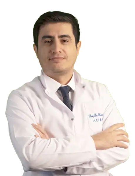









Physical Medicine & Rehabilitation
Alperen Mustafa Oğuz, Physiotherapist


Physical Medicine & Rehabilitation
Ataeollah Moghtasad Azar, Physiotherapist



Medical Technologies Used
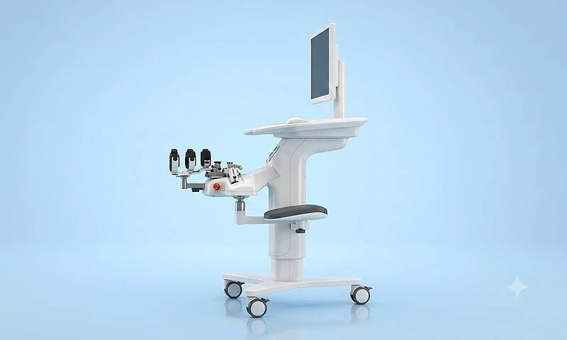
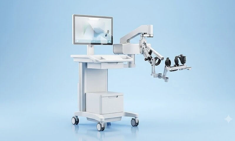
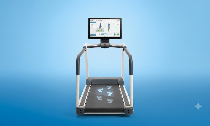
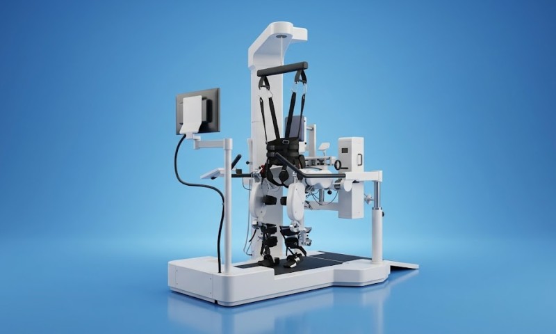
Available at these Hospitals




















What patients say about this unit
★★★★★From 2,400+ verified patient reviews“Years of worry ended once I came here for my rehabilitation programme. Prof. Güven inspired complete confidence from the first meeting.”
“From the first consultation to discharge, my full mouth rehabilitation went smoothly. Dr. Çakmakçı was honest about my options instead of pushing treatment.”
“I had been worried for months, but my physical therapy was sorted out quickly and clearly. Dr. Kasal explained the risks honestly and earned my full trust.”
“My experience with rehabilitation programme here restored my faith in medical care. Dr. Güngör went above and beyond what I expected. The follow-up care was just as good as the treatment itself.”
Patient stories from this unit
 ▶ Vasovagal Syncope: Brendan’s Diagnosis and Treatment at Acibadem
 Brendan shares his vasovagal syncope journey at Acibadem; cardiologist Prof. Dr. Duhan Fatih Bayrak explains why fainting happens and how it is diagnosed and…
Watch the story → Serbia Dušan Vlahović Completes His Beşiktaş Medical at Acibadem Altunizade Hospital
 Serbian international Dušan Vlahović completed his pre-transfer medical examination at Acibadem Altunizade Hospital in Istanbul ahead of his move to Beşiktaş JK.
Read the story →Speak with our medical team
Share your case and our international patient team will guide you to the right specialist — free of charge.