

Neurology
Headache and migraine, epilepsy, multiple sclerosis, neuropathy and movement disorders — diagnosed with EEG, EMG and dedicated neuroimaging, with free remote review of the scans and reports you already have.

Is there a lesion, where is it, and what is producing it
Those three questions in that order are most of neurology. The tempo of an illness narrows the diagnosis before any test is ordered, and the examination decides which test is worth doing at all.
Headache and facial pain
Where classification is the treatment: migraine, the neuralgias and the raised-pressure syndromes that mimic ordinary headache.
Epilepsy, MS and movement
Conditions lived with over decades, where the early treatment decision shapes how the following years go.
Nerve, memory and stroke
Neuromuscular disease investigated with electrodiagnostics, cognitive assessment, and the prevention that follows a stroke.
A neurologist does not operate — and that is the point
Most neurological conditions never need surgery, which is why the neurologist is usually the right first appointment: they establish what the problem is and where it is, and hand over only when an operation is the answer. Being sent to a surgeon first for a problem that turns out to be medical is one of the commonest detours patients describe.
Where surgery is the answer it belongs next door. Epilepsy surgery, deep brain stimulation, disc and cervical cord decompression, aneurysm and tumour surgery and shunting for hydrocephalus are covered in full by neurosurgery, and interventional treatment of head and neuropathic pain by pain management. This unit makes the diagnosis, manages the disease medically, and hands over with the reasoning rather than with a vague referral.
What we will not do
- Diagnose from a scan alone. Imaging is interpreted against the history and examination, or not at all.
- Start antiseizure medication for events that have not been shown to be seizures.
- Add a third epilepsy drug when two have failed, instead of asking whether the epilepsy is surgically treatable.
- Offer or endorse stem cell therapy for stroke, ALS, MS or neuropathy. It is not supported by evidence.
- Describe a progressive disease as treatable when what we have modifies the rate. That honesty is what makes the time available plannable.
Neurologists who lead this work
What actually happens, in order
Bring a video, or a witness
For seizures, blackouts, tremor and movement disorders, thirty seconds of phone footage routinely settles a question that months of investigation has not. These are diagnoses made by pattern recognition, and you cannot observe your own events.
Images, not reports
MRI and CT as DICOM files on a disc or drive, plus the actual EEG and EMG reports. A neuroradiological re-read of the original images changes the reading more often than any new scan would.
The full drug history
Every medication tried, at what dose, for how long, and why it was stopped — side effect, ineffectiveness or something else. Without it a reviewer can only suggest what has already failed.
Investigations as one block
Epilepsy protocol MRI, prolonged video-EEG, comprehensive electrodiagnostic testing or a lumbar puncture with the full panel — arranged together and interpreted by the people who ordered them, rather than spread across weeks.
A plan with monitoring intervals
MS therapy, drug-resistant epilepsy and advanced Parkinson care all need review at intervals. Which drug, which blood test when, who at home is responsible, and the thresholds for contact — agreed in writing before departure.
Six things worth knowing first
A normal EEG does not exclude epilepsy
A single routine recording between seizures is normal in a large proportion of people who definitely have epilepsy, and minor abnormalities appear in people who never have one. The test supports a clinical diagnosis; it does not make or exclude it.
Not every seizure is epilepsy
A substantial proportion of people referred with apparent epilepsy have syncope with a few jerks, functional seizures or a sleep disorder. Antiseizure medication does harm and no good in all three, which is why the witness account matters more than the scan.
White matter lesions are usually incidental
Small white matter changes are extremely common with age and with vascular risk factors, and in most people over fifty they mean nothing. What matters is the pattern, the location and whether they explain the symptoms you actually have.
Daily headache is often caused by the treatment
Frequent use of acute painkillers converts episodic migraine into medication overuse headache, which no longer responds to the drug producing it. It is diagnosed from a diary rather than a scan, and it is reversible — with a doctor, not alone.
MS drugs prevent relapses, not symptoms
Disease-modifying therapy reduces new relapses and new lesions. It does not repair existing damage and does not treat fatigue, spasticity or bladder symptoms, which are managed separately — and rehabilitation carries more of the daily function than most patients are told.
Nerve does not regrow
No treatment currently available regenerates degenerated nerve or replaces lost neurones. Stem cell therapy advertised for stroke, ALS, MS or neuropathy is not supported by evidence, and we say so rather than let hope be sold.
Jump to what you came for
Quick answer
Neurology is the medical specialty that diagnoses and treats disorders of the brain, spinal cord, peripheral nerves, and muscles, including conditions such as stroke, epilepsy, headaches, movement disorders, neuropathies, and dementia. At Acibadem in Turkey, neurology care involves specialist evaluation, advanced diagnostic testing, and personalized treatment planning, with medication, rehabilitation, and referral for interventional or surgical care when needed.
What our neurology unit covers — and who it is for
Neurology is the medical care of the brain, spinal cord, peripheral nerves and muscles. It is the specialty that answers three questions in order: is there a lesion, where is it, and what kind of process is producing it. Almost everything else follows from getting that sequence right, which is why the examination still matters in an era of high-resolution imaging — the scan is chosen on the basis of where the examination says to look.
It is overwhelmingly a diagnostic and long-term specialty rather than a procedural one. Most neurological conditions are managed over years, most are not cured, and the honest measure of good care is how much function and independence are preserved rather than whether a disease has been eliminated.
At Acıbadem International the work is organised into six strands.
- Headache and facial pain — migraine and its variants, the neuralgias, and the raised-pressure syndromes that mimic ordinary headache and threaten sight.
- Epilepsy and paroxysmal disorders — seizure classification, drug selection and the distinction between epileptic seizures and the several conditions that look identical from the outside.
- Neuromuscular disease — the nerves and muscles themselves, investigated with electrodiagnostic testing: neuropathy, myasthenia, motor neurone disease and the inflammatory neuropathies.
- Neuroimmunology — multiple sclerosis and the antibody-mediated diseases of the central nervous system, where treatment choice early in the disease shapes the following decades.
- Movement and cognition — Parkinson disease, tremor, dystonia and the dementias, all of which are diagnosed clinically and managed with medication, therapy and time.
- Cerebrovascular medicine — stroke, transient ischaemic attack and the investigation that determines what prevents the next one.
Where the borders sit, because this specialty has more of them than most. Operations on the brain, spine and peripheral nerves — including surgery for epilepsy, deep brain stimulation, aneurysm and tumour (tumor) surgery, disc surgery and shunting for hydrocephalus — belong with neurosurgery, which covers each of them in full. Interventional treatment of chronic pain, including nerve blocks, radiofrequency procedures and neuromodulation for pain, belongs with pain management. The rehabilitation that determines how much of a recovery is actually realised belongs with physical medicine and rehabilitation. This unit makes the diagnosis and manages the disease medically; where an operation is the answer, it says so and hands over.
What a neurologist actually does
A neurologist is a physician trained in internal medicine who has sub-specialised in disorders of the nervous system, and who does not operate. That last point is the single commonest source of confusion for patients, who frequently arrive expecting a surgeon or having been sent to a surgeon for a problem that is medical.
The consultation is mostly history and examination, and in this specialty the history carries more diagnostic weight than anywhere else in medicine. What exactly happened, in what order, over what time course. A deficit that appeared in seconds suggests a vascular event; over hours to days, inflammation or infection; over weeks to months, a tumour or a degenerative process; over years, something structural or genetic. The tempo of the illness narrows the diagnosis before any test is ordered, which is why a neurologist will ask the same question four different ways rather than move on.
The examination localises. Weakness with brisk reflexes points above the anterior horn cell; weakness with absent reflexes points below it; a sensory level points to the cord; a visual field defect points to a specific segment of the visual pathway. Localisation is what makes the imaging request meaningful — an MRI of the wrong region is a normal scan that reassures nobody and delays the diagnosis by weeks.
The third part is longitudinal management. Epilepsy, multiple sclerosis, Parkinson disease, myasthenia and the neuropathies are conditions someone lives with, and the work is adjusting treatment against how the disease is actually behaving rather than against a protocol — while being honest about what the treatment can and cannot deliver.
Headache: the classification is the treatment
Headache is the commonest reason anyone sees a neurologist, and the great majority of it is primary — meaning the headache is the disease rather than a symptom of something else. Getting the classification right is not an academic exercise: the treatments for migraine, tension headache, cluster headache and the neuralgias barely overlap, and a patient treated for the wrong one concludes that nothing works.
Two things are looked for before anything is labelled primary. A change in a long-standing headache pattern, and features that suggest raised intracranial pressure or a secondary cause — headache that is new in someone over fifty, that wakes them from sleep, that is consistently worsened by lying flat or by straining, or that comes with any neurological sign. Those features change the investigation rather than the treatment, and are why an examination including the optic discs is part of a headache consultation.
Migraine and chronic migraine
Migraine is not a severe headache; it is an episodic brain disorder in which headache is one feature. The others matter for diagnosis: sensitivity to light and sound, nausea, worsening with routine movement, and the preceding hours of yawning, mood change, neck stiffness and food craving that many patients do not connect to the attack at all. Aura, where it occurs, is a spreading cortical phenomenon lasting minutes and resolving fully — visual disturbance most often, sometimes sensory or speech.
Chronic migraine means headache on more days than not over months, with migrainous features on a substantial share of them, and it is a distinct clinical problem rather than simply more of the same. It has its own commonest cause, which is the next section, and it responds to preventive rather than acute treatment.
Treatment divides cleanly. Acute treatment aborts an attack and works better the earlier it is taken. Preventive treatment reduces attack frequency, is taken daily regardless of symptoms, and is judged over months rather than weeks — several classes exist, including drugs developed for other purposes and the newer agents that target the CGRP pathway specifically. Which drug, at what dose, is a prescribing decision made with a doctor who knows your full history, and nothing here is a schedule. Where injections or nerve blocks form part of the plan, they are delivered with pain management.
Medication overuse headache
Medication overuse headache is the commonest reason an episodic migraine becomes a daily one, and it is caused by the treatment. Frequent use of acute painkillers — simple analgesics, combination tablets, triptans, and opioids most of all — transforms the headache pattern into a persistent one that no longer responds to the drug producing it. The patient is doing exactly what they were told to do, taking something when their head hurts, and the head now hurts most days.
It is diagnosed from a headache diary rather than a scan, and it is reversible. Withdrawal of the overused medication produces a period of worse headache before improvement, which is why it is planned with a doctor and usually alongside a preventive treatment rather than attempted alone. Recognising it is one of the highest-value things a neurology consultation does, because it converts an apparently intractable daily headache into a treatable one.
Vestibular migraine
Vestibular migraine is one of the commonest causes of recurrent vertigo in adults and one of the most consistently missed, because the headache may be mild or entirely absent during the attacks. Episodes last minutes to days and produce spinning, unsteadiness or sensitivity to motion, often with visual motion sensitivity between attacks — supermarket aisles, scrolling screens, busy patterns.
The diagnosis is clinical: recurrent vestibular symptoms in someone with a history of migraine, with migrainous features during at least some episodes, and without a better explanation. Investigations are normal by definition, which is precisely why patients accumulate scans and inner-ear tests without an answer. It responds to migraine preventive treatment and to vestibular rehabilitation, which is a genuinely effective combination and is arranged with rehabilitation.
Occipital neuralgia
Occipital neuralgia is pain in the distribution of the greater or lesser occipital nerve — shooting, stabbing or electric pain running from the base of the skull over the back and side of the head, often with tenderness over the nerve and altered sensation in the scalp. It is frequently mistaken for migraine, and the distinction matters because the treatments differ.
The confusion runs in both directions. Many patients labelled with occipital neuralgia actually have migraine with prominent posterior head and neck pain, since neck symptoms are part of the migraine attack in a majority of sufferers. True occipital neuralgia is localised to the nerve territory, is paroxysmal rather than continuous, and characteristically responds — at least temporarily — to a diagnostic block of the nerve, which both confirms the diagnosis and treats it. Those blocks are performed with pain management. Where the underlying problem is upper cervical spine pathology, it is assessed with neurosurgery.
Cluster headache and the trigeminal autonomic cephalalgias
Cluster headache is one of the most severe pains in medicine and one of the most treatable, which makes delayed diagnosis particularly costly. Attacks are strictly one-sided, centred on or behind the eye, last between fifteen minutes and three hours, and are accompanied by autonomic features on the same side — a watering, red eye, a drooping lid, a blocked or running nostril. Unlike migraine, the sufferer is restless and paces rather than lying still, and attacks cluster in bouts over weeks with the same timing each day.
It has specific acute and preventive treatments that are not the migraine ones, and it is frequently misdiagnosed as sinusitis or dental disease for years. Trigeminal neuralgia is a different condition again — brief electric shocks in the face triggered by touch, chewing or wind — and where it does not respond to medical treatment the surgical options are covered by neurosurgery.
Idiopathic intracranial hypertension
Idiopathic intracranial hypertension — previously called benign intracranial hypertension, a name abandoned because it is not benign — is raised pressure inside the skull with no tumour, no clot and no hydrocephalus to explain it. It occurs most often in women of reproductive age and is strongly associated with weight gain.
It matters for one reason above all: it threatens vision. The pressure is transmitted to the optic nerves, producing swelling of the optic discs, and untreated it causes progressive and permanent visual field loss that patients often do not notice until it is advanced, because central vision is preserved until late. The headache is what brings people in; the sight is what is at stake.
The presentation is headache that is worse lying flat and on waking, pulsatile tinnitus described as hearing a heartbeat in the ear, transient greying of vision lasting seconds particularly on standing, and sometimes double vision. Diagnosis requires imaging to exclude a mass and venous sinus thrombosis, followed by lumbar puncture to measure the opening pressure and confirm normal cerebrospinal fluid content. Formal visual field testing with ophthalmology is not optional — it is the measurement that management is steered by, and it is repeated on a schedule.
Treatment addresses the pressure and the underlying driver. Weight reduction produces sustained remission in a substantial proportion and is the only intervention that treats the cause; medication reduces cerebrospinal fluid production; and where vision is deteriorating despite treatment, surgical options including shunting and optic nerve sheath fenestration exist and are decided with neurosurgery and ophthalmology together. Any medicine that may have provoked it — several classes are implicated — is reviewed by the doctor who prescribed it.
Epilepsy
Epilepsy is a tendency to recurrent unprovoked seizures, and the first task is establishing that seizures are what is actually happening. A substantial proportion of people referred with apparent epilepsy have something else: syncope with a few jerks at the end, which is extremely common and routinely misread; functional or dissociative seizures, which are real, disabling and require an entirely different treatment; sleep disorders; and in children, a list of benign paroxysmal events. Treating any of these with antiseizure medication does harm and no good.
The history from a witness is worth more than any investigation, which is why a neurologist will ask to speak to whoever saw the event, and why a phone recording of an episode is one of the most useful things a patient can bring.
Focal seizures and how seizures are classified
Modern classification starts with where the seizure begins. A focal seizure starts in one part of one hemisphere and produces symptoms that reflect that location — a rising sensation in the stomach, an intense familiarity, a smell that is not there, unilateral jerking, or speech arrest. Awareness may be preserved or impaired, and the distinction is part of the classification because it determines driving, work and safety advice. A focal seizure may spread and become a bilateral tonic-clonic seizure, which is what most people picture when they hear the word.
Generalised seizures begin in networks across both hemispheres from the outset and include absence seizures, myoclonic jerks and generalised tonic-clonic seizures. The distinction is not academic: some drugs that work well in focal epilepsy make generalised epilepsies worse, so classification precedes prescription.
Temporal lobe epilepsy
Temporal lobe epilepsy is the commonest focal epilepsy in adults. Seizures typically begin with an aura — a rising epigastric sensation, déjà vu, fear, or an unpleasant smell — followed by staring, unresponsiveness and automatisms such as lip-smacking or fumbling, then a period of confusion afterwards that may last many minutes. Patients frequently describe years of brief “funny turns” before a first convulsion brought them to attention.
It has a characteristic underlying cause in a proportion of cases, hippocampal sclerosis, which is visible on a dedicated epilepsy protocol MRI and often missed on a routine one — a normal brain scan reported elsewhere is a reason to repeat with the correct sequences rather than to abandon the diagnosis. Temporal lobe epilepsy is also the form most likely to become resistant to medication and the one with the best-established surgical outcomes, which is why persistent seizures on two appropriately chosen drugs prompt a referral for surgical evaluation rather than a third drug. That evaluation and the operation itself belong with neurosurgery.
Treatment, and what it is judged against
Around two-thirds of people with epilepsy become seizure-free on medication, most of them on the first or second drug tried. The choice depends on seizure type, sex and childbearing plans, other conditions and other medicines, and it is made with a doctor who has all of that in front of them — no page can select it. Two principles are worth stating plainly because they are so often misunderstood. Drugs are introduced slowly and one at a time, because most early side effects come from the speed of introduction rather than the drug itself. And they are never stopped abruptly, since abrupt withdrawal is a recognised cause of prolonged seizures.
Where two well-chosen drugs at adequate doses have failed, the epilepsy is defined as drug-resistant, and the chance that a third drug produces freedom from seizures is low. That is the point at which the question changes from which drug next to whether this epilepsy has a surgically treatable cause, and it is a referral rather than a further prescription. Status epilepticus — a seizure that does not stop, or repeated seizures without recovery between them — is a medical emergency and is treated in hospital immediately.
The EEG test
An EEG test records the electrical activity of the cortex through scalp electrodes, and its role is consistently misunderstood in both directions. It does not diagnose epilepsy and it does not exclude it. A single routine recording between seizures is normal in a large proportion of people who definitely have epilepsy, and minor abnormalities appear in people who never have a seizure. It is a test that supports a clinical diagnosis, classifies the epilepsy and occasionally identifies a specific syndrome.
Its yield rises with the right technique. Recording after sleep deprivation, capturing drowsiness and sleep, and the standard activation procedures of hyperventilation and photic stimulation all increase the chance of finding the abnormality that is there. Where events are frequent enough, prolonged video-EEG monitoring captures an actual episode with simultaneous video and electrical recording, and that remains the only definitive way to distinguish epileptic from non-epileptic events. Video-EEG is also the first stage of any surgical evaluation.
An EEG is painless, involves no radiation and takes around half an hour for a routine study. Patients are usually asked to arrive with clean, product-free hair and to follow specific instructions about sleep and caffeine beforehand, since those instructions are what make the recording worth doing.
The EMG test and the nerve conduction study
The EMG test and the nerve conduction study are performed together as a single examination, and they answer questions that no scan can. An MRI shows structure; these show function, and they localise a problem along the length of a nerve in a way imaging cannot.
The nerve conduction study stimulates a nerve with a small electrical pulse and records the response, measuring how fast the signal travels and how large it is. Slowing points to damage of the myelin sheath; a reduced response size points to loss of the axons themselves; and the pattern across several nerves separates a generalised neuropathy from a single trapped nerve from a nerve root problem. The needle EMG then samples the electrical activity of the muscle itself, at rest and during contraction, which distinguishes muscle disease from nerve disease and identifies which nerve root supplies an affected muscle.
The test is uncomfortable rather than painful. The stimulation feels like a series of sharp taps, and the needle examination is described by most patients as a deep ache while the muscle is being activated. It takes thirty to sixty minutes depending on how many nerves need studying, and it is worth knowing that the examination is designed during it: the neurophysiologist adds nerves and muscles as the findings direct, so the study is not a fixed protocol and its length varies.
It is the decisive test for carpal tunnel syndrome and other entrapments, for radiculopathy, for generalised neuropathies, for myasthenia using repetitive stimulation, and for motor neurone disease.
Peripheral neuropathy
Peripheral neuropathy is damage to the nerves outside the brain and spinal cord, and it presents in a characteristic pattern: numbness, tingling, burning or loss of sensation beginning in the toes and feet and progressing upwards over months to years, reaching the hands only when it has ascended to the knees. That length-dependent pattern is the signature of the commonest neuropathies and is itself a diagnostic clue, because a neuropathy that does not follow it needs a different differential.
The investigation looks for a treatable cause, and a meaningful proportion of neuropathies have one: diabetes and pre-diabetes above all, vitamin B12 deficiency, thyroid disease, kidney disease, alcohol, a paraprotein in the blood, coeliac (celiac) disease, some medications including several chemotherapy agents, and hereditary neuropathies with a family history that has often not been recognised as such. Where no cause is found after appropriate testing the neuropathy is called idiopathic, which is a common and legitimate conclusion rather than an incomplete workup.
Treatment addresses the cause where there is one and the symptoms where there is not. Nothing currently available regenerates damaged nerves, and being clear about that at the outset prevents years of expensive disappointment. Foot care matters more than most patients are told, because a foot that cannot feel injury develops ulcers silently — that work is shared with endocrinology in diabetic neuropathy. The drug treatment of neuropathic pain, and interventional options for it, belong with pain management.
Small fibre neuropathy (small fiber neuropathy)
Small fiber neuropathy affects the thin unmyelinated fibres that carry pain and temperature sensation and control autonomic function, and it is the neuropathy that gets missed, because the standard tests are normal. Nerve conduction studies examine large fibres only, so a patient with burning feet, normal strength, normal reflexes and a completely normal EMG is frequently told there is nothing wrong.
Symptoms are burning, electric or prickling pain, usually worse at night and often disproportionate to any visible sign, sometimes with autonomic features — dry eyes and mouth, altered sweating, gut symptoms, palpitations on standing. Diagnosis rests on the clinical picture supported by skin biopsy for intraepidermal nerve fibre density or by quantitative sensory and autonomic testing. Causes overlap with large-fibre neuropathy but include a higher proportion of immune and inflammatory conditions, so the investigation reaches further, and it is worth pursuing because a proportion of cases are treatable.
Guillain-Barré syndrome and CIDP
Guillain-Barré syndrome is an acute immune attack on peripheral nerves, typically following an infection, producing ascending weakness and loss of reflexes over days. It is a medical emergency because it can involve the muscles of breathing and swallowing, and it is treated in hospital with immunotherapy and close monitoring of respiratory function; most people recover substantially, though recovery is measured in months.
CIDP — chronic inflammatory demyelinating polyradiculoneuropathy — is its chronic counterpart, developing over more than eight weeks and often relapsing. The distinction from ordinary neuropathy is critical because CIDP is treatable, and it is defined by proximal as well as distal weakness, marked reduction of reflexes and demyelinating features on nerve conduction studies, with supportive cerebrospinal fluid findings. It responds to immunoglobulin, corticosteroids or plasma exchange, and it is one of the small number of neuropathies where the right diagnosis produces recovery rather than management.
Myasthenia gravis
Myasthenia gravis is an antibody-mediated disease of the neuromuscular junction, and its defining feature is fatigable weakness — muscles that work at the start of an activity and fail as it continues, recovering with rest. That fluctuation is the diagnosis, and it is also why the condition is so often dismissed: a patient examined in the morning after resting can look entirely normal.
It presents in the eyes in a majority of cases, with a drooping lid or double vision that is worse at the end of the day, and in a substantial proportion it remains ocular. Where it generalises it affects the face, swallowing, speech, neck and limb muscles, and in its severe form the muscles of breathing — a myasthenic crisis is a medical emergency treated in intensive care.
Diagnosis rests on antibody testing, on repetitive nerve stimulation and single-fibre EMG, and on imaging the chest, because a proportion of patients have a thymoma and removal of the thymus changes the course of the disease in selected patients. Treatment includes drugs that improve transmission at the junction and immunosuppression to reduce the antibodies, with newer targeted agents now available; a substantial list of common medicines can worsen myasthenia, which is why the diagnosis is declared before any anaesthesia (anesthesia) or new prescription. Thymectomy is performed with thoracic surgery.
Amyotrophic lateral sclerosis (ALS symptoms and motor neurone disease)
Amyotrophic lateral sclerosis is the commonest form of motor neurone disease, a progressive degeneration of the upper and lower motor neurones. The ALS symptoms that bring people to a neurologist are painless progressive weakness beginning in one limb, a foot that drops, a hand that loses dexterity, or — in the bulbar form — slurred speech and difficulty swallowing. Muscle wasting, visible twitching under the skin and cramps accompany it, and weakness spreads to adjacent regions over months.
The diagnosis is clinical, supported by EMG showing denervation across regions, and made by excluding the conditions that mimic it — cervical myelopathy above all, which is why an MRI of the spine is part of the workup, along with multifocal motor neuropathy, which is treatable and must not be missed.
This is the part of the specialty where honesty matters most. ALS is progressive and there is no treatment that stops it. The drugs available modify the rate of progression modestly. What genuinely changes both survival and quality of life is multidisciplinary care: non-invasive ventilation, which is the single intervention with the clearest survival benefit; nutritional support and timely feeding tube placement; secretion management; communication aids; and mobility and equipment provision arranged before they are needed rather than after. Decisions about ventilation and feeding are made in advance, calmly, with the patient leading them, and they are revisited as the person’s own view changes. We do not offer or endorse unproven therapies for this disease, including stem cell treatments advertised for it, and we say plainly that they have not been shown to help.
Multiple sclerosis symptoms, diagnosis and treatment
Multiple sclerosis is an immune-mediated disease in which the myelin of the central nervous system is attacked, producing lesions separated in space and in time. It typically begins between twenty and forty and is more common in women.
The multiple sclerosis symptoms that most often bring people in are optic neuritis — pain on eye movement with loss of vision in one eye over days — sensory disturbance in a limb or across the trunk, weakness, double vision, imbalance, and bladder symptoms. Two features are characteristic and worth knowing: symptoms typically evolve over days and recover over weeks, distinguishing them from stroke, and existing symptoms temporarily worsen with heat or fever without any new inflammation, which is a physiological effect rather than a relapse.
Diagnosis combines the clinical picture with MRI of the brain and spinal cord and, where needed, cerebrospinal fluid examination for oligoclonal bands. Modern criteria allow a diagnosis after a single clinical episode where imaging demonstrates dissemination in space and time, which matters because early treatment changes long-term outcome.
Relapsing remitting MS and the treatment decision
Relapsing remitting ms is the pattern in the large majority at onset: discrete relapses with recovery between them. Over years a proportion transition to a secondary progressive course in which disability accumulates without distinct relapses, and a minority are progressive from the start.
The central decision is which disease-modifying therapy and when. These drugs reduce relapse rate and new lesion formation and, started early, reduce long-term disability; they differ substantially in effectiveness, in route and frequency of administration, in monitoring requirements and in risk, which ranges from mild to serious. The trade-off between a highly effective therapy and its risk profile is a real one and is decided with the patient rather than for them, with the specific drugs and their monitoring belonging to that consultation.
What these drugs do not do is repair damage already present or treat existing symptoms — spasticity, fatigue, bladder dysfunction and walking difficulty are managed separately, and rehabilitation and exercise have a larger effect on day-to-day function than most patients are led to expect. Relapses themselves are treated with a short course of high-dose corticosteroid, which speeds recovery without changing the eventual level of recovery, and mild relapses are frequently not treated at all.
Neuromyelitis optica and MOG antibody disease
Two conditions that resemble MS are now recognised as distinct diseases with different treatment, and separating them is one of the more consequential distinctions in this specialty. Neuromyelitis optica spectrum disorder, associated with aquaporin-4 antibodies, produces severe optic neuritis and extensive spinal cord lesions, and several drugs used in MS make it worse. MOG antibody-associated disease is a third entity with its own pattern and outlook. Antibody testing in anyone with severe optic neuritis or extensive cord involvement is therefore standard rather than optional, and it is a test that changes treatment rather than merely labelling it.
Movement disorders
Movement disorders divide into too little movement and too much. Both are diagnosed by pattern recognition, and both are areas where a video of the problem is worth more than a description.
Parkinson disease
Parkinson disease is a clinical diagnosis based on bradykinesia — slowness with decrementing amplitude on repeated movement — together with rest tremor, rigidity, or both. No scan makes the diagnosis; imaging is used to exclude alternatives, and a dopamine transporter scan distinguishes Parkinsonian tremor from essential tremor where the examination is genuinely equivocal, not routinely.
The non-motor features often precede the motor ones by years and are frequently the more disabling: loss of smell, constipation, acting out dreams during sleep, depression, and later cognitive change and orthostatic dizziness. Asking about them is part of the diagnosis rather than a supplementary enquiry.
Treatment is symptomatic and effective, particularly in the early years. Levodopa remains the most effective drug and the long-standing advice to delay it as long as possible has not held up; timing is individualised with a specialist. Over years, response becomes less predictable, with wearing-off before the next dose and involuntary movements at peak effect, and management shifts to adjusting timing and adding agents. Where fluctuations become refractory, device-assisted therapies including deep brain stimulation and infusion treatments are considered — the surgical route is covered by neurosurgery. Exercise is not an adjunct here: structured, high-intensity exercise has evidence for slowing functional decline, and physiotherapy, speech therapy and occupational therapy through rehabilitation carry more of the outcome than patients are usually told.
Several conditions mimic Parkinson disease and behave very differently — progressive supranuclear palsy, multiple system atrophy and corticobasal syndrome among them — and a poor or absent response to levodopa, early falls, early autonomic failure or early cognitive decline are the features that prompt reconsideration.
Essential tremor and dystonia
Essential tremor is the commonest movement disorder and is routinely confused with Parkinson disease, though the two differ clearly on examination: essential tremor appears with posture and action, affects both sides relatively symmetrically, commonly involves the head and voice, often runs in families and frequently improves temporarily with alcohol. Parkinsonian tremor is present at rest, is asymmetric and is accompanied by slowness. Treatment options include several oral drugs, focal injections for head and voice tremor, and for severe refractory cases stereotactic procedures including focused ultrasound and deep brain stimulation.
Dystonia is sustained or intermittent muscle contraction producing abnormal postures and repetitive movements, most often focal in adults — cervical dystonia, blepharospasm, writer’s cramp. Its characteristic feature is the sensory trick, a light touch to the face or neck that temporarily corrects the posture, which is diagnostic and is almost never volunteered by patients unless asked. Botulinum toxin injection into the affected muscles is the treatment of choice for focal dystonia and is repeated at intervals.
Restless legs syndrome
Restless legs syndrome is defined by four features together: an urge to move the legs, usually with an unpleasant sensation; worsening at rest; relief with movement; and a clear evening or night predominance. All four are required, which is why it is both over-diagnosed in people with ordinary leg discomfort and under-diagnosed in people who never mention it.
Iron status is checked in everyone, since low iron stores are a common and correctable driver even when the blood count is normal, and the threshold used is higher than the one applied to anaemia (anemia). Several classes of medication and some antidepressants worsen it and are reviewed by the prescribing doctor. Long-term treatment with dopamine agonists carries a specific risk of augmentation, in which symptoms become earlier, more intense and more widespread over time — a complication caused by the treatment and one of the reasons prescribing in this condition has changed.
Stroke and transient ischaemic attack
Stroke is sudden neurological deficit caused by interruption of blood supply to part of the brain, or by bleeding into it. The deficit appears in seconds to minutes — that tempo is the diagnosis — and its nature reflects the territory affected: weakness or numbness down one side, speech disturbance, visual field loss, or sudden severe imbalance and double vision in posterior circulation events.
Stroke is a medical emergency and is treated in hospital immediately. The treatments that restore blood flow — thrombolysis and mechanical thrombectomy — work within a limited window measured from the moment symptoms began, and their benefit falls steeply as that window advances, which is why the time of onset is the single most important piece of information anyone can supply and why imaging is done before any treatment to distinguish an infarct from a haemorrhage (hemorrhage). Surgical management of haemorrhage and of some large infarcts belongs with neurosurgery.
Transient ischaemic attack (transient ischemic attack)
A transient ischemic attack is the same event with symptoms that resolve completely, usually within an hour. Its importance is entirely prospective: it is a warning, the risk of a completed stroke is highest in the days immediately afterwards, and that risk is substantially reduced by prompt investigation and treatment. A TIA is therefore assessed urgently in hospital rather than at a routine appointment, and it is investigated exactly as a stroke would be.
The investigation asks where the clot came from, because the answer determines the prevention. Imaging of the brain and of the neck and intracranial vessels; an ECG and prolonged cardiac rhythm monitoring, since atrial fibrillation is frequently paroxysmal and missed on a single tracing; an echocardiogram; and blood tests for glucose and lipids. In younger patients the investigation extends to arterial dissection, patent foramen ovale and clotting disorders.
Carotid artery stenosis
Carotid artery stenosis is narrowing of the internal carotid artery by atherosclerotic plaque, and it causes stroke by shedding fragments downstream rather than by restricting flow. It is graded by degree of narrowing on ultrasound, CT or MR angiography, and the crucial distinction is between symptomatic stenosis — one that has already produced a stroke or TIA in that territory — and asymptomatic stenosis found incidentally.
Symptomatic stenosis of significant degree benefits from revascularisation by endarterectomy or stenting, and the benefit is greatest when it is done soon after the event rather than weeks later. Asymptomatic stenosis is a different calculation entirely: modern medical treatment has reduced its stroke risk considerably, and intervention is reserved for selected cases rather than applied because a narrowing was found. Either way, the medical treatment — antiplatelet therapy, blood pressure control, high-intensity statin treatment, diabetes control and stopping smoking — is what does most of the work, and it applies to everyone. The procedures are performed with cardiovascular surgery and interventional radiology.
Prevention after any stroke or TIA is where this unit spends most of its time, and it is unglamorous and effective: the right antithrombotic for the mechanism — antiplatelet for atherosclerotic events, anticoagulation for atrial fibrillation, and those are different decisions — blood pressure, lipids, glucose, smoking, alcohol and physical activity.
Memory, cognition and the dementias
Memory complaints are among the commonest reasons for a neurology referral and among the least often caused by a dementia. Late-life depression can impair memory convincingly enough to earn the name pseudodementia — and it is treatable, which is why old-age psychiatry sits beside this assessment. The assessment separates three things: normal ageing, mild cognitive impairment — measurable deficit on testing without loss of independence — and dementia, in which cognitive decline interferes with day-to-day function. The distinction rests on function rather than on a test score, which is why an account from someone who knows the patient is essential and why the consultation asks about finances, medication management, cooking and navigation rather than about forgetting names.
The reversible contributors are looked for in everyone, because they are common and treatable: depression, which produces a very convincing pseudo-dementia; sleep apnoea; thyroid disease; B12 deficiency; alcohol; and medication, particularly anticholinergics, sedatives and painkillers, whose cumulative burden is one of the most underestimated causes of cognitive slowing in older people. Reviewing that list is done with the doctor who prescribed them.
Lewy body dementia and frontotemporal dementia
Where a degenerative dementia is present, distinguishing the type matters because management differs. Alzheimer disease begins with episodic memory. Lewy body dementia presents with fluctuating attention, visual hallucinations that are typically well-formed and not frightening at first, parkinsonism and acting out dreams in sleep — and it carries a critical practical consequence, because severe sensitivity to antipsychotic drugs in this group can be dangerous, and that is stated on every prescription record we can reach. Frontotemporal dementia presents in younger patients with personality change, loss of empathy, disinhibition and compulsive behaviour, or with progressive language failure, and memory is preserved early — it is frequently mistaken for a psychiatric illness for years. Vascular cognitive impairment follows the pattern of the vascular damage rather than a fixed sequence.
Investigation includes cognitive testing, blood tests, and structural imaging, with functional imaging and cerebrospinal fluid or blood biomarkers used in selected cases, particularly in younger patients and atypical presentations. Treatment is honest about its limits: the available drugs produce modest symptomatic benefit in some dementias and none in others, the newer antibody treatments for Alzheimer disease have specific eligibility criteria and monitoring requirements and are not appropriate for most patients, and the interventions with the largest effect on quality of life remain non-pharmacological — routine, environment, hearing and vision correction, exercise, treatment of depression, and support for the person doing the caring. Care is shared with psychiatry where behavioural symptoms dominate.
Lumbar puncture
A lumbar puncture samples cerebrospinal fluid through a fine needle inserted between the vertebrae in the lower back, below the level at which the spinal cord ends — a point worth stating because the commonest fear about the procedure is of the cord being damaged, and the needle does not reach it.
It is done for diagnosis and occasionally for treatment. Diagnostically it identifies infection of the nervous system, subarachnoid haemorrhage where imaging is negative, inflammatory diseases including multiple sclerosis through oligoclonal bands, some cancers involving the meninges, and the biomarkers used in dementia assessment. It also measures the opening pressure, which is the diagnostic measurement in idiopathic intracranial hypertension and where removing fluid is itself therapeutic.
The procedure takes twenty to thirty minutes with local anaesthetic and is performed sitting or lying on the side. Most patients describe pressure rather than pain. Imaging is performed first where there are features suggesting raised pressure from a mass. The most common complication is a post-dural-puncture headache — positional, worse on sitting or standing, better lying flat — which is much less frequent with modern atraumatic needles, resolves in most cases, and where persistent is treated effectively with a blood patch. Lying flat afterwards does not prevent it, despite the persistence of that advice.
Cervical radiculopathy and the neck-arm problem
Cervical radiculopathy is compression or irritation of a nerve root as it leaves the cervical spine, producing pain radiating into the arm in a recognisable distribution, with numbness, tingling and sometimes weakness in the muscles that root supplies. The distribution is what makes the diagnosis: a C6 root produces different findings from a C7 root, and the pattern usually identifies the level before any imaging.
Most cervical radiculopathy improves without surgery over weeks to months, and the initial management is non-operative. The neurologist’s contribution is confirming that the problem is a root rather than something else — carpal tunnel syndrome, ulnar neuropathy at the elbow, brachial plexus pathology, or a shoulder problem producing referred pain — and the electrodiagnostic study described under EMG and nerve conduction studies is what separates them, particularly where imaging shows degenerative change at several levels, which it does in most adults regardless of symptoms.
Two situations change the pathway. Progressive weakness rather than pain, and features of cord compression — clumsy hands, deteriorating balance and gait, brisk reflexes in the legs — which indicate cervical myelopathy, a spinal cord problem rather than a root one, with a different natural history and a surgical threshold. Both belong with neurosurgery, which covers cervical myelopathy, disc herniation and the operations for them in full. Injection treatment for persistent radicular pain is delivered with pain management.
Normal pressure hydrocephalus
Normal pressure hydrocephalus is worth its own section because it is one of the few causes of dementia and gait failure in older people that can be substantially improved. It produces a triad: a characteristic gait — broad-based, shuffling, with feet that seem stuck to the floor and marked difficulty turning — together with cognitive slowing and urinary urgency, with the gait typically first and most prominent.
It is frequently attributed to age, to Parkinson disease or to vascular dementia, and the imaging finding of enlarged ventricles is common in the elderly and does not by itself make the diagnosis. What supports it is the clinical triad with ventricular enlargement out of proportion to cortical atrophy, and above all a measured improvement in gait after removal of a volume of cerebrospinal fluid — a tap test or an extended drainage — which is the closest thing to a predictive test for who will benefit from treatment. Timed walking is recorded before and after, because the difference is objective and the impression of improvement is not.
Treatment is surgical, by shunting, and it is performed and assessed by neurosurgery. The role of this unit is recognising the syndrome, excluding the mimics and running the assessment that predicts response, because the outcome depends heavily on selecting the right patients and on not waiting until the cognitive component is advanced.
Dizziness causes: vertigo, presyncope and blackouts
Dizziness causes divide into four different complaints that share one word, and separating them is most of the diagnosis. Vertigo is an illusion of movement and points to the vestibular system. Presyncope is a feeling of impending faint and points to the circulation. Disequilibrium is unsteadiness on standing and walking without a sense of spinning and points to a sensory, cerebellar or neuropathic cause. Non-specific lightheadedness overlaps with anxiety, hyperventilation and medication effects.
Within vertigo, the timing pattern narrows it further: seconds with head position change suggests benign paroxysmal positional vertigo, treated at the bedside with repositioning manoeuvres that are highly effective and frequently not offered; minutes to hours with hearing symptoms suggests Ménière disease; hours to days with hearing loss suggests a labyrinthine cause; hours to days without hearing loss suggests vestibular neuritis or, if recurrent, vestibular migraine. Ear-related causes are assessed with otorhinolaryngology, and persistent unsteadiness responds to vestibular rehabilitation.
Blackouts are a separate problem and the distinction between syncope and seizure is one of the most consequential in this specialty, because it determines whether someone is investigated for a heart problem or started on antiseizure medication. Syncope is a transient loss of consciousness from global cerebral hypoperfusion, is usually preceded by warning, is brief, and is followed by rapid recovery — and it is frequently accompanied by a few jerks, which is the single commonest reason syncope is misdiagnosed as epilepsy. Prolonged confusion afterwards, tongue biting at the side, and a stereotyped onset point the other way. Cardiac causes are excluded with cardiology, and syncope during exertion or with a family history of sudden death is investigated urgently.
What neurology cannot do
- It cannot regenerate damaged nerve or brain. No treatment currently available regrows a degenerated nerve or replaces lost neurones, and any offer to do so — including stem cell therapy advertised for stroke, ALS, MS or neuropathy — is not supported by evidence. We say so plainly rather than allowing hope to be sold.
- It cannot make most degenerative disease stop. Treatment for Parkinson disease and the dementias is symptomatic; treatment for ALS modifies the rate modestly. Being clear about that at diagnosis is what allows the time available to be planned well.
- It cannot diagnose from a scan alone. A brain MRI reported as showing white matter changes is a finding, not a diagnosis, and in most people over fifty it is an incidental one. Imaging is interpreted against the history and examination or not at all.
- It cannot fix what a normal test has already answered. A normal EEG does not exclude epilepsy, and a normal nerve conduction study does not exclude neuropathy. Repeating a normal test rarely helps; choosing a different test usually does.
- It cannot treat a headache caused by its own treatment without withdrawal. Medication overuse headache does not respond to more medication, and the improvement requires a period of feeling worse.
- It does not operate. Where the answer is surgical it is handed to neurosurgery with the diagnosis and the reasoning, not with a vague referral.
Your multidisciplinary team
The neurologist makes the diagnosis, chooses and adjusts treatment and holds the longitudinal picture. Sub-specialisation is real in this field, and a unit of this size runs separate clinics for epilepsy, multiple sclerosis and neuroimmunology, movement disorders, neuromuscular disease, headache, stroke and cognitive disorders — which matters, because outcomes in these conditions track with how many the clinician sees. The clinical neurophysiologist performs and interprets the EEG, EMG and evoked potential studies. The neuroradiologist reads the imaging, and in this specialty the difference between a general report and a dedicated neuroradiological one is frequently the diagnosis. The specialist nurse runs the monitoring and is the practical point of contact between appointments in epilepsy, MS and Parkinson care.
Around them: neurosurgery for everything operative, physical medicine and rehabilitation with physiotherapy, occupational therapy and speech and language therapy — the specialty that converts a diagnosis into preserved function — pain management for interventional treatment of neuropathic and head pain, ophthalmology for optic neuritis and for the visual field monitoring that raised intracranial pressure requires, cardiology for syncope and for the rhythm monitoring that stroke prevention depends on, psychiatry for the mood and behavioural components that accompany most chronic neurological disease, endocrinology for diabetic neuropathy, hematology for clotting disorders in young stroke, rheumatology where a systemic inflammatory disease involves the nervous system, and medical genetics for the hereditary neuropathies, ataxias and dystonias.
The international patient journey
Neurology travels differently from surgery, because what is usually wanted is a diagnosis or a second opinion on one, not a procedure with a date.
The first and commonest pattern is a remote review of a diagnosis already made — an MS diagnosis a patient wants confirmed before committing to years of treatment, an epilepsy that has not responded to two drugs, a neuropathy with no cause found, or a diagnosis of a progressive disease that nobody has explained properly. What makes this useful is the raw material rather than the letters: imaging as DICOM files rather than reports, the actual EEG or EMG report with the traces where available, the full list of drugs tried with doses and reasons for stopping, and a description or video of the events themselves.
The second is a diagnostic visit for investigations that need to be done in one place and interpreted together — dedicated epilepsy protocol MRI, prolonged video-EEG monitoring, comprehensive electrodiagnostic testing, or a lumbar puncture with the full panel. These are arranged as a single block rather than spread across weeks, and the value is in the interpretation being done by people who ordered the tests.
The third is transfer of ongoing management for a chronic condition, which is the one that needs most planning. Disease-modifying therapy for MS, drug-resistant epilepsy and advanced Parkinson disease all require monitoring at intervals, and a plan that ends at the airport is not a plan. Which drug, which blood test at which interval, who at home is responsible for each, and what the thresholds are for contact — agreed in writing before departure.
One practical note specific to this specialty. Bring a witness account, or a recording. For seizures, blackouts, movement disorders and tremor, thirty seconds of phone video routinely settles a question that months of investigation has not, and it is the single most useful thing a patient can arrive with.
Frequently Asked Questions
What is the difference between a neurologist and a neurosurgeon?
A neurologist is a physician who diagnoses and treats disorders of the brain, spinal cord, nerves and muscles with medication and other non-surgical means, and does not operate. A neurosurgeon operates. Most neurological conditions never need surgery, which is why the neurologist is usually the right first appointment: they establish what the problem is and where it is, and hand over only when an operation is the answer. Being sent to a surgeon first for a problem that turns out to be medical is one of the commonest detours patients describe.
Why does the neurologist ask the same question so many different ways?
Because the tempo of an illness is the diagnosis. A deficit that appeared over seconds suggests a vascular event, over hours to days inflammation or infection, over weeks to months a tumour or degeneration, over years something structural or genetic. Patients naturally compress and reorder events when telling the story, so the same question asked differently is how the actual sequence is recovered. In neurology the history carries more diagnostic weight than any single test, and time spent on it is not preamble to the investigation — it is the investigation.
Do I need an MRI?
Only if the examination says where to look. An MRI of the wrong region is a normal scan that reassures nobody and delays the diagnosis by weeks, and imaging the whole nervous system to be thorough produces incidental findings that generate more anxiety and more tests than they resolve. Localising the problem clinically is what makes the imaging request meaningful. There are conditions where imaging is essential and done immediately, and there are many where a careful examination and an electrodiagnostic study answer the question better than any scan would.
My brain MRI showed white matter lesions. Should I worry?
Usually not on its own. Small white matter changes are extremely common with age and with vascular risk factors such as high blood pressure and diabetes, and in most people over fifty they are an incidental finding rather than a disease. What matters is the pattern, the location, the number, whether they enhance with contrast, and above all whether they explain the symptoms you actually have. Multiple sclerosis lesions have a characteristic distribution and are read alongside the clinical picture and often cerebrospinal fluid findings — a report of white matter lesions is a finding, not a diagnosis.
Is my headache dangerous?
The great majority of headache is primary, meaning the headache is the condition rather than a symptom of something else, and does not indicate danger. What changes the investigation is a change in a long-standing pattern, a new headache in someone over fifty, headache that wakes you from sleep or is consistently worse lying flat or on straining, and any accompanying neurological sign. Those features prompt imaging; they do not by themselves mean something serious has been found. Examining the back of the eye is part of a proper headache consultation because it detects raised pressure that the description alone would miss.
Why do my migraines suddenly happen almost every day?
The commonest reason an episodic migraine becomes a daily headache is the treatment. Frequent use of acute painkillers — simple analgesics, combination tablets, triptans, and opioids most of all — converts the pattern into medication overuse headache, which no longer responds to the drug producing it. It is diagnosed from a headache diary rather than a scan, and it is reversible: withdrawing the overused medication produces a period of worse headache before improvement, which is why it is planned with a doctor and usually alongside starting a preventive treatment rather than attempted alone.
What is vestibular migraine, and why did nobody find it?
It is one of the commonest causes of recurrent vertigo in adults, and it is missed because the headache may be mild or entirely absent during the attacks — so the patient is investigated for an inner-ear problem instead. Episodes last minutes to days and produce spinning, unsteadiness or motion sensitivity, often with visual motion sensitivity between attacks in supermarkets, on escalators or when scrolling. The investigations are normal by definition, which is precisely why scans and inner-ear tests accumulate without an answer. It responds well to migraine preventive treatment combined with vestibular rehabilitation.
Is occipital neuralgia the same as migraine?
No, though they are confused in both directions. Occipital neuralgia is pain in the territory of the occipital nerve — shooting, stabbing or electric pain from the base of the skull over the back of the head, often with scalp tenderness and altered sensation. Many patients labelled with it actually have migraine with prominent neck and posterior head pain, since neck symptoms occur in most migraine attacks. True occipital neuralgia is localised to the nerve, paroxysmal rather than continuous, and characteristically responds at least temporarily to a diagnostic nerve block, which both confirms it and treats it.
My headache is worse when I lie down and I hear my heartbeat in my ear. What is that?
That combination — headache worse lying flat and on waking, pulsatile tinnitus, and brief greying of vision on standing — is the classic presentation of idiopathic intracranial hypertension, raised pressure inside the skull without a tumour or clot to explain it. It matters because it threatens vision: the pressure damages the optic nerves and causes progressive field loss that is often not noticed until advanced, because central vision is preserved until late. It is investigated with imaging and then a lumbar puncture to measure the opening pressure, and formal visual field testing is what management is steered by thereafter.
Does a normal EEG mean I do not have epilepsy?
No. A single routine EEG between seizures is normal in a large proportion of people who definitely have epilepsy, and minor abnormalities appear in people who never have a seizure — the test supports a clinical diagnosis rather than making or excluding one. Yield improves with the right technique: recording after sleep deprivation, capturing drowsiness and sleep, and the standard activation procedures. Where events are frequent enough, prolonged video-EEG monitoring captures an actual episode with simultaneous video and electrical recording, and that remains the only definitive way to distinguish epileptic from non-epileptic events.
Could my seizures not be epilepsy at all?
Quite possibly, and this is one of the most important questions in the specialty. A substantial proportion of people referred with apparent epilepsy have something else: syncope with a few jerks at the end, which is extremely common and routinely misread as a seizure; functional or dissociative seizures, which are real and disabling and require an entirely different treatment; sleep disorders; and in children, a list of benign paroxysmal events. Antiseizure medication does harm and no good in all of these, which is why a witness account, and better still a phone recording of an episode, is worth more than any test.
Why do I need to keep taking medication if I have not had a seizure for years?
Because being seizure-free on treatment is evidence the treatment is working, not that the tendency has gone. Whether it is reasonable to try withdrawal depends on the epilepsy syndrome, the EEG, how long you have been free of seizures, the underlying cause and the practical consequences of a recurrence — driving and occupation among them — and it is a decision made with a specialist. What is never done is stopping abruptly: abrupt withdrawal is a recognised cause of prolonged seizures, so any reduction is gradual and planned.
Two epilepsy drugs have not worked. Should I try a third?
Probably not as the next step. Where two well-chosen drugs at adequate doses have failed, the epilepsy is defined as drug-resistant and the chance a third produces freedom from seizures is low. The question changes at that point from which drug next to whether this epilepsy has a surgically treatable cause, which requires a formal evaluation including dedicated epilepsy protocol imaging and prolonged video-EEG monitoring. That referral is frequently delayed by years, and the delay costs, because outcomes from epilepsy surgery are better the earlier the evaluation happens.
What does an EMG feel like, and how long does it take?
It is uncomfortable rather than painful. The nerve conduction part delivers small electrical pulses that feel like a series of sharp taps; the needle part is described by most patients as a deep ache while the muscle is being activated. It takes thirty to sixty minutes depending on how many nerves and muscles need studying, and the length varies because the examination is designed as it goes — the neurophysiologist adds territories as the findings direct rather than following a fixed protocol. No sedation is needed and you can drive afterwards.
My nerve tests were normal but my feet still burn. What now?
Normal nerve conduction studies do not exclude neuropathy, because they examine large fibres only. Burning feet with normal strength, normal reflexes and a normal EMG is the classic presentation of small fibre neuropathy, which affects the thin fibres carrying pain and temperature sensation and controlling autonomic function. It is diagnosed on the clinical picture supported by skin biopsy for nerve fibre density or by quantitative sensory and autonomic testing. It is worth pursuing rather than accepting a normal result as the answer, because a proportion of cases have a treatable immune or metabolic cause.
Can nerve damage be reversed?
Damaged nerve does not regenerate in any way current medicine can deliver, and that is worth being clear about because it prevents years of expensive disappointment. What can change is the cause: correcting B12 deficiency, controlling diabetes, stopping a causative drug or treating an underlying immune condition can halt progression and sometimes allow partial recovery over long periods. Some neuropathies — CIDP in particular — are genuinely treatable and recover substantially, which is why finding the exact type matters. Stem cell therapies advertised for neuropathy are not supported by evidence, and we say so.
What is CIDP and how is it different from ordinary neuropathy?
CIDP is chronic inflammatory demyelinating polyradiculoneuropathy, an immune attack on the myelin of peripheral nerves developing over more than eight weeks and often relapsing. The distinction from ordinary length-dependent neuropathy is critical because CIDP is treatable and the others largely are not. It is suggested by weakness that is proximal as well as distal, markedly reduced or absent reflexes, and demyelinating features on nerve conduction studies, with supportive spinal fluid findings. It responds to immunoglobulin, corticosteroids or plasma exchange, and identifying it converts a progressive disability into a managed condition.
How is myasthenia gravis diagnosed when I look normal in clinic?
By recognising that looking normal after rest is the disease rather than evidence against it. Myasthenia produces fatigable weakness — muscles that work at the start of an activity and fail as it continues, recovering with rest — so a patient examined in the morning can appear entirely well. The examination therefore tests muscles repeatedly to provoke fatigue, and the diagnosis is confirmed with antibody testing, repetitive nerve stimulation and single-fibre EMG. Imaging of the chest is part of the workup because a proportion of patients have a thymoma.
How is MS diagnosed, and how quickly should treatment start?
By demonstrating lesions separated in space and in time, combining the clinical picture with MRI of the brain and spinal cord and, where needed, spinal fluid examination for oligoclonal bands. Modern criteria allow diagnosis after a single clinical episode where imaging shows both, and that matters because starting disease-modifying treatment early reduces long-term disability. The main decision is which therapy: these drugs differ substantially in effectiveness, route, monitoring burden and risk, and the trade-off between a highly effective drug and its risk profile is genuinely a choice, made with you rather than for you.
Will disease-modifying treatment make my existing MS symptoms better?
No, and this is a frequent and painful misunderstanding. These drugs reduce the rate of new relapses and new lesions; they do not repair damage already present and they do not treat current symptoms. Fatigue, spasticity, bladder dysfunction, walking difficulty and pain are managed separately, and rehabilitation and exercise have a larger effect on day-to-day function than most patients are led to expect. Relapses themselves are treated with a short course of high-dose corticosteroid, which speeds recovery without changing how much recovery eventually occurs.
My symptoms get worse in hot weather. Is that a relapse?
Usually not. Temporary worsening of existing MS symptoms with heat, exercise or fever is a physiological effect on conduction in already-damaged nerve fibres, and it resolves when the temperature does — it does not indicate new inflammation and is not treated as a relapse. A genuine relapse means new or clearly worsened symptoms lasting more than twenty-four hours in the absence of fever or infection. Distinguishing the two prevents unnecessary steroid courses, and any fever or infection is looked for first when symptoms suddenly worsen.
Is my tremor Parkinson disease?
Often not. Essential tremor is the commonest movement disorder and differs clearly from Parkinsonian tremor on examination: it appears with posture and action rather than at rest, affects both sides relatively symmetrically, commonly involves the head and voice, frequently runs in families and often improves temporarily with alcohol. Parkinsonian tremor occurs at rest, is asymmetric, and comes with slowness of movement — the slowness, not the tremor, is what defines Parkinson disease. Where the examination is genuinely equivocal a dopamine transporter scan separates them, but it is not a routine test.
Should levodopa be delayed as long as possible?
The long-standing advice to delay it has not held up. Levodopa remains the most effective symptomatic treatment for Parkinson disease, and withholding it from someone whose function is impaired costs them years of better mobility for no demonstrated benefit. The timing is individualised with a specialist against symptoms, age, occupation and other treatments. What is true is that over years the response becomes less predictable, with wearing-off before the next dose and involuntary movements at peak effect, and that managing those fluctuations is a large part of long-term Parkinson care.
Does exercise actually help in Parkinson disease?
Yes, and it is not an adjunct to treatment. Structured, high-intensity exercise has evidence for slowing functional decline in Parkinson disease, and physiotherapy, speech therapy and occupational therapy carry more of the long-term outcome than most patients are told. Speech and swallow therapy in particular is frequently offered too late. The practical implication is that a Parkinson care plan built only around medication is an incomplete plan, and that the referrals to rehabilitation belong at diagnosis rather than at the point of visible decline.
What should I do about restless legs?
First, be sure it is restless legs syndrome, which requires four features together: an urge to move the legs usually with an unpleasant sensation, worsening at rest, relief with movement, and clear evening or night predominance. If all four are present, iron status is checked in everyone, because low iron stores are a common and correctable driver even when the blood count is normal and the threshold used is higher than the one applied to anaemia. Several medicines including some antidepressants worsen it and are reviewed by the prescribing doctor. Long-term dopamine agonist treatment carries a specific risk of the symptoms becoming earlier and more widespread over time, which is why prescribing in this condition has changed.
I had symptoms that went away in an hour. Do I still need to be seen?
Yes, urgently. Symptoms of stroke that resolve completely constitute a transient ischaemic attack, and its whole importance is prospective: the risk of a completed stroke is highest in the days immediately afterwards, and that risk is substantially reduced by prompt investigation and treatment. A TIA is assessed in hospital rather than at a routine appointment and is investigated exactly as a stroke would be — brain and vessel imaging, prolonged cardiac rhythm monitoring because atrial fibrillation is often paroxysmal and missed on a single tracing, an echocardiogram, and blood tests.
A scan found narrowing in my carotid artery but I have no symptoms. Do I need surgery?
Usually not. The crucial distinction is between symptomatic stenosis, which has already caused a stroke or TIA in that territory, and asymptomatic stenosis found incidentally. Symptomatic narrowing of significant degree benefits from revascularisation and the benefit is greatest soon after the event. Asymptomatic stenosis is a different calculation: modern medical treatment has reduced its stroke risk considerably and intervention is reserved for selected cases rather than applied because a narrowing exists. Either way the medical treatment — antiplatelet therapy, blood pressure, high-intensity statin, glucose control, stopping smoking — does most of the work.
My memory is getting worse. Does that mean dementia?
Frequently not. The assessment separates normal ageing, mild cognitive impairment — measurable deficit without loss of independence — and dementia, in which decline interferes with daily function, and that distinction rests on function rather than a test score. The reversible contributors are looked for in everyone because they are common: depression, which produces a convincing pseudo-dementia; sleep apnoea; thyroid disease; B12 deficiency; alcohol; and medication burden, particularly anticholinergics and sedatives, which is one of the most underestimated causes of cognitive slowing in older people. An account from someone who knows you well is essential to the assessment.
Is a lumbar puncture dangerous, and will it damage my spinal cord?
No. The needle is inserted in the lower back below the level at which the spinal cord ends, so it does not reach the cord — that fear is the commonest one and it is unfounded. The procedure takes twenty to thirty minutes under local anaesthetic and most patients describe pressure rather than pain. The commonest complication is a positional headache afterwards, much less frequent with modern atraumatic needles, which resolves in most cases and is treated effectively with a blood patch where it persists. Lying flat afterwards does not prevent it, despite how often that advice is still given.
What is the single most useful thing I can bring to a neurology appointment?
A witness account, or a video. For seizures, blackouts, tremor and movement disorders, thirty seconds of phone footage routinely settles a question that months of investigation has not, because these are diagnoses made by pattern recognition and patients cannot observe their own events. After that: imaging as DICOM files rather than printed reports, the actual EEG and EMG reports, and a complete list of drugs tried with doses and the reason each was stopped. That last list prevents a reviewer from suggesting something that has already failed.
Conditions We Treat
Medically reviewed by the Acıbadem International Medical Board — August 31, 2026
See our medical review board →
Update history
- PublishedJune 7, 2026
- Medical review approvedAugust 31, 2026
- Last content updateSeptember 3, 2026
Add us as a Preferred Source to see more of our trusted health content across Google Search, AI Overviews and Discover.
Treatments in Neurology
Specialists in this Unit

Prof. Dr. Elif Ilgaz Aydınlar
Neurology
Prof. Dr. Ayşe Sağduyu Kocaman
Neurology
Prof. Dr. Dilaver Kaya
Neurology
Prof. Dr. Aytekin Akyüz
Neurology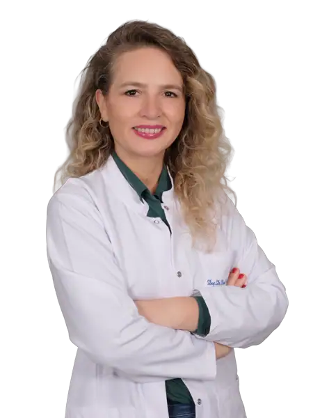
Assoc. Prof. Dr. Emel Ur Özçelik
Neurology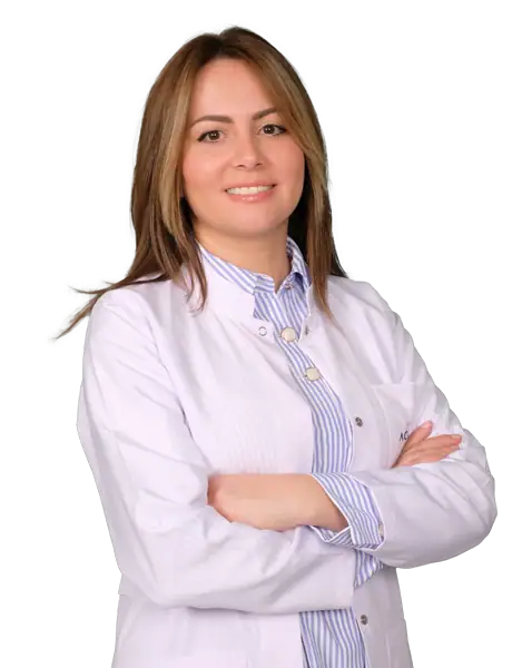
Assoc. Prof. Dr. Esma Kobak Tur
Neurology
Asst. Prof. Dr. Erkan Acar
Neurology
Asst. Prof. Dr. Ezgi Yakupoğlu
Neurology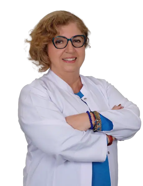
Dr. Aydan Angay
Pediatric Neurology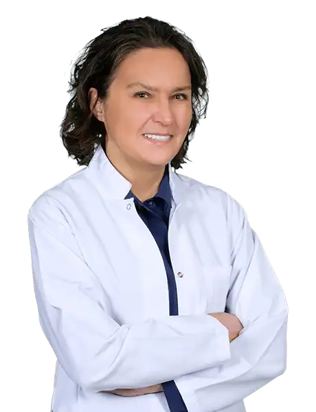
Dr. Ayla Sifoğlu
Neurology
Dr. Aytekin Ceviz
Neurology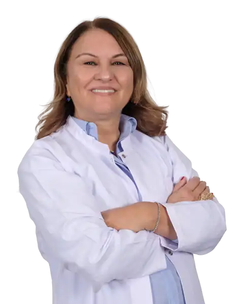
Dr. Ayşen Bıçakcı Genç
Neurology
Dr. Başak Bolluk Kılıç
Neurology
Dr. Beyza Çitçi
Neurology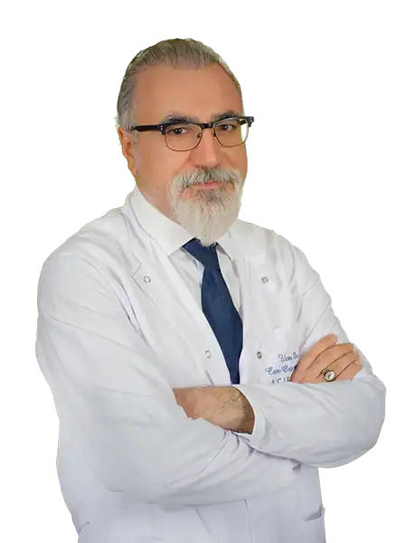
Dr. Cem Ortaçbayram
Neurology
Dr. Cennet Nalan Soyder
Neurology
Dr. Elvan Cevizci Akkılıç
Neurology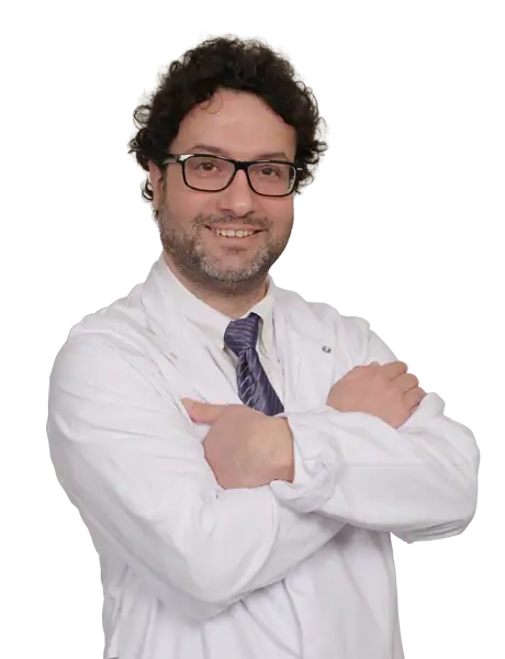
Dr. Fikri Halaçoğlu
Neurology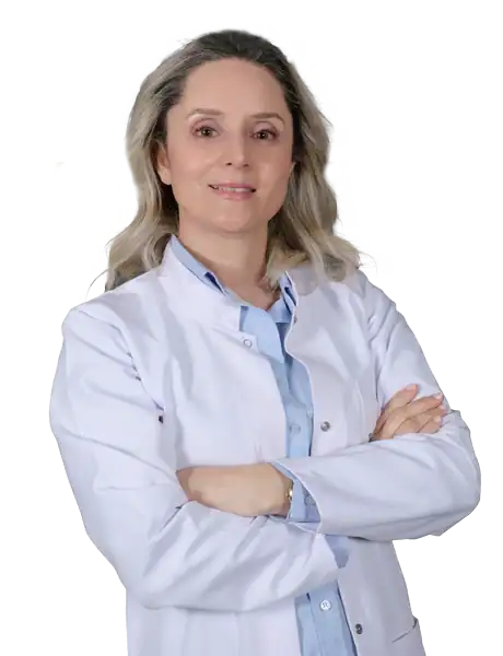
Dr. Gamze Almak
Neurology
Dr. Hafsa Hicret Bülbül Göktaş
Neurology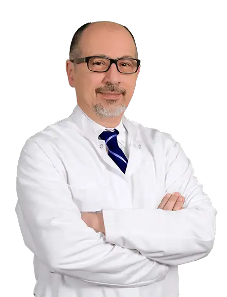
Dr. Haluk Caneroğlu
Neurology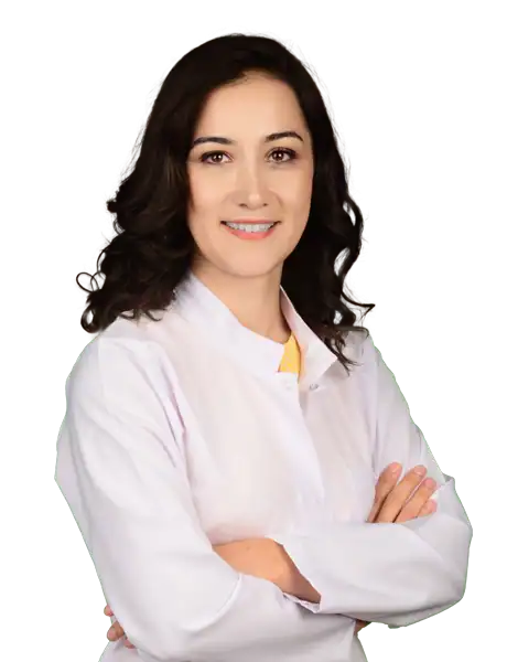
Dr. Handan Bilgin
Neurology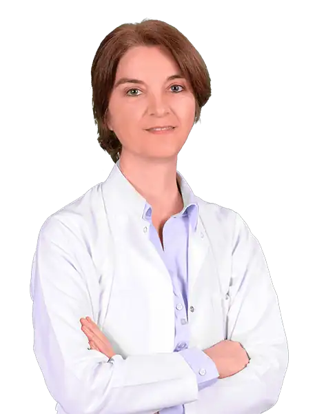
Dr. Hülya Altıntaş
Neurology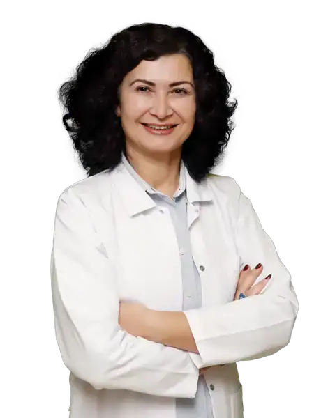
Dr. Hülya Yıldız Bayar
NeurologyMedical Technologies Used
Available at these Hospitals


















What patients say about this unit
★★★★★From 2,400+ verified patient reviews“I was so anxious about my child's developmental neurology review, but the team put us at ease. Prof. Dr. Işık answered all our worried questions without making us feel silly. The nursing staff were attentive and kind throughout.”
“Our child's developmental neurology review was explained clearly and managed beautifully. Prof. Dr. Aydinli made our child feel comfortable straight away.”
“The neurology clinic took my migraine history seriously — imaging to rule out the frightening causes, then a prevention plan that has halved my attacks. Medication choices were explained with alternatives, never dictated.”
“EMG, MRI and a long consultation gave my neurology case a name after two years of uncertainty. The consultant drew the mechanism on paper until I understood it. Treatment is working and reviews continue by video.”
Patient stories from this unit
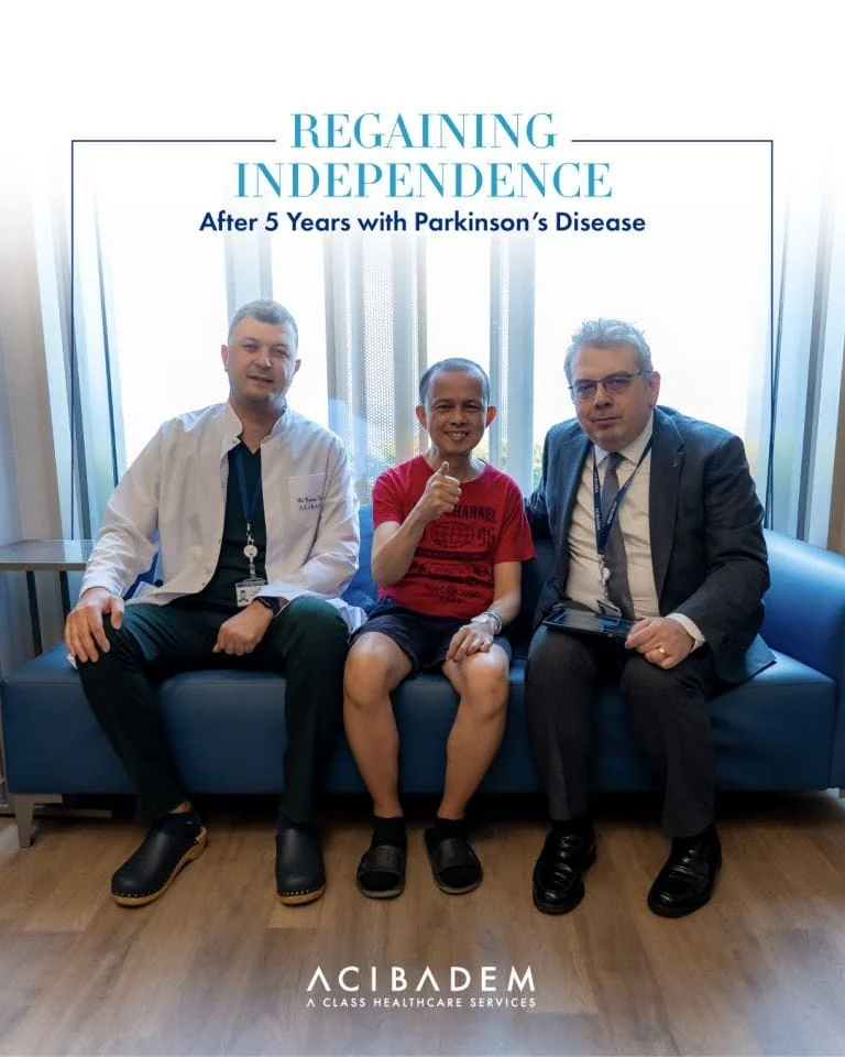
Comparison guides for this specialty
Speak with our medical team
Share your case and our international patient team will guide you to the right specialist — free of charge.