

Pulmonology
Asthma and COPD confirmed with proper breathing tests, bronchiectasis and interstitial lung disease, sleep apnoea and CPAP that people actually keep using, and occupational lung disease, with free remote review of the CT and lung function you already have.
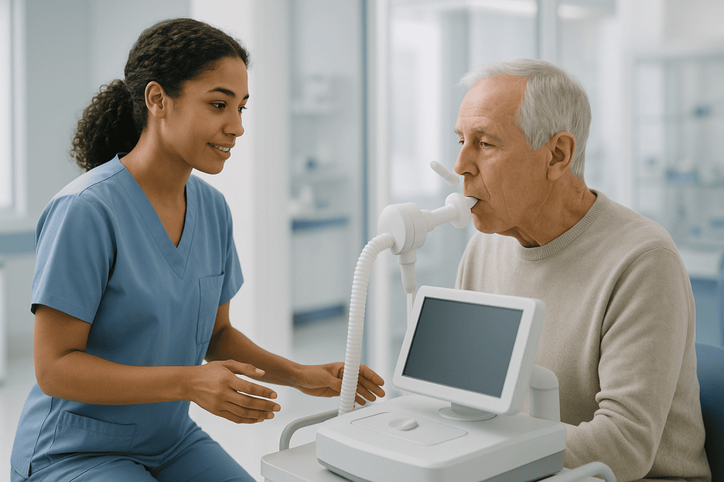
Almost every diagnosis here is confirmed by a number
A respiratory consultation without breathing tests is an incomplete one, and a substantial share of people referred here carrying a diagnosis of asthma or COPD turn out to have something else.
Airway disease and sleep
Conditions defined by what spirometry and a sleep study show, and controlled rather than cured, where treatment is judged against exacerbations rather than against how the day feels.
Lung tissue and vessels
Where the pattern on high-resolution CT carries most of the diagnosis, and separating the patterns determines whether the treatment is immunosuppression or an antifibrotic.
Infection, occupation and the pleura
What was breathed in, at work or from someone else, including the exposures this region has particular experience of.
Check the technique before escalating the drug
The commonest reason an inhaled treatment is not working is that the drug never reaches the lungs. A large share of patients make at least one critical error with their device, and most have never had their technique checked since the first demonstration. It is therefore reviewed at every appointment, with the patient's own inhaler, before anything is escalated, two minutes that frequently removes the need for a stronger prescription.
The same discipline applies to sleep. CPAP is abandoned far more often because of an unsolved mask, pressure or nasal problem than because of genuine intolerance, and adherence is largely decided in the first weeks, which is why follow-up here happens early rather than at a year.
What we will not do
- Diagnose asthma or COPD without spirometry, or accept a label applied elsewhere without it.
- Escalate an inhaled treatment before checking the technique and whether it is being taken.
- Prescribe oxygen for breathlessness in someone whose oxygen levels are normal.
- Treat snoring as though it were sleep apnoea, or silence it without testing.
- Suggest antifibrotic drugs reverse fibrosis. They slow the rate of decline, and that is what we say.
Pulmonologists who lead this work
What actually happens, in order
Send the CT, not the report
In interstitial lung disease the pattern is the diagnosis and it cannot be assessed from a description. A re-read of the high-resolution CT by a thoracic radiologist changes the diagnosis often enough to be worth doing on its own.
Every lung function test, not the last one
The trend carries more information than any single result, and a falling gas transfer with stable spirometry is a finding that only the series shows.
Bring the inhalers themselves
Technique is checked with your own device, because the commonest reason a treatment is not working is that the drug never reaches the lungs. And do not take a dose on the morning of testing unless you were told to, reversibility cannot be assessed through it.
A diagnostic block, not a series of visits
Full physiology, high-resolution CT, bronchoscopy with the right sampling technique, a sleep study or a pleural procedure, arranged over a few days and interpreted by the people who ordered them.
A plan built for continuing at home
Biologics, antifibrotics, oxygen, home ventilation and CPAP all continue after departure. The plan names the monitoring interval, who reviews it, and for equipment the servicing, consumables and what happens when a mask needs replacing abroad.
Six things worth knowing first
Asthma and COPD need a breathing test
Both are defined by what spirometry shows, and a label applied without it is wrong often enough that we check. The conditions mistaken for them, bronchiectasis, heart failure, vocal cord dysfunction, have entirely different treatments.
A normal spirometry is not a clear chest
Spirometry measures airflow. Gas transfer is the number that matters in diffuse lung and pulmonary vascular disease, and a reduced DLCO with normal spirometry is the pattern missed whenever only spirometry is done.
Most CPAP failure is fixable
Mask fit, pressure comfort, humidification and untreated nasal obstruction account for most abandonment, and adherence is largely decided in the first weeks. Genuine intolerance is far rarer than unsolved practical problems.
Silencing a snore is not treating apnoea
No snoring treatment on its own treats sleep apnoea. Removing the noise removes the warning sign while the breathing pauses and their cardiovascular consequences continue. Test first, treat second.
Oxygen is not a breathlessness drug
Long-term oxygen improves survival where levels are genuinely low, and the benefit depends on hours used. In people with normal saturations it has been shown not to relieve breathlessness, and prescribing it substitutes equipment for what does help.
Stopping smoking still works late
It is the only intervention shown to change the rate of lung function decline, and it remains effective at every age and every stage, including after a diagnosis, which is exactly when people conclude it is too late.
Jump to what you came for
Quick answer
Pulmonology is the medical unit that diagnoses, treats, and monitors diseases of the lungs and respiratory system, including conditions affecting breathing, airways, and sleep-related breathing. At Acibadem in Turkey, pulmonology care includes clinical evaluation, pulmonary function testing, imaging, bronchoscopy, and coordinated treatment plans using medication, respiratory support, and follow-up when needed.
What our pulmonology unit covers, and who it is for
Pulmonology, respiratory medicine, is the medical care of the lungs and airways, and of the breathing that depends on them. It is a specialty built around measurement: almost every diagnosis here is confirmed or excluded by a number, and almost every treatment decision is judged against how that number moves.
That has a practical consequence patients notice immediately. A respiratory consultation that does not include breathing tests is usually an incomplete one, and a diagnosis of asthma or COPD made on symptoms alone is a diagnosis that has not been made. A substantial proportion of people referred here carrying one of those labels turn out to have something else entirely.
At Acıbadem International the work is organised into six strands.
- Airway diseaseasthma, COPD and the overlap between them, where the diagnosis is confirmed with spirometry and the treatment is judged against exacerbations rather than against how the patient feels on the day.
- Sleep and breathingobstructive and central sleep apnoea, obesity hypoventilation, and the treatments that people are prescribed and then abandon.
- Diffuse and interstitial lung diseasethe scarring and inflammatory diseases of the lung tissue itself, where the pattern on high-resolution imaging carries most of the diagnosis.
- Airway and pleural proceduresbronchoscopy, sampling of pleural fluid, and the diagnostic work that answers what a scan has found.
- Respiratory failureoxygen, non-invasive ventilation and the long-term support that keeps people out of hospital.
- Occupational and environmental lung diseasedust, fume and mould exposure, including the silica exposure this region has particular experience of.
Where the borders sit. Operations on the chest, lung cancer resection, surgery for pneumothorax, decortication and surgical management of pleural disease, mediastinal surgery and airway surgery, belong with thoracic surgery, which also owns the assessment of a lung nodule found on a scan. Surgery for snoring and sleep apnoea, and assessment of the nose and palate, belongs with otorhinolaryngology. Drug treatment of lung cancer belongs with medical oncology, and the exercise programmes that convert lung function into daily capacity belong with physical medicine and rehabilitation. This unit diagnoses, treats medically, and performs the diagnostic procedures; where an operation is the answer it says so.
What a pulmonologist actually does
A pulmonologistin British and European usage a respiratory physician or respiratory consultant, is a doctor trained in internal medicine who has sub-specialised in the lungs. The work has three components.
The first is separating causes of the same symptom. Breathlessness is produced by the lungs, the heart, anaemia (anemia), obesity, deconditioning, anxiety and neuromuscular weakness, and the commonest error in this field is treating one when the answer is another. A cough lasting months is asthma, reflux or post-nasal drip in the great majority of cases, three conditions with three unrelated treatments, and getting the order of testing right is what shortens the process from years to weeks.
The second is measurement and its interpretation. Spirometry, lung volumes, gas transfer, exhaled nitric oxide, oximetry, sleep studies and exercise testing each answer a specific question, and a test ordered without a question produces a number nobody can act on. Interpreting them well means knowing what makes them wrong: poor technique, the wrong reference values, a patient who took their inhaler that morning.
The third is long-term management of conditions that are controlled rather than cured. Asthma, COPD, bronchiectasis and interstitial disease are lived with, and the honest measures of good care are how few exacerbations happen, how much hospital is avoided and how much the person can still do, not whether a disease has been eliminated.
Breathlessness and chronic cough
These are the two symptoms that bring most people to a respiratory clinic, and both are approached by systematic exclusion rather than by pattern alone.
Breathlessness is assessed against what it is triggered by, how quickly it developed and what accompanies it. Sudden onset points towards pulmonary embolism, pneumothorax or acute cardiac disease; over days towards infection, heart failure or an asthma exacerbation; over months towards COPD, interstitial disease, anaemia, deconditioning or pulmonary hypertension. Orthopnoea, breathlessness lying flat, and swelling point towards the heart, and that assessment is shared with cardiology, because the two systems produce the same complaint and frequently coexist.
The chronic cough causes that account for the overwhelming majority of cases in non-smokers with a normal chest radiograph are three: cough variant asthma and related eosinophilic airway inflammation, gastro-oesophageal (gastroesophageal) reflux, and upper airway cough syndrome from post-nasal drip. A fourth cause is iatrogenic: ACE inhibitor cough, which resolves after the drug is changed by the prescribing doctor and which is diagnosed by asking what someone takes for blood pressure. Because these overlap and coexist, cough is often treated sequentially, and a treatment trial that fails is diagnostic information rather than a wasted month.
Vocal cord dysfunction
Vocal cord dysfunctioninducible laryngeal obstruction, is paradoxical closure of the vocal cords during breathing, and it is the single commonest reason someone carries a diagnosis of severe asthma that has never responded to treatment. The distinction is learnable: the difficulty is in breathing in rather than out, the noise is at the throat rather than in the chest, onset and resolution are abrupt rather than gradual, exercise triggers it within the first minutes rather than after them, and inhalers do not help.
It is diagnosed by direct visualisation during an episode or during provocation, with otorhinolaryngology, and it is treated with speech and language therapy and breathing retraining rather than with drugs. Recognising it removes years of escalating asthma treatment, including in some cases long-term oral corticosteroid that was never going to work.
Haemoptysis (hemoptysis)
Hemoptysiscoughing up blood, is the respiratory symptom that most reliably brings people in quickly, and rightly so, because it is one of the few that is investigated in essentially everyone rather than watched.
The first task is confirming where the blood came from. Blood from the nose or the upper gastrointestinal tract is misattributed to the lungs regularly, and the distinction changes the entire investigation. True haemoptysis is typically bright red and frothy, mixed with sputum, and preceded by an urge to cough rather than by nausea.
The causes divide by setting. In a lifelong non-smoker with a normal chest radiograph, infection, bronchitis, pneumonia, and in this region tuberculosis, accounts for most of it, along with bronchiectasis, which is the commonest cause of recurrent haemoptysis over years. In a smoker over forty, or where the bleeding is persistent or unexplained, malignancy has to be excluded regardless of how well the person feels, and a normal chest radiograph does not achieve that: CT and bronchoscopy do. Pulmonary embolism, mitral valve disease, some vasculitides and anticoagulant treatment make up much of the remainder.
The investigation is therefore a CT of the chest and, in most cases, bronchoscopy, which localises the bleeding segment even when imaging does not. Massive haemoptysis is a medical emergency treated in hospital, where the immediate priorities are protecting the airway and identifying the bleeding source, with bronchial artery embolisation performed by interventional radiology as the definitive treatment in most cases; surgery is reserved for those it does not control and is performed by thoracic surgery.
The pulmonary function test
A pulmonary function test is not one test but a set, and knowing which part answers which question prevents both over-testing and misdiagnosis.
Spirometry, FEV1 and the ratio
Spirometry measures how much air can be blown out and how fast. Two numbers carry the diagnosis: FEV1, the volume exhaled in the first second, and FVC, the total volume exhaled. Their ratio separates the two fundamental patterns. A reduced ratio means obstruction: the airways are narrowed, as in asthma and COPD. A preserved or high ratio with reduced volumes suggests restriction: the lungs cannot expand fully, as in interstitial disease, chest wall abnormality, neuromuscular weakness or obesity.
Reversibility testing repeats spirometry after a bronchodilator. Significant improvement supports asthma; persistent obstruction after bronchodilator is what defines COPD. Two cautions matter more than patients are told: a single normal spirometry does not exclude asthma, because asthma is variable by definition and lung function between attacks is frequently normal; and spirometry is effort-dependent, so a poor-quality manoeuvre produces a result that looks like disease. Technicians reject and repeat tests for exactly that reason, and being asked to blow again is quality control rather than failure.
Lung volumes and gas transfer: DLCO
Full lung volumes, measured by body plethysmography or gas dilution, confirm restriction and identify hyperinflation and gas trapping that spirometry alone underestimates. Gas transferDLCOmeasures how efficiently gas crosses from the air spaces into the blood, and it is the most informative single number in diffuse lung disease. A reduced DLCO with normal spirometry is an important pattern that points towards early interstitial disease, pulmonary vascular disease or anaemia, and it is the finding most often missed when only spirometry is performed.
Exercise testing and oximetry
The six minute walk test measures how far someone walks on the flat in six minutes with continuous oxygen saturation monitoring, and it answers the question resting numbers cannot: what happens when this person actually moves. Desaturation on walking with normal resting saturation is a common and clinically important finding in interstitial disease and in COPD, and it is what determines whether ambulatory oxygen is appropriate. The test is also repeated over time as an objective measure of whether treatment is working. Cardiopulmonary exercise testing goes further and separates a cardiac from a respiratory from a deconditioning cause of breathlessness when the resting tests are equivocal.
Asthma
Asthma is variable airflow obstruction with airway inflammation, and the word variable is the diagnosis. It is confirmed by demonstrating that variability, reversibility on spirometry, variation on serial peak flow, or a positive challenge test, rather than by symptoms alone, and a substantial proportion of people carrying an asthma label have never had it confirmed.
Treatment has changed fundamentally and the change has not reached most patients. Reliever-only treatment with a short-acting bronchodilator is no longer recommended even in mild disease, because it relieves symptoms while leaving the inflammation that causes attacks untreated, and frequent reliever use is itself a marker of risk. Modern regimens are built around inhaled corticosteroid, taken regularly or combined with a fast-onset long-acting bronchodilator in a single inhaler used both regularly and as needed. Which regimen and which device is a prescribing decision made with your own doctor, and nothing here is a schedule.
Inhaler technique
The most common reason an asthma treatment is not working is that the drug is not reaching the lungs. Studies of inhaler use consistently find that a large share of patients make at least one critical error, and the majority have never had their technique checked after the first demonstration. Inhaler technique is therefore reviewed at every review here, with the patient’s own device, before any treatment is escalated: a step that costs two minutes and frequently removes the need for a stronger drug. Spacers substantially improve delivery from pressurised inhalers and are underused in adults.
Severe and eosinophilic asthma
Before asthma is called severe, three things are checked, because most apparently severe asthma is not: whether the diagnosis is right at all, whether the inhaler is being used correctly and regularly, and whether something else is driving it, reflux, obesity, smoking, a nasal condition, or vocal cord dysfunction.
Genuine severe asthma is then characterised by type, because that determines treatment. Eosinophilic asthma, driven by type 2 inflammation, is identified from the blood eosinophil count and from the FeNO testexhaled nitric oxide, a simple breath test that measures eosinophilic airway inflammation directly and helps both to confirm the type and to detect patients who are not taking their inhaled steroid. This matters because it opens the door to asthma biologics: monoclonal antibodies targeting IgE, interleukin-5 and the interleukin-4 receptor have transformed outcomes in the right patients, substantially reducing exacerbations and allowing many people to come off long-term oral corticosteroid. Eligibility is defined by specific criteria, and selecting the right agent depends on the phenotype rather than on preference.
Aspirin-exacerbated respiratory disease: the combination of asthma, nasal polyps and reactions to aspirin and related anti-inflammatory drugs, is worth naming, because it is frequently unrecognised and the drugs that trigger it are sold without prescription.
COPD
Chronic obstructive pulmonary disease is persistent airflow obstruction that does not fully reverse with a bronchodilator, and it is confirmed by spirometry. That confirmation is not a formality: a diagnosis of COPD made without spirometry is wrong often enough that it is one of the first things checked here, and the conditions mistaken for it, asthma, bronchiectasis, heart failure, interstitial disease, have entirely different treatments.
Smoking is the dominant cause, but not the only one. Biomass fuel exposure from indoor cooking and heating is a major cause worldwide and is under-recognised in people who have never smoked, and occupational dust and fume exposure contributes substantially. Alpha-1 antitrypsin deficiency is an inherited cause worth excluding in anyone with emphysema at a young age, with lower-lobe predominant disease, or with a family history.
COPD stages and what they actually predict
The gold criteria copd framework grades COPD stages by the degree of airflow obstruction on spirometry, and the numbers are useful but frequently over-interpreted. Modern assessment combines that grade with two other things: how breathless the person actually is on a standard scale, and how many exacerbations they have had in the past year. Exacerbation history is the strongest single predictor of future exacerbations and drives the treatment choice more than the spirometry number does, two people with identical FEV1 can need quite different treatment.
On copd life expectancy, the honest answer is that it varies enormously and that population figures do not predict individuals. What does change the trajectory is documented and limited: stopping smoking, which is the only intervention shown to alter the rate of lung function decline; vaccination; pulmonary rehabilitation; long-term oxygen where the criteria are met; and treatment that reduces exacerbations, because each severe exacerbation is associated with a step down in function that is not always regained.
The COPD exacerbation
A copd exacerbation is a sustained worsening of breathlessness, cough and sputum beyond day-to-day variation, and preventing them is the central goal of long-term treatment. Management combines increased bronchodilator treatment, a short course of oral corticosteroid where indicated, and antibiotics only where the sputum has changed in colour and volume or where the person is severely unwell: the antibiotic decision is a clinical one and is not made from a leaflet. Severe exacerbations with respiratory failure are treated in hospital with non-invasive ventilation, which is covered under respiratory failure.
An action plan agreed in advance, with a clear description of what worsening looks like and what to do about it, reduces hospital admission. That plan is written with the doctor who prescribes it, in the patient’s own words, and reviewed each year. For selected patients with severe emphysema, lung volume reduction surgeryor the bronchoscopic valve equivalent, performed with thoracic surgeryimproves breathlessness and exercise capacity, and eligibility is assessed against strict criteria including the pattern of emphysema and the absence of collateral ventilation.
Bronchiectasis
Bronchiectasis is permanent abnormal widening of the airways, which lose their ability to clear mucus and become chronically colonised with bacteria. The result is a self-sustaining cycle of infection, inflammation and further damage. It is diagnosed on high-resolution CT, and it is substantially under-diagnosed: a large share of patients carry a label of difficult asthma or COPD for years before a CT is performed.
The clinical picture is distinctive once recognised: a daily productive cough over years rather than an intermittent one, sputum that is present every day and increases with infections, recurrent chest infections requiring repeated antibiotic courses, and recurrent haemoptysis. Anyone with that pattern needs a CT rather than another antibiotic.
The investigation looks for a cause, because a proportion are treatable or change management: previous severe infection including tuberculosis and childhood pneumonia, which is the commonest identifiable cause in this region; immune deficiency, which is checked in everyone because immunoglobulin replacement changes the course; allergic bronchopulmonary aspergillosis; cystic fibrosis and primary ciliary dyskinesia, including in adults presenting late; and rheumatoid arthritis and inflammatory bowel disease.
Bronchiectasis treatment
Bronchiectasis treatment rests on airway clearance more than on drugs, and this is the part most often skipped. Daily physiotherapy techniques taught by a respiratory physiotherapist, active cycle of breathing, positive expiratory pressure devices, postural drainage, do more for exacerbation frequency than any prescription, and they have to be done daily rather than during infections. Nebulised saline improves clearance in many patients.
Around that: sputum culture at intervals, because the organism guides antibiotic choice and because Pseudomonas colonisation changes the outlook and the treatment; prompt and adequately long antibiotic courses for exacerbations, which are longer than the courses given for ordinary chest infections; long-term macrolide therapy in selected patients with frequent exacerbations, which requires screening for mycobacterial infection first and monitoring thereafter; vaccination; and pulmonary rehabilitation. Inhaled corticosteroid is not routine treatment for bronchiectasis and is frequently prescribed inappropriately when the label of asthma persists alongside it. Microbiological management is shared with infectious diseases.
Interstitial lung disease
Interstitial lung disease is a group of more than a hundred conditions affecting the lung tissue between and around the air sacs, producing inflammation, scarring or both. They present alike, breathlessness on exertion over months, a dry cough, fine crackles at the lung bases, sometimes finger clubbing, and separating them is what determines whether the treatment is immunosuppression, an antifibrotic drug, or removing an exposure.
The single most important investigation is a high-resolution CT of the chest, and the pattern it shows carries most of the diagnosis. A usual interstitial pneumonia pattern, basal and peripheral reticulation with honeycombing and traction bronchiectasis, is characteristic of idiopathic pulmonary fibrosis and, where the pattern is definite in the right clinical setting, can establish that diagnosis without a biopsy. A nonspecific interstitial pneumonia pattern, with ground-glass change and less honeycombing, points instead towards connective tissue disease or drug-induced disease, both of which behave and are treated differently. An hrct chest reported as showing fibrosis without describing the pattern is an incomplete report, and re-reading by a thoracic radiologist frequently changes the diagnosis.
The workup is broader than the lungs. Autoimmune serology and a careful review of joints, skin, muscles and swallowing, because a proportion of interstitial disease is the first manifestation of a connective tissue disease and is managed with rheumatology. A detailed exposure history covering birds, feather bedding, mould, hot tubs and occupation. And a full drug history, since a long list of medicines including some cardiac, rheumatological and chemotherapy agents cause interstitial disease that resolves when the drug is changed by the prescriber.
Diagnosis is made at a multidisciplinary meeting rather than by one person, respiratory physician, thoracic radiologist and, where a biopsy has been taken, pathologist reviewing the case together. That format is not administrative: it changes the diagnosis in a meaningful proportion of cases, and it is the standard this unit works to.
Idiopathic pulmonary fibrosis and antifibrotic treatment
Idiopathic pulmonary fibrosis is progressive scarring with no identifiable cause, and it is the diagnosis in this group with the poorest outlook. Two antifibrotic drugspirfenidone and nintedanibslow the rate of decline in lung function. They do not reverse fibrosis, do not repair the lung and do not make people feel better in the short term, and they have real gastrointestinal and skin side effects that require management rather than tolerance. Being clear about all of that before starting is what allows someone to decide properly.
Nintedanib is also used in other progressive fibrosing interstitial diseases and in systemic sclerosis-associated disease. Around the drugs, the interventions that change day-to-day life are oxygen assessment including on exertion, pulmonary rehabilitation, treatment of reflux and, in appropriate patients, early referral for transplant assessment, early, because the assessment takes months and being referred late is one of the commonest avoidable failures in this disease. Palliative treatment of breathlessness runs alongside disease-directed treatment from the beginning rather than at the end.
Hypersensitivity pneumonitis
Hypersensitivity pneumonitis is an immune reaction to an inhaled organic antigen, and it is the interstitial disease most worth identifying because removing the exposure can halt it. The antigens are domestic and occupational: birds, including feather duvets and pillows in people who own no bird, mould in damp housing, humidifiers and air conditioning, hot tubs, and agricultural dusts. The exposure history has to be taken deliberately, because patients do not volunteer a feather duvet as medical information, and the fibrotic form is otherwise indistinguishable from idiopathic pulmonary fibrosis on imaging.
Sarcoidosis
Sarcoidosis is a multisystem granulomatous disease of unknown cause that involves the lungs and mediastinal lymph nodes in the great majority of cases. It is unusual among the conditions this unit treats in that a large proportion of patients need no treatment at all: many cases resolve spontaneously over months to years, and treatment is directed by organ involvement and symptoms rather than by the presence of the diagnosis.
It is staged by the pattern on imaging, from isolated lymph node enlargement through to fibrosis. Diagnosis usually requires tissue showing non-caseating granulomas, obtained by bronchoscopic needle sampling of mediastinal nodes, and excluding the conditions that produce identical granulomas, tuberculosis above all, which matters greatly in this region because the treatments are opposite: immunosuppression given to someone with undiagnosed tuberculosis is dangerous.
Involvement outside the chest determines urgency rather than the lung findings: the eyes, the heart, the nervous system and the kidneys, along with hypercalcaemia. Cardiac and neurological sarcoidosis are treated regardless of how mild the lung disease is, and screening for them is part of the initial assessment rather than something done if symptoms appear. Where treatment is needed, corticosteroid remains first line with steroid-sparing agents added for prolonged disease, and Löfgren syndrome: the acute presentation with erythema nodosum, joint swelling and hilar lymphadenopathy, carries a good prognosis and usually needs only symptomatic treatment.
Sleep apnoea (sleep apnea)
Obstructive sleep apnea is repeated collapse of the upper airway during sleep, producing breathing pauses, oxygen dips and arousals that fragment sleep without waking the person properly. It is extremely common, substantially under-diagnosed, and it matters beyond tiredness: untreated moderate to severe disease is associated with hypertension that is difficult to control, cardiovascular disease, and road traffic accidents.
The sleep apnea symptoms that patients themselves report are unrefreshing sleep, waking with a dry mouth or headache, nocturia, and daytime sleepiness, but the most useful history usually comes from a partner describing loud snoring, witnessed pauses and gasping. The Epworth sleepiness scale is a short questionnaire scoring the likelihood of dozing in eight everyday situations; it is useful for tracking response to treatment but is a poor screening test on its own, because many people with severe disease score low, particularly those whose main complaint is fatigue rather than sleepiness.
Diagnosis, the Epworth sleepiness scale and the AHI
A home sleep apnea testa portable device recording airflow, effort, oximetry and heart rate in the patient’s own bed, is appropriate for most adults with a high probability of straightforward obstructive disease, is more comfortable and less expensive than laboratory testing, and is what this unit uses first in that group. Full laboratory polysomnography with EEG is reserved for the situations a home study cannot answer: suspected central apnoea, significant cardiac or neuromuscular disease, other sleep disorders, and a negative home study in someone whose history remains convincing, because home testing tends to underestimate severity.
Severity is graded by the apnea hypopnea index, the number of apnoeas and hypopnoeas per hour of sleep. The number is useful and it is not the whole picture: how far the oxygen falls and for how long, how fragmented the sleep is and how symptomatic the person is all matter, and treatment decisions in mild disease are driven by symptoms and cardiovascular risk rather than by the index alone.
Central sleep apnoea (central sleep apnea) and obesity hypoventilation
Central sleep apnea is a different mechanism entirely: the airway is open but the drive to breathe pauses. It occurs in heart failure, often as Cheyne-Stokes breathing, after stroke, with opioid use, at altitude, and sometimes as a result of CPAP treatment itself. It is important to distinguish because treatment differs and because some devices used for obstructive apnoea are not appropriate in central apnoea associated with reduced cardiac function.
Obesity hypoventilation syndrome is under-recognised and more serious than obstructive apnoea alone: daytime carbon dioxide retention in a person with obesity, in whom breathing is inadequate while awake as well as asleep. It is suspected from a raised bicarbonate on routine blood tests, confirmed by blood gas measurement, and treated with non-invasive ventilation rather than CPAP alone in many patients. Missing it is the reason some patients labelled as having simple sleep apnoea do not improve on treatment.
The CPAP machine, and what to do when it is not tolerated
A CPAP machine delivers continuous positive airway pressure through a mask, splinting the upper airway open during sleep. In moderate to severe obstructive sleep apnoea it is the most effective treatment available, and in people who use it consistently it improves sleepiness, quality of life and blood pressure control.
The problem is that a large share of people stop using it, and much of the advice available online ignores that. Adherence is determined mostly in the first weeks, and it is determined by fixable things: mask fit and type, pressure comfort and the use of pressure-relief or auto-titrating modes, humidification for dryness, nasal obstruction treated properly with otorhinolaryngology, and follow-up that happens early rather than at a year. Most cpap side effectsdryness, congestion, mask marks, air leak, aerophagia, have a specific and simple fix, and abandoning treatment is far more often a set of unsolved practical problems than a genuine intolerance.
CPAP alternatives
Where CPAP is genuinely not tolerated after proper optimisation, the cpap alternatives are real and are chosen by mechanism rather than preference. A mandibular advancement device, fitted by a dentist experienced in sleep medicine, holds the lower jaw forward and is effective in mild to moderate disease and in positional apnoea; the over-the-counter versions sold online are not equivalent and can damage the bite and the jaw joint. Weight reduction genuinely reduces severity and in some patients resolves the condition, and it is discussed as treatment rather than as advice. Positional therapy helps the subgroup whose apnoea occurs almost entirely when supine. Sleep apnea surgerynasal, palatal and tongue-base procedures, is assessed by otorhinolaryngology, and hypoglossal nerve stimulation, an implanted device that activates the tongue muscles in time with breathing and is widely known by the trade name of the Inspire implant, is an option for selected patients meeting defined criteria who cannot use CPAP.
Two points are worth stating plainly because they are widely marketed otherwise. No snoring treatment on its own treats sleep apnoea, and silencing the snoring without testing for apnoea removes the warning sign while leaving the disease. And CPAP is treatment rather than cure: the apnoea returns on any night it is not used.
Pulmonary embolism
Pulmonary embolism is obstruction of the pulmonary arteries by clot, usually travelling from the deep veins of the legs or pelvis. It is a medical emergency and is treated in hospital, and it belongs in any respiratory account because it is both common and commonly missed: its presentation ranges from sudden collapse to nothing more than unexplained breathlessness or a mildly fast pulse in someone who feels reasonably well.
Diagnosis works by combining clinical probability with testing rather than by testing everyone. Where probability is low, a D-dimer that is normal excludes it reliably; where probability is high, imaging is done regardless of the D-dimer, because a normal result in that setting does not exclude anything. The imaging test is a ct pulmonary angiogram, with a ventilation-perfusion scan used in pregnancy, in renal impairment and in contrast allergy.
Treatment is anticoagulation, with thrombolysis or catheter-directed treatment reserved for those with circulatory compromise. Two questions follow every diagnosis and are frequently neglected. How long to anticoagulate depends on whether the clot was provoked by a temporary factor, unprovoked, or associated with cancer or a thrombophilia, and that decision is made deliberately with hematology rather than defaulted to a fixed period. And a proportion of patients develop chronic thromboembolic pulmonary hypertension, which is potentially curable by surgery and which is why persistent breathlessness months after a pulmonary embolism is investigated rather than attributed to deconditioning.
Pulmonary hypertension
Pulmonary hypertension is raised pressure in the pulmonary circulation, and the word covers several very different diseases with different treatments, which is the single most important thing to understand about it. It is classified into groups: pulmonary arterial hypertension, which is rare and has specific drug therapy; pulmonary hypertension due to left heart disease, which is by far the commonest and is treated by treating the heart; pulmonary hypertension due to lung disease and hypoxia; chronic thromboembolic disease, which is potentially curable surgically; and a miscellaneous group.
Giving pulmonary arterial hypertension drugs to someone whose raised pressure is caused by left heart disease can be harmful, which is why the classification is established before treatment rather than after. Echocardiography estimates the pressure and is the screening test; right heart catheterisation measures it directly and is required to confirm the diagnosis and assign the group. That work is shared with cardiology.
Cor pulmonale
Cor pulmonale is the term for right heart failure resulting from lung disease, leg swelling, raised jugular venous pressure and liver congestion in someone with advanced COPD, interstitial disease or untreated sleep-disordered breathing. Its appearance marks a change in the trajectory of the underlying lung disease and prompts a review of oxygenation, including overnight, because untreated nocturnal hypoxaemia is a correctable driver.
Bronchoscopy and airway sampling
Bronchoscopy passes a flexible camera into the airways under sedation and local anaesthesia (anesthesia), and it is both a diagnostic and a therapeutic tool. Diagnostically it answers what imaging cannot: it inspects the airways directly, samples abnormal areas, washes and samples the alveolar spaces in diffuse disease and in immunocompromised patients with infiltrates, and localises bleeding in haemoptysis.
Sampling has become considerably more capable. Endobronchial ultrasound allows needle sampling of mediastinal lymph nodes and central masses through the airway wall, replacing surgical mediastinoscopy for most diagnostic and staging purposes. Radial ultrasound and navigational techniques reach peripheral lesions that were previously accessible only by CT-guided needle or surgery. Transbronchial cryobiopsy obtains larger, better-preserved samples of lung tissue in interstitial disease than conventional forceps, with a lower complication rate than surgical biopsy in appropriately selected patients.
The procedure takes twenty to forty minutes, most patients remember little of it with sedation, and the throat is numbed so nothing is eaten or drunk until the local anaesthetic wears off. Its main risks are bleeding and pneumothorax, both uncommon and both higher with biopsy than with inspection alone. Where the answer is surgical staging or resection, the case passes to thoracic surgery, which also owns the assessment of a lung nodule found incidentally on a scan.
Thoracentesis and pleural sampling
Thoracentesispleural aspiration, removes fluid from the space between the lung and the chest wall through a needle under local anaesthetic, with ultrasound guidance. Ultrasound is not optional: guided aspiration is substantially safer than the landmark technique and is the standard here.
It does two jobs. Diagnostically, analysis of the fluid separates a transudate, usually heart failure, liver disease or low protein, where the treatment is of the underlying condition, from an exudate, which requires an explanation: infection, malignancy, pulmonary embolism, tuberculosis or an inflammatory disease. Tuberculous pleural effusion is common in this region and is frequently missed, because the fluid contains few organisms; adenosine deaminase measurement and pleural biopsy substantially improve the yield, and a pleural biopsy is taken where an exudate remains unexplained after aspiration.
Therapeutically, drainage relieves breathlessness immediately, and how much is removed at once is limited to avoid re-expansion problems. Where fluid recurs, most often in malignancy, the options are repeated drainage, an indwelling tunnelled catheter managed at home, or pleurodesis, and that choice and the surgical management of infected or loculated collections belong with thoracic surgery, which covers pleural effusion and pneumothorax in full.
Respiratory failure, oxygen and ventilation
Respiratory failure means the lungs can no longer maintain adequate gas exchange, and it comes in two types that are treated differently. Type 1 is low oxygen with normal or low carbon dioxide, seen in pneumonia, pulmonary embolism and interstitial disease. Type 2 adds carbon dioxide retention, hypercapnia, and occurs in COPD, obesity hypoventilation, neuromuscular weakness and chest wall disease, where the problem is inadequate ventilation rather than gas transfer.
The distinction governs oxygen prescribing, and this is one of the few areas where well-meant treatment does harm. In patients at risk of type 2 failure, high-flow oxygen given without monitoring can worsen carbon dioxide retention, which is why target saturation ranges are specified rather than oxygen simply being turned up, and why blood gases are checked. Oxygen alert cards exist for exactly this reason.
Long-term and ambulatory oxygen
Home oxygen therapy is prescribed against defined criteria, and it is worth being clear about what it is for. Long-term oxygen improves survival in patients with chronic severe hypoxaemia when used for the majority of the day: the survival benefit depends on the number of hours, which is why the prescription specifies them. It is assessed on blood gases measured when the person is stable, not during an exacerbation, and it is not a treatment for breathlessness in people whose oxygen levels are normal, where it has been shown not to help. Ambulatory oxygen is a separate assessment for people who desaturate on exertion, based on a walk test.
Non-invasive ventilation
Non invasive ventilation delivers pressure support through a mask, assisting each breath rather than simply supplying oxygen. In acute type 2 respiratory failure from a COPD exacerbation it reduces the need for intubation and reduces mortality, and it is among the best-evidenced treatments in respiratory medicine. Long term, it is used at home in obesity hypoventilation, neuromuscular disease including motor neurone disease, chest wall deformity and selected patients with severe COPD and persistent hypercapnia. BiPAP is the common name for the two-pressure mode used for this, and it is a different treatment from CPAP with a different purpose, despite the machines looking similar.
Occupational lung disease, and this region
Occupational lung disease is caused by what people breathe at work, and it is the group most often missed, because the question is not asked. The exposure history has to cover every job held, not the current one, and it has to ask what was actually in the air rather than what the job was called. Latency is long: asbestos-related disease appears decades after exposure, which is why a retired worker with breathlessness is asked about work done in their twenties.
Silicosis is where this region has particular and hard-won experience. Turkey saw a well-documented outbreak of silicosis among young workers sandblasting denim to distress it: an occupation with intense crystalline silica exposure in poorly ventilated workshops, which produced accelerated and acute forms of the disease in men in their twenties and thirties rather than the classical form after decades, and which led to the practice being banned. The consequence is that respiratory units here recognise accelerated silicosis in a young patient with an upper-zone nodular pattern, where units elsewhere may not consider it at all. Silica exposure continues in stone cutting, engineered stone worktop fabrication, mining, quarrying, foundries and construction, and the engineered stone problem is now emerging internationally.
Silicosis has no cure and no treatment that reverses it, and saying so plainly is part of the consultation. What management can do is stop further exposure, which is the only thing that alters the course; screen for and treat the complications, particularly tuberculosis, to which silicosis substantially predisposes and for which these patients are screened; treat airflow obstruction and hypoxaemia; and refer appropriately for transplant assessment in advanced disease. Documentation for occupational compensation is part of the clinical work rather than an administrative afterthought. Asbestos-related disease, coal workers’ pneumoconiosis, occupational asthma, which resolves if identified early and the exposure stops, and becomes permanent if it does not, and hypersensitivity pneumonitis from agricultural and industrial organic dusts are assessed the same way.
Smoking, and what actually helps
Stopping smoking is the only intervention shown to change the rate of lung function decline in COPD, and it remains effective at every age and at every stage of disease, including after a diagnosis has been made, which is when many people conclude it is too late.
What the evidence supports is combining behavioural support with pharmacological treatment, because together they perform considerably better than either alone. Which medication is appropriate depends on medical history and previous attempts and is a prescribing decision. Two practical points worth stating: relapse is part of the process rather than a failure, and most people who eventually stop have made several attempts, so a relapse is a reason to restart rather than to stop trying. And electronic cigarettes are not a neutral subject: they are less harmful than continued smoking and are not harmless, dual use maintains the exposure, and the evidence on long-term effects is still accumulating. We give that answer rather than a simpler one in either direction.
Pulmonary rehabilitation deserves the same emphasis. It is a structured exercise and education programme, and in COPD and interstitial disease it improves breathlessness, exercise capacity and quality of life more than most drugs do, while reducing hospital admission after an exacerbation. It is delivered with physical medicine and rehabilitation and it is under-referred everywhere, including here, which is why it is offered at diagnosis rather than at the point of severe limitation.
What pulmonology cannot do
- It cannot reverse fibrosis or emphysema. Antifibrotic drugs slow decline; they do not repair scarred lung. Destroyed alveoli do not regenerate. Treatment preserves what remains.
- It cannot cure asthma or COPD. Both are controlled. Well-controlled asthma looks like no asthma, which is precisely why people stop their inhaler and the attacks return.
- It cannot make oxygen a treatment for breathlessness. Oxygen helps people whose oxygen levels are low. In people with normal saturations it has been shown not to relieve breathlessness, and prescribing it anyway substitutes equipment for the things that do help.
- It cannot fix a treatment that is not being taken. The commonest reason an inhaler is not working is technique or adherence, and escalating the drug without checking both produces stronger prescriptions and no improvement.
- It cannot treat sleep apnoea with a device left in a cupboard. CPAP works only on the nights it is used, and unaddressed practical problems are the reason most people stop.
- It cannot undo an exposure. In silicosis, asbestos-related disease and occupational asthma the damage already done is permanent; stopping the exposure is what protects the future.
Your multidisciplinary team
The pulmonologist makes the diagnosis, orders and interprets the physiology, performs the bronchoscopic and pleural procedures, and manages the disease long term. The respiratory physiologist performs the lung function testing, and the quality of that testing determines whether the numbers mean anything: a poorly performed spirometry produces a confident diagnosis of the wrong condition. The respiratory physiotherapist teaches airway clearance in bronchiectasis and runs pulmonary rehabilitation, and in this specialty carries more of the outcome than most patients expect. The sleep technologist runs the studies and, more importantly, the mask fitting and troubleshooting that decide whether CPAP is still being used in six months. The thoracic radiologist reads the high-resolution CT, and in interstitial disease that reading is the diagnosis.
Around them: thoracic surgery for lung nodules, resection, pneumothorax and pleural surgery, medical oncology and radiation oncology for lung cancer, cardiology for the breathlessness that turns out to be cardiac and for pulmonary hypertension, infectious diseases for tuberculosis, resistant organisms and infection in the immunocompromised, rheumatology for connective tissue disease with lung involvement, otorhinolaryngology for nasal obstruction, vocal cord dysfunction and upper airway surgery, endocrinology and dietetics for the weight management that changes sleep apnoea and hypoventilation, rehabilitation for pulmonary rehabilitation, and allergy assessment where inhaled allergen drives the airway disease.
The international patient journey
Respiratory medicine travels differently from surgery. What is usually wanted is a diagnosis that has not been reached, or a second opinion on one that has, and a large share of it can be done before anyone books a flight.
The first pattern is a remote review of imaging and physiology. Interstitial lung disease is the clearest example: a re-read of the high-resolution CT by a thoracic radiologist, combined with the lung function trend and the serology, changes the diagnosis often enough to be worth doing on its own. What is needed is the CT as DICOM files rather than a report, the pattern cannot be assessed from a description, plus every lung function test performed rather than the most recent one, because the trend carries more information than any single result.
The second is a diagnostic block on site: full lung function including gas transfer, high-resolution CT, bronchoscopy with the appropriate sampling technique, a sleep study, or a pleural procedure, arranged together over a few days and interpreted by the people who ordered them. This is the pattern for undiagnosed interstitial disease, for suspected bronchiectasis in someone treated for years as asthma, and for difficult airway disease where the question is whether the diagnosis is right at all.
The third is starting a treatment that then continues at homea biologic for severe asthma, an antifibrotic, long-term oxygen, home ventilation or CPAP. These need the plan written for continuation rather than for initiation: the drug and its monitoring schedule, who at home reviews it, what the thresholds are, and for equipment the practical questions of servicing, consumables and what happens when a mask or a filter needs replacing in another country.
Two practical notes. Do not use your inhalers on the morning of lung function testing unless you have been told to, because reversibility cannot be assessed through a dose taken an hour earlier, and bring the actual devices with you, since technique is checked with your own inhaler. And for anything sleep-related, bring the partner or the account: witnessed pauses and gasping are the most useful diagnostic information there is, and the patient by definition cannot supply them.
Frequently Asked Questions
What does a pulmonologist do that a general physician does not?
Three things mainly. They separate causes of the same symptom, breathlessness comes from the lungs, the heart, anaemia, obesity, deconditioning, anxiety and neuromuscular weakness, and treating the wrong one is the commonest error in this field. They order and interpret the physiology properly, knowing which test answers which question and what makes a result wrong. And they perform the diagnostic procedures, bronchoscopy, pleural sampling, that answer what a scan has found. In British and European usage the same doctor is called a respiratory physician or respiratory consultant.
Can asthma be diagnosed without breathing tests?
It should not be. Asthma is defined by variable airflow obstruction, and confirming that variability, reversibility on spirometry, variation on serial peak flow, or a positive challenge test, is what makes the diagnosis. A substantial proportion of people carrying an asthma label have never had it confirmed, and a proportion of those have something else: COPD, bronchiectasis, heart failure or vocal cord dysfunction. That said, a single normal spirometry does not exclude asthma either, because lung function between attacks is frequently normal, which is why the testing may be repeated or a challenge test used.
My spirometry was normal but I am still breathless. What now?
Spirometry measures airflow and misses several important things. Gas transfer, DLCO, is the number that matters in diffuse lung disease and pulmonary vascular disease, and a reduced DLCO with normal spirometry is an important pattern that is missed whenever only spirometry is done. Full lung volumes detect restriction and gas trapping. And a six-minute walk with oximetry answers what resting tests cannot: desaturation on exertion with a normal resting saturation is common and clinically significant. Cardiac causes are assessed in parallel, since the heart and lungs produce the same complaint.
Why do they keep making me blow again?
Because spirometry is entirely effort-dependent and a poor manoeuvre produces a result that looks like disease. Quality criteria require a sharp start, a sustained blow of adequate length and reproducibility between attempts, and a technician who accepts a substandard effort produces a number that will be acted on wrongly for years. Being asked to repeat it is quality control, not failure. It is also why you may be asked not to use your inhalers beforehand: a dose taken an hour earlier makes reversibility impossible to assess.
I have a cough that has lasted months and my chest X-ray is normal. What causes that?
In a non-smoker with a normal chest radiograph, three conditions account for the overwhelming majority: cough-variant asthma and related eosinophilic airway inflammation, gastro-oesophageal reflux, and upper airway cough syndrome from post-nasal drip. A fourth is the ACE inhibitor cough, which is why you will be asked what you take for blood pressure. These overlap and often coexist, so cough is treated sequentially, and a treatment trial that fails is diagnostic information rather than a wasted month.
My asthma inhalers have never worked. Could it be something else?
Quite possibly. The single commonest reason for a diagnosis of severe asthma that has never responded is vocal cord dysfunction, paradoxical closure of the vocal cords, in which the difficulty is breathing in rather than out, the noise is at the throat rather than the chest, onset and resolution are abrupt, and inhalers do not help. It is treated with speech therapy and breathing retraining rather than drugs. Before asthma is called severe we also check that the diagnosis was confirmed at all, that the inhaler technique is correct, and whether reflux, obesity, smoking or nasal disease is driving it.
Is my inhaler technique really that important?
It is the most common reason an asthma treatment is not working. Studies consistently find that a large share of patients make at least one critical error, and most have never had their technique checked since the first demonstration. Technique is therefore reviewed at every appointment here, with your own device, before any treatment is escalated: it takes two minutes and frequently removes the need for a stronger drug. Spacers substantially improve delivery from pressurised inhalers and are underused in adults, not just children.
What is eosinophilic asthma and why does it matter?
It is asthma driven by type 2 inflammation, identified from the blood eosinophil count and the FeNO test: a simple breath test measuring exhaled nitric oxide. It matters because it determines treatment: patients with this phenotype and frequent exacerbations may be eligible for biologic therapy, monoclonal antibodies targeting IgE, interleukin-5 or the interleukin-4 receptor, which substantially reduce exacerbations and allow many people to come off long-term oral steroid. FeNO has a second use: it detects patients who are not actually taking their inhaled steroid, which changes the conversation before the treatment is escalated.
Should I still be using a blue reliever inhaler on its own?
Current practice has moved away from that, and the reason is mechanical rather than fashionable: a short-acting reliever relieves symptoms while leaving the inflammation that causes attacks untreated, and frequent reliever use is itself a marker of risk rather than of good control. Modern regimens are built around inhaled corticosteroid, taken regularly or combined with a fast-onset long-acting bronchodilator in a single inhaler used both regularly and as needed. Which regimen suits you is a decision for the doctor prescribing it, with your history in front of them, do not change your own inhalers on the basis of a general description.
Can COPD be diagnosed without spirometry?
No, and a diagnosis made without it is wrong often enough that it is one of the first things checked here. COPD is defined by airflow obstruction that persists after a bronchodilator, and confirming that requires spirometry with reversibility testing. The conditions mistaken for it, asthma, bronchiectasis, heart failure, interstitial disease, have entirely different treatments, so the confirmation is not a formality. Smoking is the dominant cause but not the only one: biomass fuel exposure from indoor cooking and heating, occupational dust, and alpha-1 antitrypsin deficiency all cause it too.
What does my COPD stage actually tell me?
Less than most people assume. The stage grades the degree of airflow obstruction on spirometry, but modern assessment combines it with how breathless you actually are and how many exacerbations you had in the past year, and exacerbation history is the strongest predictor of future exacerbations, driving treatment more than the spirometry number does. Two people with identical FEV1 can need quite different treatment. On life expectancy the honest answer is that it varies enormously and population figures do not predict individuals; what changes the trajectory is stopping smoking, vaccination, rehabilitation, oxygen where criteria are met, and reducing exacerbations.
Is it too late to stop smoking now that I already have COPD?
No. Stopping smoking is the only intervention shown to change the rate of lung function decline, and it remains effective at every age and every stage of disease, including after a diagnosis, which is exactly when many people conclude it is too late. What works is behavioural support combined with medication, which performs considerably better than either alone. Relapse is part of the process rather than a failure; most people who eventually stop have made several attempts, so a relapse is a reason to restart rather than to stop trying.
I cough up sputum every single day. Is that just smoker’s cough?
It may be bronchiectasis, which is substantially under-diagnosed and frequently carried for years under a label of difficult asthma or COPD. The pattern is distinctive: a daily productive cough over years rather than an intermittent one, sputum present every day and increasing with infections, recurrent chest infections needing repeated antibiotics, and sometimes coughing up blood. That pattern needs a high-resolution CT rather than another antibiotic course, because plain chest radiographs miss it.
What is the most important part of bronchiectasis treatment?
Daily airway clearance, and it is the part most often skipped. Physiotherapy techniques taught by a respiratory physiotherapist, active cycle of breathing, positive expiratory pressure devices, postural drainage, do more for exacerbation frequency than any prescription, and they have to be done every day rather than only during infections. Nebulised saline helps clearance. Around that sits regular sputum culture, prompt and adequately long antibiotic courses, long-term macrolide therapy in selected patients, and vaccination. Inhaled steroid is not routine treatment for bronchiectasis and is often continued inappropriately from an old asthma label.
My CT report says pulmonary fibrosis. Does that mean idiopathic pulmonary fibrosis?
Not necessarily, and the distinction determines the treatment. Fibrosis is a finding; the pattern is the diagnosis. A usual interstitial pneumonia pattern, basal and peripheral reticulation with honeycombing, supports idiopathic pulmonary fibrosis. A nonspecific interstitial pneumonia pattern points instead towards connective tissue disease or drug-induced disease, which are treated with immunosuppression rather than antifibrotics. A report describing fibrosis without describing the pattern is incomplete, and re-reading by a thoracic radiologist changes the diagnosis often enough to be worth requesting.
Do the antifibrotic drugs cure pulmonary fibrosis?
No. Pirfenidone and nintedanib slow the rate of lung function decline; they do not reverse fibrosis, do not repair the lung and do not make people feel better in the short term. They also have real gastrointestinal and skin side effects that need managing rather than enduring. Being clear about all of that before starting is what lets someone decide properly. Around the drugs, the things that change daily life are oxygen assessment including on exertion, pulmonary rehabilitation, treatment of reflux, and early referral for transplant assessment where appropriate, early, because the assessment takes months.
Why are you asking about my pillows and my old jobs?
Because both cause interstitial lung disease and neither is volunteered as medical information. Hypersensitivity pneumonitis is an immune reaction to inhaled organic antigens, birds including feather duvets and pillows in people who own no bird, mould in damp housing, humidifiers and air conditioning, hot tubs, agricultural dusts, and removing the exposure can halt the disease, which makes it the interstitial disease most worth identifying. Occupational exposures have long latency, so a retired worker with breathlessness is asked about jobs held decades ago; asbestos-related disease appears thirty or more years after exposure.
Does sarcoidosis always need treatment?
No, and a large proportion of patients need none. Many cases resolve spontaneously over months to years, and treatment is directed by organ involvement and symptoms rather than by the diagnosis existing. What determines urgency is involvement outside the chest, the eyes, heart, nervous system and kidneys, and raised calcium, which is why screening for those is part of the initial assessment rather than something done if symptoms appear. Cardiac and neurological sarcoidosis are treated regardless of how mild the lung findings are.
How do I know if I have sleep apnoea if I am asleep when it happens?
Mostly from someone else. The most useful history comes from a partner describing loud snoring, witnessed breathing pauses and gasping, information the patient cannot supply. What you may notice yourself is unrefreshing sleep, waking with a dry mouth or headache, getting up at night to pass urine, and daytime sleepiness. The Epworth sleepiness scale is useful for tracking treatment response but is a poor screening test alone, because many people with severe disease score low, particularly those whose main complaint is fatigue rather than sleepiness.
Do I need a hospital sleep study or is a home test enough?
For most adults with a high probability of straightforward obstructive sleep apnoea, a home test is enough: it records airflow, effort, oximetry and heart rate in your own bed, is more comfortable and less expensive, and is what we use first in that group. Full laboratory polysomnography is reserved for what a home study cannot answer: suspected central apnoea, significant cardiac or neuromuscular disease, other sleep disorders, and a negative home study in someone whose history remains convincing, because home testing tends to underestimate severity rather than overestimate it.
What does my AHI number mean?
The apnoea-hypopnoea index is the number of breathing pauses and partial obstructions per hour of sleep, and it grades severity. It is useful and it is not the whole picture: how far your oxygen falls and for how long, how fragmented the sleep is, and how symptomatic you are all matter. In mild disease the treatment decision is driven by symptoms and cardiovascular risk rather than by the index alone, which is why two people with the same number can be given different advice.
I cannot tolerate CPAP. What are my options?
First, whether it has genuinely been optimised, because most abandonment is a set of unsolved practical problems rather than true intolerance, mask fit and type, pressure comfort and pressure-relief modes, humidification for dryness, and nasal obstruction treated properly. Adherence is largely decided in the first weeks, which is why early follow-up matters. Where it is genuinely not tolerated, the alternatives are chosen by mechanism: a mandibular advancement device fitted by a dentist experienced in sleep medicine, weight reduction, positional therapy for those whose apnoea is almost entirely supine, upper airway surgery assessed by ENT, and hypoglossal nerve stimulation for selected patients meeting defined criteria.
Are the anti-snoring devices sold online the same thing?
No. Over-the-counter mandibular devices are not equivalent to a custom device fitted by a dentist experienced in sleep medicine, and they can damage the bite and the jaw joint. More importantly, no treatment for snoring alone treats sleep apnoea, silencing the snoring removes the warning sign while leaving the disease and its cardiovascular consequences untreated. If you snore and have any of the other features, the sensible order is to test first and treat second.
If I lose weight, can I stop CPAP?
Sometimes, and it is a legitimate goal rather than a consolation. Weight reduction genuinely reduces the severity of obstructive sleep apnoea and in some patients resolves it, which is why it is discussed as treatment rather than as general advice, with dietetic and endocrine support. What is not safe is stopping CPAP on the assumption that it has worked: the decision to stop is made after a repeat sleep study off treatment, because the apnoea returns on any night the device is not used and the symptoms of its return are easy to miss.
What is the difference between CPAP and BiPAP?
The machines look similar and the purposes are different. CPAP delivers one continuous pressure that splints the upper airway open, and it treats obstructive sleep apnoea. BiPAP delivers two pressures, higher on inspiration and lower on expiration, so it actively assists each breath: that is ventilation rather than splinting, and it treats inadequate breathing: obesity hypoventilation, neuromuscular disease, chest wall deformity and selected severe COPD with carbon dioxide retention. Using CPAP where ventilation is needed is one reason some patients do not improve on treatment.
Is home oxygen going to help my breathlessness?
Only if your oxygen levels are actually low. Long-term oxygen improves survival in people with chronic severe hypoxaemia when used for the majority of the day, and the benefit depends on the number of hours, which is why the prescription specifies them. In people whose saturations are normal it has been shown not to relieve breathlessness, and prescribing it anyway substitutes equipment for the things that do help, pulmonary rehabilitation, treatment of the underlying disease, breathing techniques and, where appropriate, medication for the sensation of breathlessness itself.
I coughed up a small amount of blood once. Does it need investigating?
Yes. Haemoptysis is one of the few respiratory symptoms investigated in essentially everyone rather than watched, and how much came up correlates poorly with how serious the cause is. The first question is whether the blood came from the lungs at all, since bleeding from the nose or the stomach is misattributed regularly. After that the investigation depends on the setting: in a smoker over forty, or where bleeding is persistent or unexplained, malignancy must be excluded regardless of how well you feel, and a normal chest X-ray does not achieve that, which is why CT and usually bronchoscopy follow.
What is a bronchoscopy like?
A flexible camera is passed into the airways under sedation with the throat numbed by local anaesthetic. It takes twenty to forty minutes and most patients, with sedation, remember little of it. You will not eat or drink until the anaesthetic wears off, because the swallow is temporarily unprotected. The main risks are bleeding and a small chance of pneumothorax, both uncommon and both higher when a biopsy is taken than with inspection alone. Modern sampling has become considerably more capable, ultrasound-guided needle sampling of mediastinal nodes has replaced surgical mediastinoscopy for most diagnostic purposes.
Why do I need silicosis screening if I only did that work for a couple of years?
Because intensity matters as much as duration. The silicosis outbreak among denim sandblasting workers in Turkey produced accelerated and acute disease in men in their twenties and thirties after relatively short exposure in poorly ventilated workshops, rather than the classical form after decades, which is why respiratory units here consider it in a young patient with an upper-zone nodular pattern where units elsewhere might not. The same intensity problem is now appearing internationally in engineered stone worktop fabrication. Silicosis also substantially raises the risk of tuberculosis, which is why screening for that accompanies it.
What should I bring for a remote respiratory opinion?
Imaging as DICOM files rather than reports, in interstitial disease the pattern cannot be assessed from a description, and a re-read by a thoracic radiologist changes the diagnosis often enough to be worth doing on its own. Then every lung function test you have had rather than only the most recent, because the trend carries more information than any single result. Then the full drug list including inhalers with the device names, any sleep study reports with the raw summary rather than a sentence, sputum culture results, and the autoimmune serology if it has been done.
Conditions We Treat
- Acute Bronchitis
- Acute Respiratory Distress Syndrome
- Allergic Asthma
- Asbestosis
- Asthma
- Bronchiectasis
- Bronchitis
- Chronic Bronchitis
- Chronic Cough
- Chronic Obstructive Pulmonary Disease
- Coal Pneumoconiosis
- COPD Disease
- Cystic Fibrosis
- Emphysema
- Hemoptysis
- Hypersensitivity Pneumonitis
- Interstitial Lung Disease
- Kartagener Syndrome
- Lung Abscess
- Lung Disease
- Lung Infection
- Lung Nodules
- Middle Lobe Syndrome
- Occupational Lung Disease
- Pleural Effusion
- Pleurisy
- Pneumonia
- Pneumothorax
- Pulmonary Aspergillosis
- Pulmonary Disease
- Pulmonary Embolism
- Pulmonary Fibrosis
- Pulmonary Hypertension
- Pulmonary Nodule
- Respiratory Failure
- Sarcoidosis
- Silicosis
No condition matches your search.
Select a condition to read its guide
All conditions A–ZMedically reviewed by the Acıbadem International Medical Board September 26, 2026
See our medical review board →
Update history
- PublishedJune 7, 2026
- Medical review approvedSeptember 26, 2026
- Last content updateSeptember 25, 2026
Add us as a Preferred Source to see more of our trusted health content across Google Search, AI Overviews and Discover.
Treatments in Pulmonology
Specialists in this Unit





















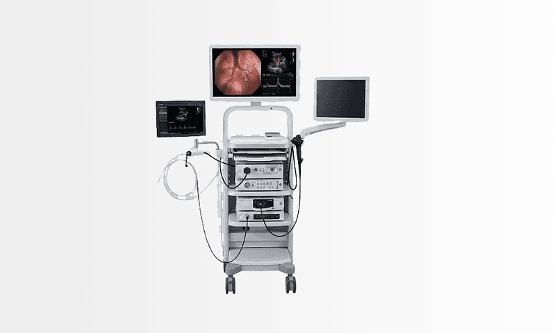
Medical Technologies Used
Available at these Hospitals



















Speak with our medical team
Share your case and our international patient team will guide you to the right specialist, free of charge.