

Radiology
Sub-specialist MRI, CT and ultrasound reporting, image-guided biopsy, and interventional treatment through a puncture rather than an incision, plus a formal second read of imaging performed anywhere else, which needs no travel at all.
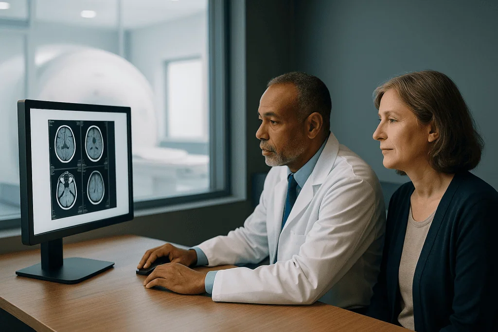
Two specialties sharing one name
Diagnostic radiology produces and interprets the images other specialties depend on. Interventional radiology uses those same images to treat, reaching a tumour, a bleeding vessel or a blocked duct through a puncture rather than an incision.
What the imaging finds
Where the scan and its reading determine the diagnosis, the stage and the treatment, and where a sub-specialist read differs from a general one.
Liver, kidney and abdomen
The area where non-invasive measurement has replaced biopsy for monitoring, and where structured scoring can establish a diagnosis from imaging alone.
What we treat through a needle
Embolisation, ablation and drainage performed under imaging guidance, frequently replacing an operation or converting an emergency into a planned procedure.
In this unit, the data travels instead of the patient
Our largest single service to international patients is not producing new images but re-reading existing ones. A formal sub-specialist second read changes something more often than people expect, not because the first radiologist was careless, but because pattern recognition in imaging is built on volume within a narrow domain, and because the second reader usually has the prior imaging that the first did not.
Two honest limits go with that. A re-read cannot recover information the original examination never captured: if the protocol was wrong or the patient moved, the answer is that the study needs repeating rather than reinterpreting. And a second opinion that agrees with the first is not wasted, independent confirmation is frequently what allows someone to stop searching and start treatment.
What we will not do
- Sell a whole-body screening scan to someone with no symptoms and no risk factor.
- Refuse a necessary scan because of a shellfish allergy or a mildly abnormal creatinine.
- Escalate an incidental finding that published criteria say needs no follow-up.
- Report a scan without knowing the clinical question, because the protocol and the reading both depend on it.
- Ablate a lesion because it is technically reachable. Sometimes the honest answer is that surgery is better.
Radiologists who lead this work
What actually happens, in order
Send the data, not yourself
A formal sub-specialist re-read of imaging performed anywhere else is entirely remote. It changes something more often than people expect, and in the large majority of cases it ends without a journey.
DICOM files, not reports
On a disc, a drive or through a transfer link. A report is a description of the images and cannot be re-read; photographs of a screen are worse. This is the single most common obstacle to a remote opinion.
Include the old imaging
Even from years ago. A finding unchanged over four years and one that appeared six months ago are different diagnoses, and comparison changes interpretation more than any other single factor.
State the clinical question
The protocol is chosen from it, and so is the reading. A scan reviewed without knowing what is being asked produces a confident answer to something else.
If you travel, bring the implant card
Most implants are MR conditional rather than unsafe, which means the scan is possible provided the exact device is known. A study cancelled because nobody could identify a stent is a wasted journey a piece of card would have prevented.
Six things worth knowing first
Shellfish allergy is not a contrast allergy
The connection is a persistent myth with no basis, iodinated contrast has nothing to do with the proteins that cause shellfish allergy. What matters is a previous reaction to contrast itself, which is documented so premedication can be arranged.
Hyperintense does not mean abnormal
It describes brightness relative to surrounding tissue, and normal structures are described that way constantly. The same finding looks white on one sequence and black on another without anything having changed.
Most incidental findings mean nothing
Simple cysts, small adrenal and thyroid nodules, small lung nodules and mild spinal degeneration are found in a large share of adults. Published criteria say which need follow-up and which need none, and applying them prevents a cascade that does real damage.
Whole-body screening is usually a bad idea
In people with no symptoms and no risk factor it generates incidental findings at a rate that outweighs the benefit. Whole-body MRI has genuine indications, myeloma, selected staging, hereditary cancer syndromes, and those are not the marketed package.
An open scanner is not always better
It solves confinement, usually at lower field strength, longer scans and lower resolution. For a knee that rarely matters; for a brain or prostate it can be the difference between a diagnostic and a non-diagnostic study.
Ultrasound depends on who holds the probe
Unlike CT and MRI it largely disappears once the probe is lifted, so nobody can review it afterwards. A hurried negative ultrasound is worth less than a negative CT, and repeating one is legitimate rather than duplication.
Jump to what you came for
Quick answer
Radiology is the medical unit that uses imaging methods to diagnose, monitor, and sometimes guide the treatment of disease and injury throughout the body. At Acibadem in Turkey, radiology services include imaging such as X-ray, ultrasound, CT, MRI, and interventional procedures, with scans interpreted by radiologists and coordinated with other specialties for treatment planning.
What our radiology unit covers, and who it is for
Radiology is two specialties sharing a name. Diagnostic radiology produces and interprets the images that most other specialties depend on: the scan is only as good as the reading, and a substantial share of diagnostic disagreement in medicine is disagreement about what an image shows. Interventional radiology uses those same images to treat, reaching a tumour, a bleeding vessel, a blocked duct or a collection of pus through a needle or a catheter, with no incision.
The unit is unusual in that most of its patients arrive through another specialty rather than directly. That has one consequence worth stating: our largest single service to international patients is not producing new images but re-reading existing ones. A second opinion on a scan already performed changes the diagnosis often enough to be worth requesting before anyone books a flight.
At Acıbadem International the work is organised into five strands.
- Cross-sectional imagingMRI and CT across every body region, including the dedicated protocols that a general scan does not use.
- Ultrasoundthe most operator-dependent modality in medicine, including elastography for liver stiffness and structured thyroid and breast assessment.
- Image-guided biopsyobtaining tissue from anywhere in the body through a needle, accurately and safely.
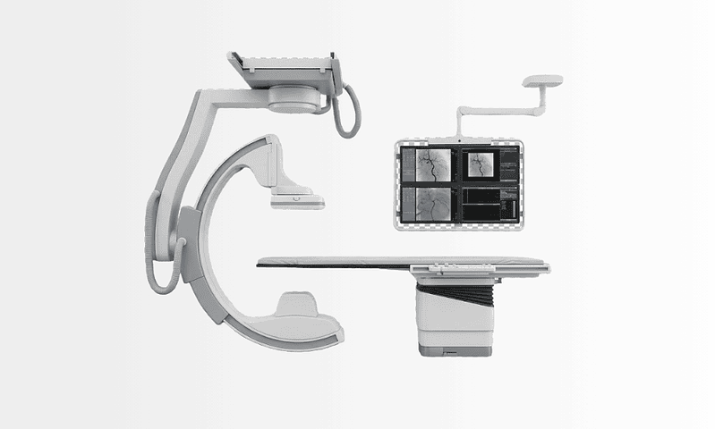
- Interventional radiologyembolisation, ablation, drainage, stenting and vascular access, performed through a puncture rather than an incision.
- Second-opinion reportingsub-specialist re-reading of imaging performed elsewhere, which is the service most international patients actually need.
Where the borders sit. Nuclear medicine, PET-CT, bone scans, thyroid uptake studies and radionuclide therapy, belongs with nuclear medicine, which covers functional imaging and theranostics in full. Mammography, breast ultrasound and breast MRI belong within breast health. Radiotherapy planning belongs with radiation oncology. The MRI lumbar spine report language and the choice of test for a spinal problem are covered by neurosurgery; this unit covers the general principles that apply everywhere else. Vertebral cement augmentation for fracture is performed with neurosurgery.
What a radiologist actually does
A radiologist is a doctor who has trained in interpreting medical images and, increasingly, in treating through them. The work is not looking at pictures; it is answering a clinical question with a probability attached, and the quality of the answer depends heavily on the quality of the question.
Three things separate a good report from a poor one. The first is protocol selection: which sequences, which phases of contrast, which coverage. A scan performed with the wrong protocol produces a confident report about something nobody asked. The second is comparison. A finding that has not changed in four years and a finding that appeared six months ago are different diagnoses, and a radiologist who cannot see the previous imaging is working with one hand tied, which is precisely why old scans matter more than old reports. The third is sub-specialisation. Neuroradiology, musculoskeletal, abdominal, cardiothoracic and paediatric (pediatric) imaging are separate disciplines in practice, and the difference between a general report and a sub-specialist one is frequently the diagnosis itself.
An interventional radiologist does something different again: a procedural specialty performed under imaging guidance, in which the same anatomical knowledge is used to navigate a catheter rather than to describe a finding.
MRI vs CT: which test answers which question
The mri vs ct question is the most common one patients ask, and the answer is not that one is better. They measure different physical properties and each is superior for specific tissues.
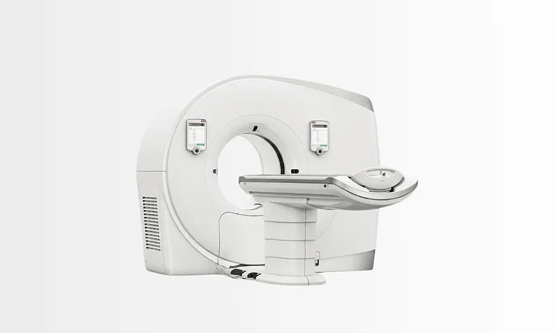
CT uses X-rays and reconstructs a three-dimensional dataset in seconds. It is unmatched for bone detail, for acute haemorrhage (hemorrhage), for lung parenchyma, for the acutely unwell patient who cannot lie still, and for anything urgent: a CT of the abdomen takes moments while an MRI takes half an hour. Its cost is ionising radiation.
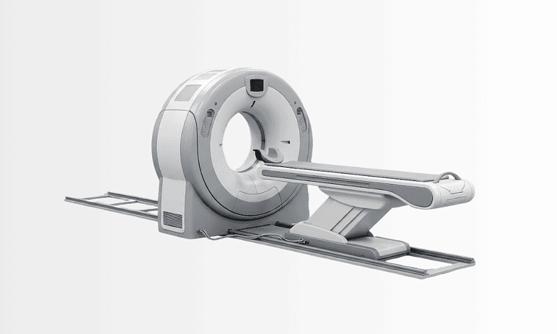
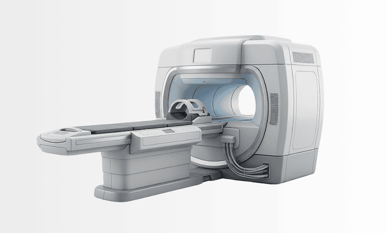
MRI uses a strong magnetic field and radio waves, with no ionising radiation, and produces far superior contrast between soft tissues. It is the test for the brain and spinal cord, for ligaments, cartilage and bone marrow, for the liver, prostate, uterus and rectum, and for anything where distinguishing one soft tissue from another matters. Its costs are time, noise, confinement, and susceptibility to movement: a patient who cannot keep still produces an uninterpretable study.
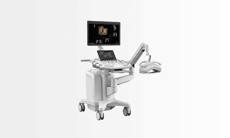
Ultrasound is often the correct first test and is under-used by comparison: no radiation, real-time, cheap, portable, excellent for the gallbladder, kidneys, thyroid, testes, soft-tissue lumps and vessels, and the only modality that can watch something move. Its limitation is that it cannot see through bone or gas and that it depends more on the person holding the probe than any other test in medicine.
The practical rule is that the question determines the test. A radiologist who is told what is actually being asked will frequently suggest a different and cheaper examination than the one requested, and that conversation is part of the service rather than an obstruction of it.
MRI safety, implants and claustrophobia
MRI safety is not a formality. The magnet is always on, including when no scan is running, and it exerts force on ferromagnetic objects with enough energy to cause serious injury. That is why the screening questionnaire is asked repeatedly by different people and why nothing metallic enters the room.
Implants divide into three groups: MR safe, MR conditional, safe only under specified conditions of field strength and scanning parameters, and MR unsafe. Most modern implants including joint replacements, most stents, most surgical clips and increasingly most pacemakers and defibrillators are conditional rather than unsafe, which means the scan is usually possible provided the exact device is known. Bring the implant card. A cochlear implant, certain older aneurysm clips and some neurostimulators remain genuine contraindications, and metallic fragments in the eye are excluded by a plain radiograph in anyone with a history of metal working.
Tattoo MRI concerns are largely overstated: some older inks contain iron oxide and can warm slightly or cause tingling, and this is monitored rather than a reason to cancel. MRI in pregnancy carries no known harmful effect and is performed where the information is needed, though gadolinium contrast is avoided; CT scan in pregnancy is likewise performed when the clinical need justifies it, with dose minimised, because a missed pulmonary embolism is far more dangerous than the radiation.
Claustrophobia and open MRI
Around one patient in ten finds the tunnel difficult and a smaller number cannot complete a scan. Claustrophobia MRI is managed in a sequence rather than by jumping straight to sedation: an explanation of exactly what will happen and how long, prone positioning or feet-first entry where the anatomy allows, a mirror, a companion in the room, music and a call button that is genuinely answered. Wide-bore scanners with a larger aperture solve the problem for the large majority.
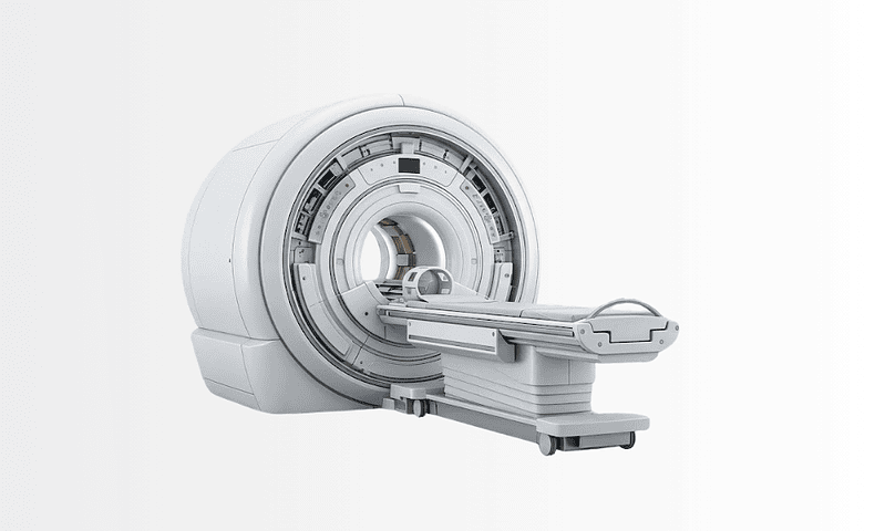
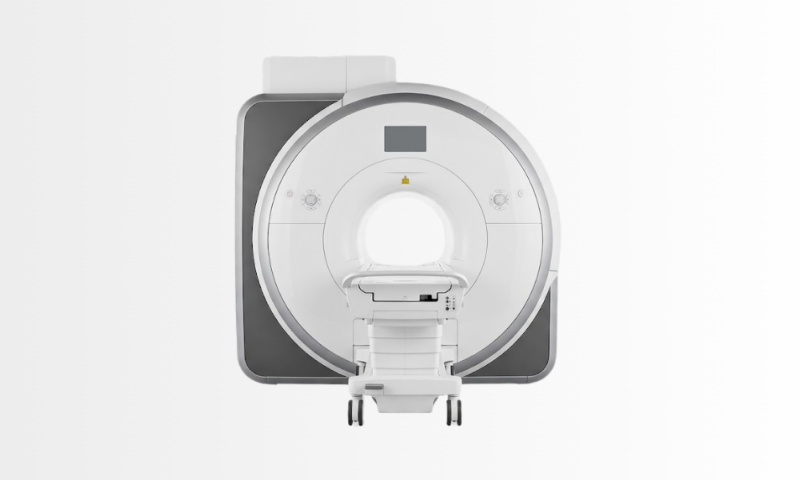
An open MRIan upright or open-sided scanner, solves confinement but usually at a lower field strength, which means longer scans and lower image quality. That trade-off is worth stating plainly rather than presenting open scanners as simply nicer: for a knee it rarely matters, and for a brain or a prostate it can be the difference between a diagnostic and a non-diagnostic study. Sedation is available where all else fails and is arranged with anesthesiology. On field strength, a 3 tesla MRI gives higher signal and finer resolution and is preferred for the brain, prostate and small joints; 1.5 tesla remains better in some situations, particularly around metal implants where higher fields produce worse artefact.
Contrast agents, and the questions worth asking about them
Contrast is given in most CT and many MRI examinations because it separates tissues that otherwise look identical, and the two contrast families are unrelated chemically despite being described interchangeably by patients.
Iodinated contrast is used for CT, injected intravenously and excreted by the kidneys. Reactions are usually mild, warmth, a metallic taste, a sensation of having passed urine, which is expected rather than a reaction at all. True allergic-type reactions are uncommon and severe ones rare, and a previous reaction is documented so that premedication or an alternative examination is arranged. A contrast dye allergy to iodinated contrast does not predict a reaction to gadolinium, and neither predicts a reaction to shellfish: the shellfish connection is a persistent myth with no basis, and it should not be the reason a scan is refused.
Contrast induced nephropathy has been substantially reconsidered. The risk in patients with normal kidney function is now understood to be far smaller than was believed, and the practice of withholding a necessary scan on the basis of a mildly abnormal creatinine has caused more harm than the contrast would have. Kidney function is checked in those with risk factors, hydration is optimised, and the decision weighs the risk against the cost of not knowing. Metformin is not a contraindication to contrast; the guidance concerns when it is restarted afterwards, and that is a decision for the prescribing doctor.
Gadolinium contrast is used for MRI. Nephrogenic systemic fibrosis, a serious complication in patients with severe kidney impairment, has become extremely rare with modern macrocyclic agents and appropriate screening. Small amounts of gadolinium are retained in tissue including the brain after repeated administration; no clinical harm has been demonstrated, and the honest position is that it is used when it adds information and avoided when it does not. Contrast extravasationleakage into the tissues at the injection site, causes swelling and discomfort, usually settles with elevation and cold, and is recognised early precisely because the injection is watched.
Reading your own radiology report
A radiology report is written for the referring doctor rather than for the patient, and that produces predictable alarm. A few conventions are worth knowing.
Hyperintense and hypointense describe brightness on MRI relative to surrounding tissue; hypodense and hyperdense do the same on CT. None of these words means abnormal: they describe appearance, not pathology. On MRI, T1 vs T2 weighting is the most useful thing a patient can understand: broadly, fat is bright and fluid dark on T1, while fluid is bright on T2, which is why the same structure looks opposite on two images of the same scan. Diffusion weighted imaging measures how freely water molecules move and is exquisitely sensitive to acute stroke, to abscess and to some tumours, which is why a lesion described as showing restricted diffusion carries specific meaning.
Hedged language is the norm rather than evasion. Radiologists write in probabilities because images show appearances and not diagnoses, and a report that reads as certain about something images cannot establish is a worse report rather than a better one.
Incidental findings
An incidental finding is something discovered on a scan performed for an unrelated reason, and it is extremely common: the more of the body is imaged and the better the resolution, the more will be found. Simple kidney and liver cysts, small adrenal nodules, benign thyroid nodules, small lung nodules, uterine fibroids and mild spinal degeneration are found in a large share of adults and are frequently of no significance whatsoever.
The problem is what follows: a cascade of further scans, biopsies and anxiety triggered by something that was never going to cause harm. Published criteria exist for most common incidental findings, specifying which need follow-up, at what interval, and which need none at all, and applying them is one of the most valuable and least visible things this unit does. This is also the strongest argument against whole-body screening scans in people without symptoms, which are marketed heavily and generate incidental findings at a rate that reliably outweighs their benefit for most people.
BI-RADS, LI-RADS, PI-RADS and the other scores
Structured reporting systems convert an impression into a category with a defined management recommendation attached, and they exist because free-text reports were interpreted inconsistently. Knowing what the number means removes most of the fear attached to receiving one.
- BI-RADS classifies breast imaging from 0 to 6. Categories 1 and 2 are normal and benign; category 3 is probably benign with short-interval follow-up; 4 is suspicious and subdivided; 5 is highly suggestive of malignancy. Breast imaging is delivered within breast health.
- LI-RADS classifies liver observations in patients at risk of hepatocellular carcinoma, and it matters because in this context imaging alone can establish the diagnosis without a biopsy, one of the few places in medicine where that is true.
- PI-RADS scores prostate MRI from 1 to 5 for the likelihood of clinically significant cancer, and it determines whether and where a biopsy is taken. Work is shared with urology.
- Lung RADS (Lung-RADS) classifies findings on low-dose CT lung cancer screening, standardising which nodules need what follow-up. Nodule management belongs with thoracic surgery.
- TIRADS (TI-RADS) scores thyroid nodules on ultrasound by composition, echogenicity, shape, margin and calcification, producing a recommendation on whether to biopsy at all, which has substantially reduced unnecessary thyroid biopsies.
- O-RADS does the same for ovarian and adnexal lesions, with gynecology.
A category is a management recommendation rather than a verdict, and the number in the report is the beginning of a conversation rather than the end of one.
Ultrasound and elastography
Ultrasound deserves more respect than it receives. It is the first test for the gallbladder and biliary tree, for the kidneys and bladder, for the thyroid, for the testes, for soft-tissue lumps, for the neonatal brain and hips, and for anything vascular. It answers many questions completely, at no radiation cost, in minutes.
Its weakness is operator dependence, and this is the single most important thing to understand about it: unlike CT and MRI, which produce a dataset that can be reviewed by anyone afterwards, an ultrasound is largely gone once the probe is lifted. A negative ultrasound performed hurriedly by an inexperienced operator is worth considerably less than a negative CT, and repeating a study rather than accepting a previous negative is a legitimate decision rather than duplication.
Liver elastography and FibroScan
Ultrasound elastography measures tissue stiffness rather than appearance, and in the liver stiffness correlates with fibrosis. FibroScantransient elastography, and shear wave elastography built into standard ultrasound machines both estimate fibrosis without a biopsy, and their arrival changed hepatology: patients with fatty liver disease and viral hepatitis who once needed serial biopsies are now monitored non-invasively. Liver elastography is quick, painless and repeatable, which is what makes surveillance practical.
Its limits are worth knowing. Acute inflammation, heart failure, a recent meal and severe obesity all raise the reading independently of fibrosis, so it is performed fasted and interpreted alongside blood tests. A single high reading is not cirrhosis; the trend and the clinical picture are. Fatty liver ultrasound detects steatosis but grades it crudely, which is why quantitative techniques are increasingly used alongside. This work is shared with gastroenterology.
Carotid ultrasound and musculoskeletal ultrasound
Carotid ultrasound grades narrowing of the carotid arteries and is the first-line test after a stroke or transient ischaemic attack, with the degree of stenosis determining whether intervention is considered: that decision belongs with neurology and cardiovascular surgery. Doppler studies of the leg veins remain the test for deep vein thrombosis.
Musculoskeletal ultrasound examines tendons, ligaments, bursae and small joints dynamically, moving the joint while watching, which no static scan can do, and it guides injections into the exact space rather than approximately, which is why ultrasound-guided injection outperforms landmark technique.
MRCP, cardiac MRI and the other MRI examinations by name
Most patients are sent for “an MRI” without being told that the protocol differs enormously by question. Several are worth naming because requesting the wrong one wastes a journey.
MRCPmagnetic resonance cholangiopancreatography, images the bile ducts and pancreatic duct without contrast and without instrumentation, and it has largely replaced diagnostic ERCP, which is now reserved for cases needing treatment at the same sitting. It is the test for a suspected bile duct stone, for pancreatic duct abnormalities and for biliary strictures, arranged with gastroenterology.
MRI prostate, performed multiparametrically and reported with PI-RADS, is now done before biopsy rather than after, so that biopsies are targeted at suspicious areas, and so that men with a normal scan may avoid biopsy altogether. Cardiac MRI assesses heart muscle characteristics that echocardiography cannot: scar, infiltration, inflammation and iron loading, which makes it the reference test for cardiomyopathy, with cardiology. MR enterography images the small bowel for Crohn disease without radiation, which matters in young patients facing decades of surveillance.
An MRI arthrogram injects dilute contrast into a joint before scanning, distending the capsule so that subtle labral and cartilage tears become visible: it is the test for shoulder and hip labral pathology and is considerably more sensitive than a plain MRI for that specific question, with orthopedics. A whole body MRI has genuine indications, myeloma, cancer staging in selected tumours, and screening in defined hereditary cancer syndromes, and is a different proposition entirely from the commercially marketed screening scan in a person with no symptoms and no risk factor.
CT angiography, the coronary calcium score and the other CT examinations
CT angiography images arteries anywhere in the body after a timed contrast injection, and it has replaced diagnostic catheter angiography almost entirely, catheters are now reserved for treatment. It is the test for pulmonary embolism, aortic dissection and aneurysm, and for planning vascular intervention.
The coronary calcium score is a fast, contrast-free, low-dose CT that quantifies calcified plaque in the coronary arteries, and it is one of the most useful tests in preventive cardiology because it reclassifies risk in people whose calculated risk sits in the middle: a score of zero is strongly reassuring. It is interpreted with cardiology.
CT colonography images the colon after gas insufflation and is the alternative for patients who cannot tolerate or complete colonoscopy; it still requires bowel preparation, and anything found needs colonoscopy to remove it. CT urography images the kidneys, ureters and bladder in a delayed excretory phase and is the standard investigation for visible haematuria, with urology.
On radiation, the honest framing is proportion rather than reassurance. CT scan radiation dose varies by examination, and the increase in lifetime cancer risk from a single clinically indicated CT is very small in an adult and is outweighed by the harm of missing what it was ordered to find. Dose matters most in children and in people having repeated scans, which is where dose reduction, alternative modalities and asking whether the scan is needed at all carry real weight.
Image-guided biopsy
Obtaining tissue is where diagnosis usually ends, and imaging has made that possible almost anywhere in the body through a needle rather than an operation. A CT guided biopsy reaches lesions in the lung, abdomen, retroperitoneum and bone; ultrasound guides biopsies of the liver, kidney, thyroid, lymph nodes and superficial masses in real time, watching the needle move.
Liver biopsy is performed under ultrasound guidance rather than blindly, which reduced complications substantially, and where clotting is abnormal or ascites is present a transjugular approach through the neck vein allows biopsy that would otherwise be unsafe. Thyroid biopsyfine needle aspiration under ultrasound, is quick, needs only local anaesthetic and is directed by the TI-RADS category rather than by nodule size alone.
Fusion biopsy of the prostate overlays the MRI onto live ultrasound so that the needle targets the area the MRI identified, rather than sampling systematically and hoping. It detects clinically significant cancer more reliably and finds fewer of the insignificant ones, which is exactly the right direction, and it is performed with urology.
Two principles govern all of it. The sample must reach the laboratory in the form the pathologist needs, some tests require fresh tissue, some require specific fixation, and agreeing that with pathology before the needle goes in prevents a repeat procedure. And the safest route is not always the shortest: a longer path that avoids bowel, pleura or major vessels is the correct one.
Drainage, nephrostomy, stenting and vascular access
This is the least glamorous and most consistently valuable part of interventional radiology: it replaces operations.
Percutaneous drainage of an abscess or collection, abdominal, pelvic, chest or liver, is performed under CT or ultrasound guidance through a small puncture, and it has changed the management of intra-abdominal sepsis fundamentally. A collection that once required laparotomy is now drained through a catheter with the patient awake, often avoiding surgery entirely or converting an emergency operation into a planned one. It is done jointly with general surgery and infectious diseases.
Nephrostomy places a tube directly into an obstructed kidney through the back, relieving obstruction and preserving renal function when a stent cannot be passed from below: it is frequently urgent, since an obstructed and infected kidney is a medical emergency treated in hospital. Biliary drainage does the same for an obstructed bile duct where ERCP has failed or is not possible, and a biliary stent can then be placed internally so the patient carries no external bag. This work is shared with urology and gastroenterology.
Vascular access is the quiet backbone of oncology and long-term treatment. Tunnelled central lines, PICC lines and implanted ports are placed under imaging guidance with ultrasound for the vein and fluoroscopy for the tip position, which is safer and more accurate than landmark insertion. Port a cath insertion is a day-case procedure under local anaesthetic and sedation, and the device can stay for years, for someone facing months of chemotherapy with medical oncology, it is the difference between treatment being tolerable and being dreaded.
Embolisation: interventional radiology without an incision
Embolisation blocks a blood vessel deliberately, and it treats an unexpectedly wide range of conditions because so much pathology depends on its blood supply. The catheter is passed from the wrist or groin, the target vessel is selected, and particles, coils, glue or drug-loaded beads are delivered precisely into it.
Uterine fibroid embolization blocks the arteries supplying fibroids, shrinking them and improving bleeding and pressure symptoms while preserving the uterus. It avoids surgery, has a shorter recovery than hysterectomy or myomectomy, and is a genuine alternative rather than a lesser option, though it is less suitable for women planning pregnancy and for certain fibroid positions. The choice is made with gynecology, with both options presented rather than one.
Prostate artery embolization reduces prostate volume in benign enlargement and is an option for men who are unsuitable for or wish to avoid surgery, particularly with very large glands or on anticoagulation. Its trade-off is honest: it preserves ejaculatory function better than standard surgical resection and produces a smaller improvement in flow, and it is discussed with urology rather than presented as equivalent.
Varicocele embolization treats a varicocele through a vein in the neck or groin with no scrotal incision, with comparable results to surgery and faster recovery. Genicular artery embolization targets the abnormal vessels that accompany inflammation in knee osteoarthritis, reducing pain in selected patients with mild to moderate disease who have not responded to conservative treatment: the evidence base is still developing, and it is described that way rather than as established, with orthopedics.
Transarterial chemoembolization delivers chemotherapy directly into the arteries feeding a liver tumour and blocks them at the same time, concentrating the drug where it is needed and sparing the rest of the body. It is a standard treatment for intermediate-stage hepatocellular carcinoma and is decided at a multidisciplinary meeting rather than by any single specialty. Embolisation also stops bleeding, after trauma, from a bleeding ulcer or tumour, in postpartum haemorrhage and in massive haemoptysis, and in that setting it is genuinely life-saving and frequently faster than surgery.
Tumour ablation: radiofrequency ablation and microwave ablation
Radiofrequency ablation and microwave ablation destroy tumours with heat delivered through a needle placed under imaging guidance. Microwave heats faster, reaches higher temperatures and is less affected by nearby blood vessels carrying heat away, which is why it has largely replaced radiofrequency for liver work. Cryoablation freezes instead, and its advantage is that the ice ball is visible during the procedure, which helps near critical structures.
These are curative-intent treatments in the right patients, not palliative ones. Small hepatocellular carcinomas, selected liver metastases, small renal tumours in patients unsuitable for surgery, lung metastases and painful bone metastases are the main indications. In small renal cancers ablation offers comparable cancer control to surgery with better preservation of kidney function in appropriately selected patients.
Selection is everything, and it is made at a multidisciplinary meeting: size, number, position relative to bile ducts, bowel and large vessels, and the underlying liver or kidney function. Ablation of a lesion adjacent to a bile duct or the bowel is not attempted simply because it is technically reachable, and the honest answer in some cases is that surgery is the better option.
Vascular intervention
A TIPS proceduretransjugular intrahepatic portosystemic shunt, creates a channel through the liver between the portal and hepatic veins, relieving the high pressure that causes variceal bleeding and refractory ascites in cirrhosis. It is performed entirely through a neck vein with no incision, and it is one of the clearest examples of interventional radiology replacing major surgery. Its main trade-off is that diverting blood past the liver can precipitate encephalopathy, which is why selection and post-procedure monitoring with gastroenterology matter as much as the technique.
An IVC filter is placed in the inferior vena cava to catch clot travelling from the legs towards the lungs, and it is indicated in a narrow situation: venous thromboembolism in a patient who genuinely cannot be anticoagulated. The important development is retrievability. Filters left in place indefinitely cause their own complications, and the single most common failure in this area is a temporary filter that nobody ever removed, so a retrieval date is set when the filter is inserted, and it is tracked with hematology.
Angioplasty and stenting of narrowed arteries, thrombolysis and thrombectomy for acute limb ischaemia, and treatment of vascular malformations complete the vascular work, shared with cardiovascular surgery. Vertebroplasty and kyphoplasty stabilise painful osteoporotic and malignant vertebral fractures with injected cement; patient selection is the whole of the debate in this area and the procedure is performed with neurosurgery.
Second-opinion reporting on imaging done elsewhere
This is the service most international patients actually need, and the one they are least often offered. A radiology second opinion is a formal sub-specialist re-read of imaging already performed, producing a new written report rather than an informal comment.
It changes something more often than people expect, and for identifiable reasons rather than because the first radiologist was careless. A general radiologist and a sub-specialist reading the same prostate MRI, liver CT or musculoskeletal study genuinely see different things, because pattern recognition in imaging is built on volume within a narrow domain. Prior imaging is frequently unavailable to the first reader and available to the second, and comparison changes interpretation more than any other single factor. And the clinical question sometimes changes after the scan, which means the images are being asked a question nobody asked when they were reported.
What it needs is the images themselves in DICOM format on a disc, a drive or through a transfer link, not a report, and not photographs of a screen. The original report should come too, along with the clinical question and any previous imaging, however old. Teleradiology makes the process remote end to end: nothing has to travel except the data, and a written report follows.
Two honest limits. A re-read cannot recover information the original examination did not capture, if the protocol was wrong, if the phases of contrast were missed, or if the patient moved, the answer may be that the study needs repeating rather than reinterpreting. And a second opinion that agrees with the first is not a wasted exercise: confirmation from an independent sub-specialist is frequently what allows a patient to proceed with treatment rather than continue searching.
What radiology cannot do
- It cannot answer a question nobody asked. The protocol is chosen from the clinical question, and a scan requested without one produces a confident report about something else.
- It cannot make a diagnosis from appearance alone. Images show appearances; diagnoses come from combining them with the history, examination and laboratory results. Hedged language is accuracy rather than evasion.
- It cannot interpret an ultrasound it did not perform. Unlike CT and MRI, an ultrasound largely disappears when the probe is lifted, which is why repeating one is legitimate rather than duplication.
- It cannot recover a poor examination by re-reading it. Wrong protocol, missed contrast phase, patient movement: a second opinion on those concludes that the study needs repeating.
- It cannot make screening the whole body a good idea. Whole-body scans in people without symptoms or risk factors generate incidental findings at a rate that reliably outweighs the benefit for most people, and we say so rather than sell them.
- It cannot treat what it cannot reach safely. A lesion adjacent to bile duct or bowel is not ablated because it is technically reachable; sometimes the honest answer is that surgery is better.
Your multidisciplinary team
The diagnostic radiologist selects the protocol, interprets the images and writes the report, sub-specialised by body region, because the difference between a general and a sub-specialist read is frequently the diagnosis. The interventional radiologist performs the procedures and, importantly, runs a clinic: modern interventional radiology sees patients before and after rather than only during, because consent, selection and follow-up belong to whoever holds the catheter. The radiographer and sonographer acquire the images, and in ultrasound the operator largely determines the result. The medical physicist manages radiation dose optimisation and MRI safety.
Around them: nuclear medicine for PET-CT and functional imaging, pathology for every sample obtained, medical oncology and radiation oncology at the tumour boards where imaging drives staging, general surgery for drainage and for the operations that imaging avoids, gastroenterology for biliary and portal work, urology for prostate and renal imaging and intervention, gynecology for fibroid treatment, cardiology for cardiac imaging and calcium scoring, neurosurgery for spinal and neurovascular work, orthopedics for musculoskeletal imaging and guided injection, and anesthesiology for sedation.
The international patient journey
Radiology travels better than any other specialty on this site, because in most cases the data travels instead of the patient.
The first and largest pattern is the remote second opinion set out under second-opinion reporting. It needs DICOM files rather than reports, the original report, the clinical question, and any prior imaging however old, and it concludes without a journey in the large majority of cases.
The second is a diagnostic visit where the imaging needed does not exist locally or was performed with the wrong protocol: multiparametric prostate MRI, cardiac MRI, MR enterography, MRCP, dedicated musculoskeletal protocols, or CT with a specific phase of contrast. These are arranged as a block over a few days with the interpretation done by the people who set the protocol.
The third is interventional treatmentfibroid or prostate embolisation, tumour ablation, TIPS, complex drainage or port insertion. These are planned rather than booked, because the assessment determines whether the procedure is appropriate at all, and because most need overnight admission and a defined interval before flying.
Three practical notes. Bring the discs, not the reports: the single most common frustration in this unit is a patient who has travelled with a folder of paper. Bring the old imaging too, even from years ago, because comparison changes interpretation more than any other factor. And if you are having an MRI, bring the implant card for any device you carry: a scan cancelled because nobody could identify a stent is a wasted journey that a piece of card would have prevented.
Frequently Asked Questions
Is an MRI better than a CT?
Neither is better; they measure different physical properties and each wins for specific tissues. CT is unmatched for bone detail, acute bleeding, the lungs and anything urgent, and it takes seconds, at the cost of ionising radiation. MRI has no radiation and far superior soft-tissue contrast, making it the test for the brain and spinal cord, ligaments and cartilage, the liver, prostate, uterus and rectum, at the cost of time, noise and sensitivity to movement. Ultrasound is often the right first test and is under-used. The question determines the test, which is why telling the radiologist what is actually being asked frequently produces a different and cheaper examination.
Can I have an MRI with a metal implant?
Usually yes. Implants are classified as MR safe, MR conditional, safe under specified conditions, or MR unsafe, and most modern implants including joint replacements, most stents and clips, and increasingly most pacemakers and defibrillators are conditional rather than unsafe. That means the scan is generally possible provided the exact device is known, so bring the implant card. Genuine contraindications remain, including cochlear implants, certain older aneurysm clips and some neurostimulators, and anyone with a history of metal working has a plain radiograph first to exclude metallic fragments in the eye.
Are tattoos a problem for MRI?
Far less than the internet suggests. Some older inks contain iron oxide and can warm slightly or produce a tingling sensation during scanning; this is monitored rather than treated as a reason to cancel, and modern inks are largely inert. Tell the radiographer where the tattoos are and say immediately if you feel warmth so the sequence can be adjusted. Permanent makeup is handled the same way. A tattoo is not a reason to refuse a scan you need.
I am claustrophobic. What are my options?
Several, and sedation is the last of them rather than the first. A clear explanation of exactly what happens and how long each sequence lasts resolves a great deal. Feet-first entry or prone positioning works where the anatomy allows; a mirror, a companion in the room, music and a call button that is genuinely answered all help. Wide-bore scanners with a larger aperture solve the problem for the large majority. An open scanner solves confinement but usually at lower field strength, which means longer scans and lower image quality: a real trade-off rather than simply a nicer machine.
Is an open MRI as good as a closed one?
Usually not, and it depends entirely on what is being scanned. Open and upright scanners generally operate at lower field strength, which means less signal, longer acquisitions and lower resolution. For a knee that rarely matters. For a brain, a prostate or a small joint it can be the difference between a diagnostic and a non-diagnostic study. If confinement is the only obstacle, a wide-bore high-field scanner is usually the better answer than a low-field open one, and that is a choice worth asking about specifically rather than accepting whichever machine is available.
I am allergic to shellfish. Can I have contrast?
Yes. The shellfish connection is a persistent myth with no basis, iodinated contrast has nothing to do with the proteins that cause shellfish allergy, and no link has ever been demonstrated. What does matter is a previous reaction to contrast itself, which is documented so that premedication or an alternative examination can be arranged. It is also worth knowing that a reaction to CT contrast does not predict a reaction to MRI contrast; they are chemically unrelated families.
Is contrast bad for my kidneys?
The risk has been substantially reconsidered and is far smaller than was believed for people with normal kidney function. Withholding a necessary scan because of a mildly abnormal creatinine has caused more harm than the contrast would have. Kidney function is checked in those with risk factors, hydration is optimised, and the decision weighs the risk against the cost of not knowing. Metformin is not a contraindication to contrast either: the guidance concerns when it is restarted afterwards, and that is a decision for the doctor who prescribed it.
Does gadolinium stay in the body?
Small amounts are retained in tissue including the brain after repeated administration, and that is established. What has not been demonstrated is any clinical harm from it. Nephrogenic systemic fibrosis, the serious complication in patients with severe kidney impairment, has become extremely rare with modern macrocyclic agents and appropriate screening. The honest position is the one we apply: gadolinium is used when it adds information the scan cannot give without it, and left out when it does not.
My report says hyperintense. Does that mean something is wrong?
No. Hyperintense and hypointense describe brightness on MRI relative to surrounding tissue, and hypodense and hyperdense do the same on CT. They describe appearance, not pathology, normal structures are described this way constantly. The related point is that a structure looks opposite on different sequences of the same scan: broadly, fluid is dark on T1 and bright on T2, which is why the same finding appears white on one image and black on another without anything having changed.
Why is the report so hedged? Can they not just say what it is?
Because images show appearances and not diagnoses, and a report that sounds certain about something imaging cannot establish is a worse report rather than a better one. Radiologists write in probabilities and differential lists deliberately. What resolves the uncertainty is context the images do not contain: your history, your examination, your blood results and, above all, comparison with previous imaging. That is why a report addressed to your doctor reads differently from what you might want it to say.
They found something unrelated on my scan. What now?
Very often nothing. Incidental findings are extremely common, simple kidney and liver cysts, small adrenal and thyroid nodules, small lung nodules, fibroids and mild spinal degeneration are found in a large share of adults and are frequently of no significance. Published criteria exist for most of them, specifying which need follow-up, at what interval, and which need none at all, and applying those criteria rather than escalating automatically is one of the most valuable things this unit does. The cascade of further scans and biopsies triggered by a harmless finding does real damage.
Should I have a whole-body screening scan?
For most people with no symptoms and no specific risk factor, no, and we will say so rather than sell one. Whole-body screening generates incidental findings at a rate that reliably outweighs the benefit, producing anxiety, further scans and occasionally biopsies for things that were never going to cause harm. Whole-body MRI does have genuine indications: myeloma, staging in selected cancers and surveillance in defined hereditary cancer syndromes. Those are different from a marketed screening package, and the distinction is worth insisting on.
What does my BI-RADS or PI-RADS number mean?
These are structured scores that attach a defined management recommendation to an impression, and they exist because free-text reports were read inconsistently. Broadly, low numbers mean normal or benign, the middle means short-interval follow-up, and high numbers mean suspicious enough to sample. PI-RADS scores prostate MRI from 1 to 5 for the likelihood of clinically significant cancer and determines whether and where to biopsy; TI-RADS does the same for thyroid nodules and has substantially reduced unnecessary biopsies. The number is a management recommendation, not a verdict.
How reliable is ultrasound compared with a scan?
It answers many questions completely and is the correct first test for the gallbladder, kidneys, thyroid, testes, soft-tissue lumps and vessels. Its distinctive weakness is operator dependence: unlike CT and MRI, which produce a dataset anyone can review afterwards, an ultrasound largely disappears once the probe is lifted. A negative ultrasound performed hurriedly by an inexperienced operator is worth considerably less than a negative CT, which is why repeating one is a legitimate decision rather than duplication.
What is a FibroScan and can it replace a liver biopsy?
It measures liver stiffness, which correlates with fibrosis, using either transient elastography or shear wave elastography built into a standard ultrasound machine. For monitoring fatty liver disease and viral hepatitis it has largely replaced serial biopsies, because it is quick, painless and repeatable. Its limits matter: acute inflammation, heart failure, a recent meal and severe obesity all raise the reading independently of fibrosis, so it is done fasted and read alongside blood tests. A single high reading is not cirrhosis: the trend and the clinical picture are.
What is MRCP and why was I sent for one instead of an ERCP?
MRCP images the bile ducts and pancreatic duct with MRI, without contrast injection into the ducts and without passing an endoscope. It has largely replaced diagnostic ERCP precisely because it carries none of the procedural risk: ERCP is now reserved for cases where something needs treating at the same sitting, such as removing a stone or placing a stent. If MRCP shows a duct stone that needs removal, an ERCP follows; if it shows a normal duct, you have avoided a procedure with a real complication rate.
Why do I need an MRI before a prostate biopsy?
Because it changes both whether you are biopsied and where. Multiparametric prostate MRI reported with PI-RADS identifies areas suspicious for clinically significant cancer, so biopsies can be targeted at them rather than taken systematically and hoping. Men with a reassuring scan may avoid biopsy altogether. Fusion biopsy overlays the MRI onto live ultrasound so the needle reaches the identified area, which detects more significant cancers and fewer insignificant ones, which is the right direction on both counts.
Can a biopsy be done through a needle instead of an operation?
Almost anywhere in the body, yes. CT guidance reaches lung, abdominal, retroperitoneal and bone lesions; ultrasound guides liver, kidney, thyroid, lymph node and superficial biopsies in real time with the needle visible throughout. Two things determine success beyond technique: the sample must reach the laboratory in the form the pathologist needs, which is agreed before the needle goes in, and the safest route is not always the shortest: a longer path avoiding bowel, pleura or major vessels is the correct one.
Can an abscess be drained without surgery?
Usually. Percutaneous drainage under CT or ultrasound guidance places a catheter through a small puncture with the patient awake, and it has changed the management of intra-abdominal infection fundamentally, collections that once required an open operation are now drained through a tube, frequently avoiding surgery entirely or converting an emergency operation into a planned one. It is done jointly with the surgical and infection teams rather than instead of them, and where drainage is not enough the operation still happens.
What is uterine fibroid embolisation and is it as good as surgery?
It blocks the arteries supplying the fibroids so they shrink, improving bleeding and pressure symptoms while preserving the uterus, and it is performed through a puncture at the wrist or groin with a shorter recovery than hysterectomy or myomectomy. It is a genuine alternative rather than a lesser option, but it is less suitable for women planning pregnancy and for certain fibroid positions, and it does not remove the fibroids. The right approach is decided with gynaecology, with both options presented properly rather than whichever the person you are sitting in front of performs.
What is prostate artery embolisation for?
Reducing prostate size in benign enlargement, particularly for men who are unsuitable for surgery, who are on anticoagulation, or who have a very large gland. Its trade-off should be stated plainly: it preserves ejaculatory function better than standard surgical resection, and it produces a smaller improvement in urinary flow. That is not equivalence, and it is discussed with urology rather than presented as the same operation without the surgery.
Can a tumour be destroyed with a needle?
In selected cases, with curative intent rather than as palliation. Microwave and radiofrequency ablation destroy tissue with heat delivered through a needle placed under imaging guidance; cryoablation freezes instead, with the advantage that the ice ball is visible during the procedure. The main indications are small liver cancers, selected liver metastases, small renal tumours in patients unsuitable for surgery, lung metastases and painful bone metastases. Selection is everything and is made at a multidisciplinary meeting: a lesion is not ablated simply because it is technically reachable.
What is a TIPS procedure?
It creates a channel through the liver connecting the portal and hepatic veins, relieving the high portal pressure that causes variceal bleeding and refractory ascites in cirrhosis. It is performed entirely through a vein in the neck with no incision, and it is one of the clearest cases of interventional radiology replacing major surgery. Its main trade-off is that diverting blood past the liver can precipitate encephalopathy, which is why patient selection and follow-up with the hepatology team matter as much as the technical result.
I had an IVC filter put in years ago. Does it need to come out?
Very possibly, and this is worth pursuing rather than assuming someone is tracking it. Filters left in place indefinitely cause their own complications, and a temporary filter that nobody removed is the single most common failure in this area. Modern practice sets a retrieval date at the moment of insertion. If you have one and do not know its status, that is a reasonable thing to ask about specifically, because retrieval becomes more difficult the longer a filter stays.
How much radiation is a CT scan?
It varies by examination, and the honest framing is proportion rather than reassurance. The increase in lifetime cancer risk from a single clinically indicated CT in an adult is very small, and it is outweighed by the harm of missing what the scan was ordered to find. Dose matters most in children, in young adults and in anyone having repeated scans, and that is where dose-reduction techniques, choosing ultrasound or MRI instead, and asking whether the scan is needed at all carry real weight rather than being reflexive caution.
Can I have a scan while pregnant?
MRI carries no known harmful effect and is performed when the information is needed, though gadolinium contrast is avoided. CT is also performed when the clinical need justifies it, with dose minimised, because a missed pulmonary embolism or appendicitis is considerably more dangerous to both mother and baby than the radiation involved. Ultrasound is the first choice wherever it can answer the question. What is never appropriate is refusing a necessary investigation on principle, and the decision is made case by case rather than by rule.
Is it worth getting a second opinion on a scan I have already had?
Frequently, and it is the service international patients most often need and least often get. A sub-specialist reading the same prostate MRI, liver CT or musculoskeletal study genuinely sees different things from a general radiologist, because pattern recognition is built on volume within a narrow domain. Prior imaging is often available to the second reader and not the first, and comparison changes interpretation more than anything else. It requires no travel, only the data.
What exactly do you need for a remote second opinion?
The images themselves in DICOM format, on a disc, a drive or through a transfer link, not a report and not photographs of a screen. Alongside them: the original report, the clinical question being asked, and any previous imaging however old, because comparison is the single most powerful factor in interpretation. Where all of that is available, the process is entirely remote and a written report follows.
What if the second opinion just agrees with the first?
That is a useful outcome rather than a wasted one. Independent confirmation from a sub-specialist is frequently what allows someone to stop searching and proceed with treatment, and that has real value. The other honest limit is different: a re-read cannot recover information the original examination never captured. If the protocol was wrong, a contrast phase was missed or the patient moved, the answer will be that the study needs repeating rather than reinterpreting, and saying so is part of the service.
What should I bring if I am travelling for imaging?
Discs rather than paper: the most common frustration in this unit is a patient who has travelled with a folder of reports and no images. Bring the old imaging too, even from years ago, because comparison changes interpretation more than any other factor. Bring the implant card for any device you carry, since a scan cancelled because nobody could identify a stent is a wasted journey a piece of card would have prevented. And bring the clinical question in writing, because the protocol is chosen from it.
Scan & check-up costsledger-based guide ranges, or browse the full Turkey Medical Price Index.
Medically reviewed by the Acıbadem International Medical Board September 26, 2026
See our medical review board →
Update history
- PublishedJune 7, 2026
- Medical review approvedSeptember 26, 2026
- Last content updateSeptember 25, 2026
Add us as a Preferred Source to see more of our trusted health content across Google Search, AI Overviews and Discover.
Treatments in Radiology
Specialists in this Unit










Medical Technologies Used
Available at these Hospitals























What patients say about this unit
★★★★★From 2,400+ verified patient reviews“I travelled from abroad for interventional radiology procedure and the whole experience was seamless. Prof. Kara inspired complete confidence from the first meeting. The whole experience was organised and reassuring.”
“I was nervous about my interventional radiology procedure, but I felt safe at every single step. I cannot thank Dr. Özgür enough for the skill and kindness shown.”
“My interventional radiology procedure was handled with real skill and genuine care. The expertise of Prof. Dr. Parildar put my mind completely at ease. The follow-up care was just as good as the treatment itself.”
“Choosing Acibadem for my interventional radiology procedure was the best decision I made. Prof. Saçan inspired complete confidence from the first meeting. I would happily travel here again for any treatment.”
Speak with our medical team
Share your case and our international patient team will guide you to the right specialist, free of charge.